
Fabrication of Ciprofloxacin-Immobilized Calcium Phosphate Particles for Dental Drug Delivery
 , , , , and
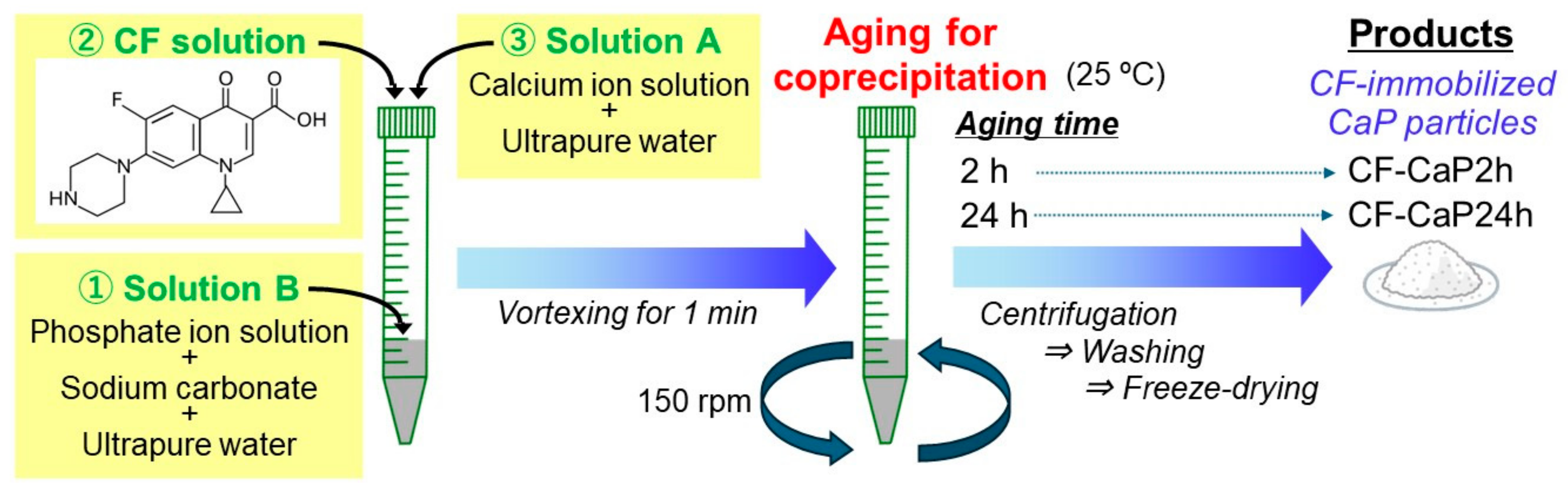
Abstract
:1. Introduction
2. Materials and Methods
2.1. Preparation of CF-Immobilized CaP Particles
2.2. Characterization of the Products
2.3. Determination of CF-Immobilization Efficiency
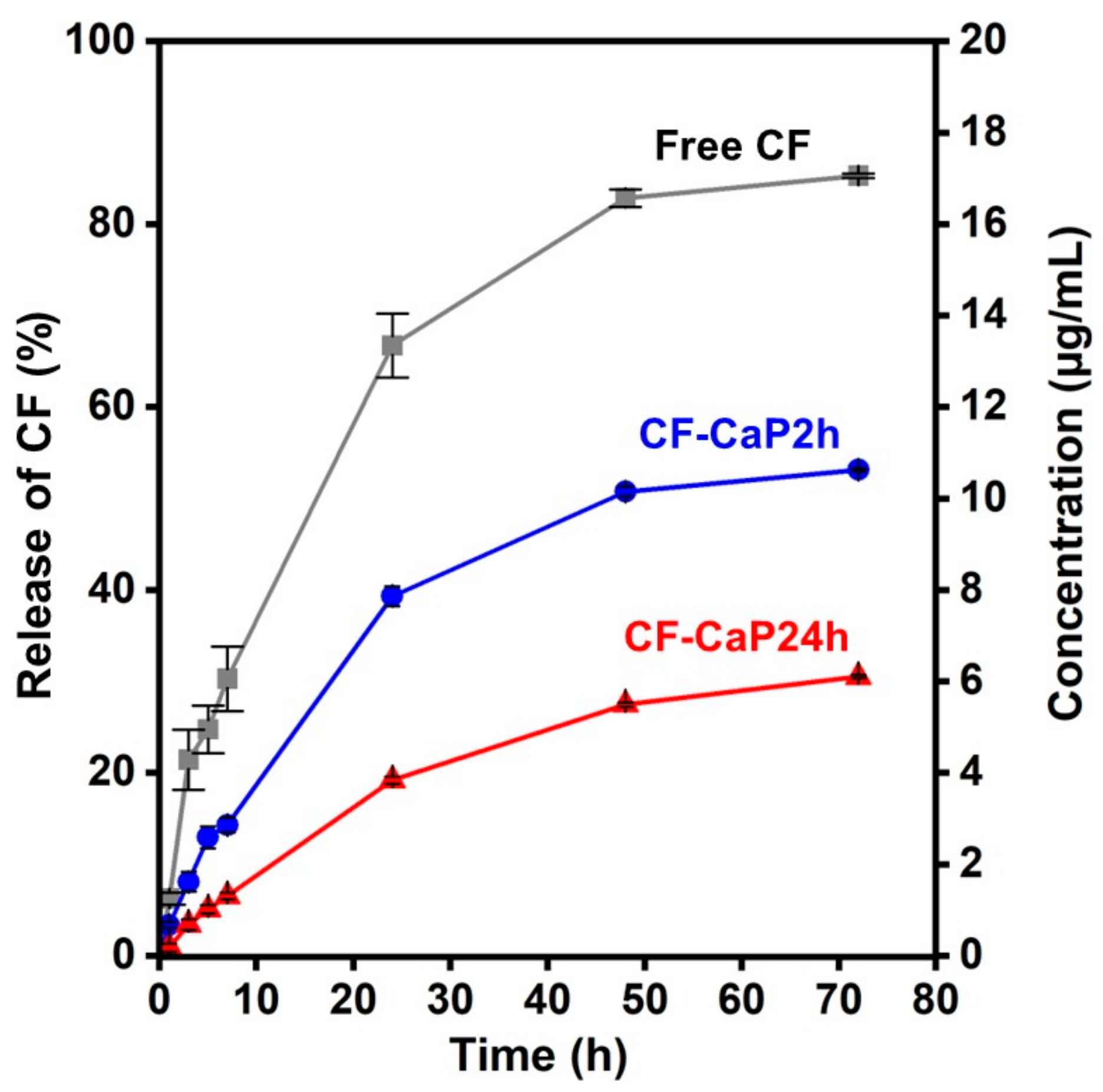
2.4. CF-Release Assay
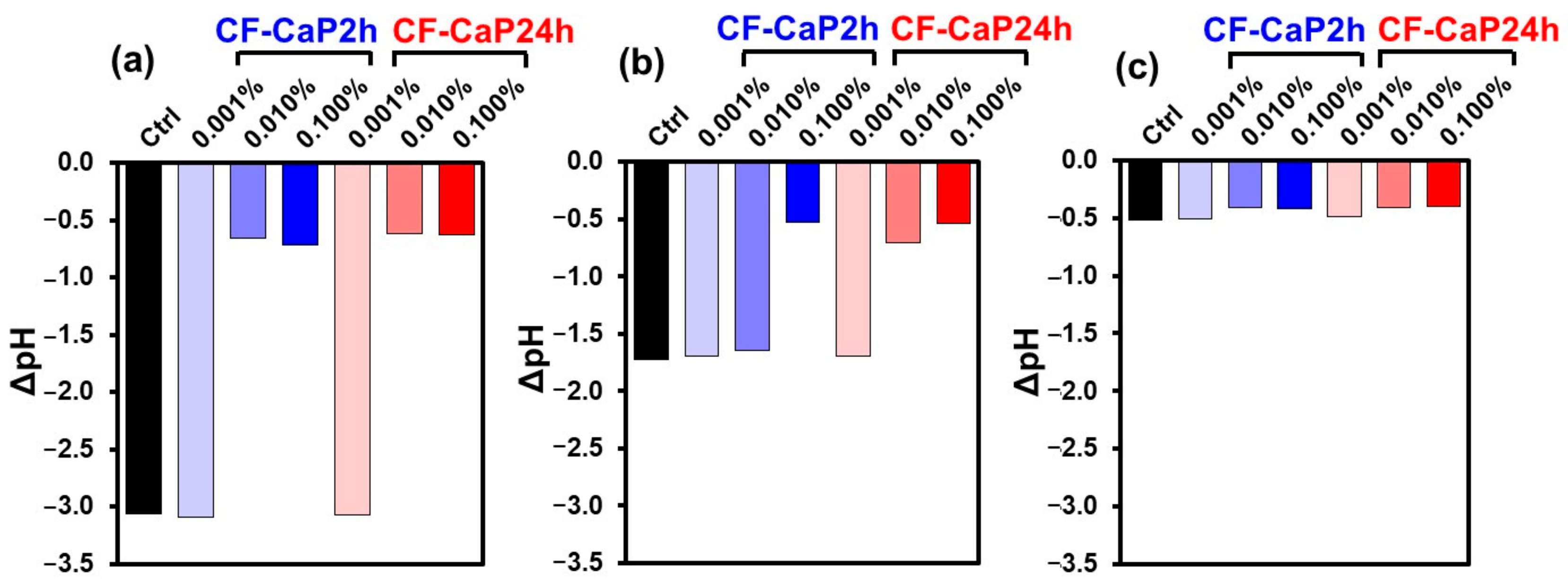
2.5. Antibacterial Assay
2.6. Biofilm Formation Assay
2.7. Statistical Analysis
3. Results
3.1. Morphological Analysis
3.2. Chemical Analysis
3.3. Crystalline Structural Analysis
3.4. CF-Release Assay
3.5. Antibacterial Assay
3.6. Biofilm Formation Assay
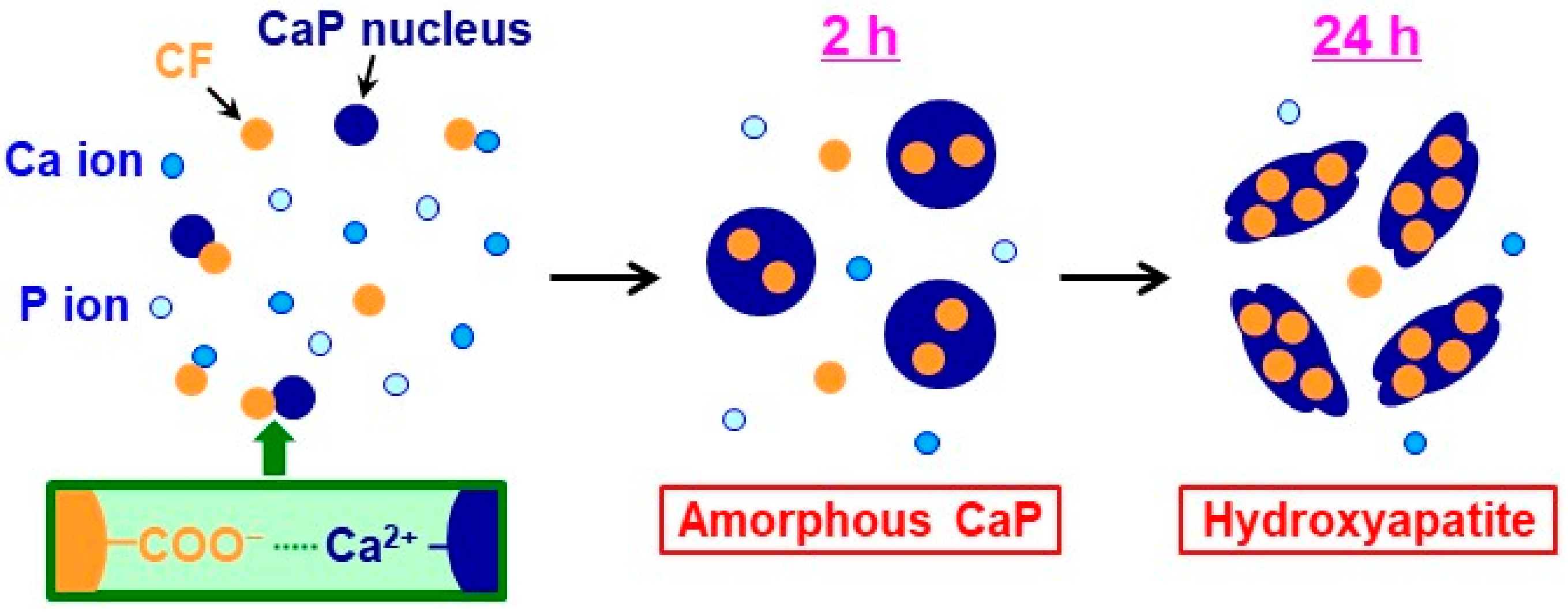
4. Discussion
5. Conclusions
Author Contributions
Funding
Institutional Review Board Statement
Informed Consent Statement
Data Availability Statement
Acknowledgments
Conflicts of Interest
References
- Li, X.; Liu, Y.; Yang, X.; Li, C.; Song, Z. The oral microbiota: Community composition, influencing factors, pathogenesis, and interventions. Front. Microbiol. 2022, 13, 895537. [Google Scholar] [CrossRef] [PubMed]
- Matsumoto-Nakano, M. Role of Streptococcus mutans surface proteins for biofilm formation. Jpn. Dent. Sci. Rev. 2018, 54, 22–29. [Google Scholar] [CrossRef] [PubMed]
- Hussain, M.; Stover, C.M.; Dupont, A. P. gingivalis in periodontal disease and atherosclerosis–Scenes of action for antimicrobial peptides and complement. Front. Immunol. 2015, 6, 45. [Google Scholar] [CrossRef] [PubMed]
- Dige, I.; Raarup, M.K.; Nyengaard, J.R.; Kilian, M.; Nyvad, B. Actinomyces naeslundii in initial dental biofilm formation. Microbiology 2009, 155, 2116–2126. [Google Scholar] [CrossRef] [PubMed]
- Rajeshwari, H.R.; Dhamecha, D.; Jagwani, S.; Rao, M.; Jadhav, K.; Shaikh, S.; Puzhankara, L.; Jalalpure, S. Local drug delivery systems in the management of periodontitis: A scientific review. J. Control. Release 2019, 307, 393–409. [Google Scholar]
- Bapat, R.A.; Joshi, C.P.; Bapat, P.; Chaubal, T.V.; Pandurangappa, R.; Jnanendrappa, N.; Gorain, B.; Khurana, S.; Kesharwani, P. The use of nanoparticles as biomaterials in dentistry. Drug Discov. Today 2019, 24, 85–98. [Google Scholar] [CrossRef]
- Sokolova, V.; Epple, M. Biological and medical applications of calcium phosphate nanoparticles. Chem. Eur. J. 2021, 27, 7471–7488. [Google Scholar] [CrossRef]
- Dorozhkin, S.V. Nanosized and nanocrystalline calcium orthophosphates. Acta Biomater. 2010, 6, 715–734. [Google Scholar] [CrossRef] [PubMed]
- Zalite, V.; Lungevics, J.; Vecstaudza, J.; Stipniece, L.; Locs, J. Nanosized calcium deficient hydroxyapatites for tooth enamel protection. J. Biomed. Mater. Res. 2022, 110, 1354–1367. [Google Scholar] [CrossRef]
- Xu, J.; Shi, H.; Luo, J.; Yao, H.; Wang, P.; Li, Z.; Wei, J. Advanced materials for enamel remineralization. Front. Bioeng. Biotechnol. 2022, 10, 985881. [Google Scholar] [CrossRef]
- Xie, C.; Lu, H.; Li, W.; Chen, F.M.; Zhao, Y.M. The use of calcium phosphate-based biomaterials in implant dentistry. J. Mater. Sci. Mater. Med. 2012, 23, 853–862. [Google Scholar] [CrossRef] [PubMed]
- Nakamura, M.; Oyane, A.; Shimizu, Y.; Miyata, S.; Saeki, A.; Miyaji, H. Physicochemical fabrication of antibacterial calcium phosphate submicrospheres with dispersed silver nanoparticles via coprecipitation and photoreduction under laser irradiation. Acta Biomater. 2016, 46, 299–307. [Google Scholar] [CrossRef] [PubMed]
- Cao, G.; Jiang, Y.; Chen, F.; Lu, B.; Tan, S.; Feng, V.; Qi, S.; He, S.; Xu, Y.; Chen, X. Antibacterial silver-doped calcium phosphate synthesized by an enzymatic strategy for initial caries treatment. Ceram. Int. 2020, 46, 22466–22473. [Google Scholar] [CrossRef]
- Yang, M.; Ren, J.; Zhang, R. Novel gallium-doped amorphous calcium phosphate nanoparticles: Preparation, application and structure study. J. Non-Cryst. Solids 2017, 466–467, 15–20. [Google Scholar] [CrossRef]
- Chen, X.; Tang, Q.L.; Zhu, Y.J.; Zhu, C.L.; Feng, X.P. Synthesis and antibacterial property of zinc loaded hydroxyapatite nanorods. Mater. Lett. 2012, 89, 233–235. [Google Scholar] [CrossRef]
- Kovtun, A.; Kozlova, D.; Ganesan, K.; Biewald, C.; Seipold, N.; Gaengler, P.; Arnold, W.H.; Epple, M. Chlorhexidine-loaded calcium phosphate nanoparticles for dental maintenance treatment: Combination of mineralising and antibacterial effects. RSC Adv. 2012, 2, 870–875. [Google Scholar] [CrossRef]
- Ghosh, S.; Wu, V.; Pernal, S.; Uskoković, V. Self-setting calcium phosphate cements with tunable antibiotic release rates for advanced antimicrobial applications. ACS Appl. Mater. Interfaces 2016, 8, 7691–7708. [Google Scholar] [CrossRef] [PubMed]
- Kumar, G.S.; Govindan, R.; Girija, E.K. In situ synthesis, characterization and in vitro studies of ciprofloxacin loaded hydroxyapatite nanoparticles for the treatment of osteomyelitis. J. Mater. Chem. B 2014, 2, 5052–5060. [Google Scholar] [CrossRef]
- Sangeetha, K.; Ashok, M.; Girija, E.K.; Vidhya, G.; Vasugi, G. Strontium and ciprofloxacin modified hydroxyapatites as functional grafts for bone prostheses. Ceram. Int. 2018, 44, 13782–13789. [Google Scholar] [CrossRef]
- Sharma, D.; Patel, R.P.; Zaidi, S.T.R.; Sarker, M.M.R.; Lean, Q.Y.; Ming, L.C. Interplay of the quality of ciprofloxacin and antibiotic resistance in developing countries. Front. Pharmacol. 2017, 21, 546. [Google Scholar] [CrossRef]
- Carreira, C.d.M.; dos Santos, S.S.F.; Jorge, A.O.C.; Lage-Marques, J.L. Antimicrobial effect of intracanal substances. J. Appl. Oral Sci. 2007, 15, 453–458. [Google Scholar] [CrossRef] [PubMed]
- Chotitumnavee, J.; Parakaw, T.; Srisatjaluk, R.L.; Pruksaniyom, C.; Pisitpipattana, S.; Thanathipanont, C.; Amarasingh, T.; Tiankhum, N.; Chimchawee, N.; Ruangsawasdi, N. In vitro evaluation of local antibiotic delivery via fibrin hydrogel. J. Dent. Sci. 2019, 14, 7–14. [Google Scholar] [CrossRef]
- Nalawade, T.M.; Bhat, K.G.; Sogi, S. Antimicrobial activity of endodontic medicaments and vehicles using agar well diffusion method on facultative and obligate anaerobes. Int. J. Clin. Pediatr. Dent. 2016, 9, 335–341. [Google Scholar] [PubMed]
- Eick, S.; Schmitt, A.; Sachse, S.; Schmidt, K.H.; Pfister, W. In vitro antibacterial activity of fluoroquinolones against Porphyromonas gingivalis strains. J. Antimicrob. Chemother. 2004, 54, 553–556. [Google Scholar] [CrossRef]
- Elshikh, M.; Moya-Ramiırez, I.; Moens, H.; Roelants, S.; Soetaert, W.; Marchant, R.; Banat, I.M. Rhamnolipids and lactonic sophorolipids: Natural antimicrobial surfactants for oral hygiene. J. Appl. Microbiol. 2017, 123, 1111–1123. [Google Scholar] [CrossRef]
- Barberis, C.; Budia, M.; Palombarani, S.; Rodriguez, C.H.; Ramírez, M.S.; Arias, B.; Bonofiglio, L.; Famiglietti, A.; Mollerach, M.; Almuzara, M.; et al. Antimicrobial susceptibility of clinical isolates of Actinomyces and related genera reveals an unusual clindamycin resistance among Actinomyces urogenitalis strains. J. Glob. Antimicrob. Resist. 2017, 8, 115–120. [Google Scholar] [CrossRef] [PubMed]
- Shubhra, Q.T.H.; Oyane, A.; Nakamura, M.; Puentes, S.; Marushima, A.; Tsurushima, H. Rapid one-pot fabrication of magnetic calcium phosphate nanoparticles immobilizing DNA and iron oxide nanocrystals using injection solutions for magnetofection and magnetic targeting. Mater. Today Chem. 2017, 6, 51–61. [Google Scholar] [CrossRef]
- Bigi, A.; Boanini, E. Calcium phosphates as delivery systems for bisphosphonates. J. Funct. Biomater. 2018, 9, 6. [Google Scholar] [CrossRef]
- Nakamura, M.; Bunryo, W.; Narazaki, A.; Oyane, A. High immobilization efficiency of basic protein within heparin-immobilized calcium phosphate nanoparticles. Int. J. Mol. Sci. 2022, 23, 11530. [Google Scholar] [CrossRef]
- Kadkhodaie-Elyaderani, A.; de Lama-Odría, M.d.C.; Rivas, M.; Martínez-Rovira, I.; Yousef, I.; Puiggalí, J.; del Valle, L.J. Medicated scaffolds prepared with hydroxyapatite/streptomycin nanoparticles encapsulated into polylactide microfibers. Int. J. Mol. Sci. 2022, 23, 1282. [Google Scholar] [CrossRef]
- Pal, A.; Oyane, A.; Nakamura, M.; Koga, K.; Nishida, E.; Miyaji, H. Fluoride-incorporated apatite coating on collagen sponge as a carrier for basic fibroblast growth factor. Int. J. Mol. Sci. 2024, 25, 1495. [Google Scholar] [CrossRef] [PubMed]
- Nugrahani, I.; Tjengal, B.; Gusdinar, T.; Horikawa, A.; Uekusa, H. A comprehensive study of a new 1.75 hydrate of ciprofloxacin salicylate: SCXRD structure determination, solid characterization, water stability, solubility, and dissolution study. Crystals 2020, 10, 349. [Google Scholar] [CrossRef]
- Sahoo, S.; Chakraborti, C.K.; Mishra, S.C. Qualitative analysis of controlled release ciprofloxacin/carbopol 934 mucoadhesive suspension. J. Adv. Pharm. Technol. Res. 2011, 2, 195–204. [Google Scholar] [CrossRef] [PubMed]
- Jin, B.; Liu, Z.; Shao, C.; Chen, J.; Liu, L.; Tang, R.; De Yoreo, J.J. Phase transformation mechanism of amorphous calcium phosphate to hydroxyapatite investigated by liquid-cell transmission electron microscopy. Cryst. Growth Des. 2021, 21, 5126–5134. [Google Scholar] [CrossRef]
- Manoj, M.; Mangalaraj, D.; Ponpandian, N.; Viswanatan, C. Core–shell hydroxyapatite/Mg nanostructures: Surfactant free facile synthesis, characterization and their in vitro cell viability studies against leukaemia cancer cells (K562). RSC Adv. 2015, 5, 48705–48711. [Google Scholar] [CrossRef]
- Kumar, K.C.V.; Subha, T.J.; Ahila, K.G.; Ravindran, B.; Chang, S.W.; Mahmoud, A.H.; Mohammed, O.B.; Rathi, M.A. Spectral characterization of hydroxyapatite extracted from black sumatra and fighting cock bone samples: A comparative analysis. Saudi J. Biol. Sci. 2021, 28, 840–846. [Google Scholar] [CrossRef] [PubMed]
- Dorozhkin, S.V. Synthetic amorphous calcium phosphates (ACPs): Preparation, structure, properties, and biomedical applications. Biomater. Sci. 2021, 9, 7748–7798. [Google Scholar] [CrossRef]
- Pleshko, N.; Boskey, A.; Mendelsohn, R. Novel infrared spectroscopic method for the determination of crystallinity of hydroxyapatite minerals. Biophys. J. 1991, 60, 786–793. [Google Scholar] [CrossRef]
- Tao, J. FTIR and raman studies of structure and bonding in mineral and organic–mineral composites. In Methods in Enzymology; De Yoreo, J.J., Ed.; Academic Press: Cambridge, MA, USA, 2013; Volume 532, pp. 533–556. [Google Scholar]
- Moreau, J.L.; Sun, L.; Chow, L.C.; Xu, H.H.K. Mechanical and acid neutralizing properties and bacteria inhibition of amorphous calcium phosphate dental nanocomposite. J. Biomed Mater. Res. Part B Appl. Biomater. 2011, 98B, 80–88. [Google Scholar] [CrossRef]
- Nardecchia, S.; Gutierrez, M.C.; Serrano, M.C.; Dentini, M.; Barbetta, A.; Ferrer, M.L.; del Monte, F. In situ precipitation of amorphous calcium phosphate and ciprofloxacin crystals during the formation of chitosan hydrogels and its application for drug delivery purposes. Langmuir 2012, 28, 15937–15946. [Google Scholar] [CrossRef]
- Ikawa, N.; Kimura, T.; Oumi, Y.; Sano, T. Amino acid containing amorphous calcium phosphates and the rapid transformation into apatite. J. Mater. Chem. 2009, 19, 4906–4913. [Google Scholar] [CrossRef]
- Astasov-Frauenhoffer, M.; Varenganayil, M.M.; Decho, A.W.; Waltimo, T.; Braissant, O. Exopolysaccharides regulate calcium flow in cariogenic biofilms. PLoS ONE 2017, 12, e0186256. [Google Scholar] [CrossRef] [PubMed]
- Mekky, A.E.M.; Sanad, S.M.H. Novel bis(pyrazole-benzofuran) hybrids possessing piperazine linker: Synthesis of potent bacterial biofilm and MurB inhibitors. Bioorg. Chem. 2020, 102, 104094. [Google Scholar] [CrossRef] [PubMed]


Disclaimer/Publisher’s Note: The statements, opinions and data contained in all publications are solely those of the individual author(s) and contributor(s) and not of MDPI and/or the editor(s). MDPI and/or the editor(s) disclaim responsibility for any injury to people or property resulting from any ideas, methods, instructions or products referred to in the content. |
© 2024 by the authors. Licensee MDPI, Basel, Switzerland. This article is an open access article distributed under the terms and conditions of the Creative Commons Attribution (CC BY) license (https://creativecommons.org/licenses/by/4.0/).
Share and Cite
Pal, A.; Oyane, A.; Inose, T.; Nakamura, M.; Nishida, E.; Miyaji, H. Fabrication of Ciprofloxacin-Immobilized Calcium Phosphate Particles for Dental Drug Delivery. Materials 2024, 17, 2035. https://0-doi-org.brum.beds.ac.uk/10.3390/ma17092035
Pal A, Oyane A, Inose T, Nakamura M, Nishida E, Miyaji H. Fabrication of Ciprofloxacin-Immobilized Calcium Phosphate Particles for Dental Drug Delivery. Materials. 2024; 17(9):2035. https://0-doi-org.brum.beds.ac.uk/10.3390/ma17092035
Chicago/Turabian StylePal, Aniruddha, Ayako Oyane, Tomoya Inose, Maki Nakamura, Erika Nishida, and Hirofumi Miyaji. 2024. "Fabrication of Ciprofloxacin-Immobilized Calcium Phosphate Particles for Dental Drug Delivery" Materials 17, no. 9: 2035. https://0-doi-org.brum.beds.ac.uk/10.3390/ma17092035