Neuroglia 2024, 5(2), 105-118; https://0-doi-org.brum.beds.ac.uk/10.3390/neuroglia5020008 - 28 Apr 2024
Abstract
►
Show Figures
Brain tumors necessitate swift detection and classification for optimal patient outcomes. Deep learning has been extensively utilized to recognize complex tumor patterns in magnetic resonance imaging (MRI) images, aiding in tumor diagnosis, treatment, and prognostication. However, model complexity and limited generalizability with unfamiliar
[...] Read more.
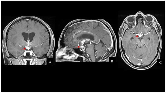
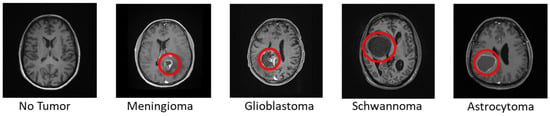
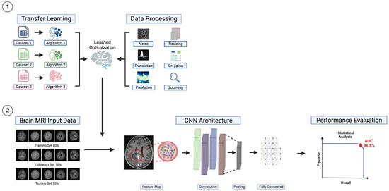
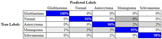
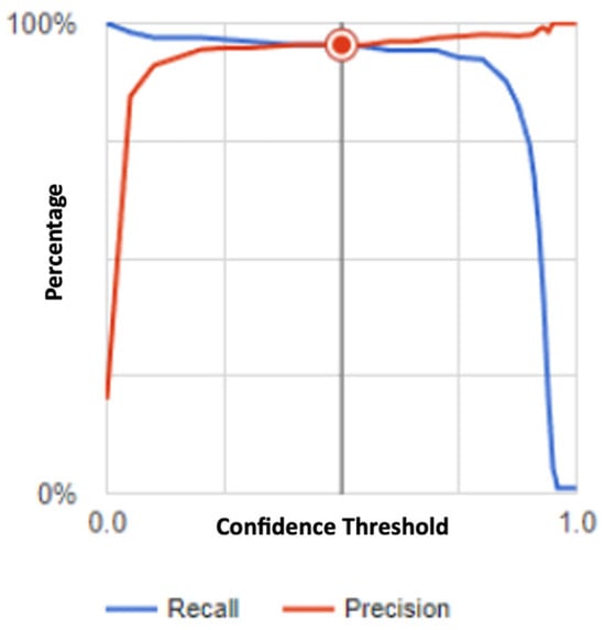
Brain tumors necessitate swift detection and classification for optimal patient outcomes. Deep learning has been extensively utilized to recognize complex tumor patterns in magnetic resonance imaging (MRI) images, aiding in tumor diagnosis, treatment, and prognostication. However, model complexity and limited generalizability with unfamiliar data hinder appropriate clinical integration. The objective of this study is to develop a clean-energy cloud-based deep learning platform to classify brain tumors. Three datasets of a total of 2611 axial MRI images were used to train our multi-layer convolutional neural network (CNN). Our platform automatically optimized every transfer learning and data augmentation feature combination to provide the highest predictive accuracy for our classification task. Our proposed system identified and classified brain tumors successfully and efficiently with an overall precision value of 96.8% [95% CI; 93.8–97.6]. Using clean energy supercomputing resources and cloud platforms cut our workflow to 103 min, $0 in total cost, and a negligible carbon footprint (0.0014 kg eq CO2). By leveraging automated optimized learning, we developed a cost-effective deep learning (DL) platform that accurately classified brain tumors from axial MRI images of different levels. Although studies have identified machine learning tools to overcome these obstacles, only some are cost-effective, generalizable, and usable regardless of experience.
Full article
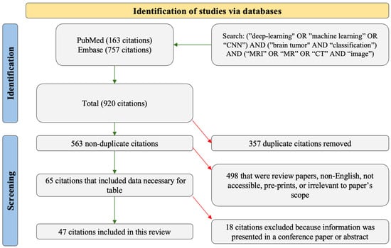
Figure 1
