Article

Computational Insights into the Interaction between Neprilysin and α-Bisabolol: Proteolytic Activity against Beta-Amyloid Aggregates in Alzheimer’s Disease

by
Jonathan Elias Rodrigues Martins
1,
José Ednésio da Cruz Freire
1,*,
Francisco Sérgio Lopes Vasconcelos-Filho
1,2,
Diego da Silva de Almeida
3,
Vânia Marilande Ceccatto
1 and
Bruno Lopes de Sousa
3
1
Biochemistry and Gene Expression Laboratory, Superior Institute of Biomedical Sciences, State University of Ceará, Fortaleza 60714-903, Ceará, Brazil
2
Dean of Culture, Federal University of Cariri, Juazeiro do Norte, Fortaleza 63048-080, Ceará, Brazil
3
Faculty of Philosophy Dom Aureliano Matos, State University of Ceará, Limoeiro do Norte, Fortaleza 62930-000, Ceará, Brazil
*
Author to whom correspondence should be addressed.
Submission received: 18 March 2024 / Revised: 25 April 2024 / Accepted: 26 April 2024 / Published: 27 April 2024
(This article belongs to the Section Pharmaceutical Processes)

Abstract

:
(1) Background: Alzheimer’s disease (AD) is an irreversible disorder of the central nervous system associated with beta-amyloid protein (Aβ) deposition and accumulation. Current treatments can only act on symptoms and not the etiologic agent. Neprilysin and α-bisabolol have been shown to reduce the aggregation of Aβ, suggesting a potential interaction between both molecules, leading to increased proteolytic activity on Aβ aggregates. (2) Methods: Computational simulations were conducted to explore the interaction between murine neprilysin [NEP(m)] and α-bisabolol and their effects on enzymatic activity. NEP(m) structure was predicted using comparative modeling, and the binding pattern to α-bisabolol and its effects on leu-enkephalin binding were explored through docking calculations and molecular dynamics simulations, respectively. (3) Results: The findings suggest that α-bisabolol stabilizes the Val481-Pro488 segment of NEP2(m), which directly interacts with the peptide substrate, enabling an optimized alignment between the catalytic residue Glu525 and leu-enkephalin. (4) Conclusions: This computational evidence strongly supports the notion that α-bisabolol stabilizes peptide substrates at the NEP2(m) catalytic site, leading to the positive modulation of enzymatic activity.

1. Introduction

Alzheimer’s disease (AD) is a prevalent and irreversible disorder of the central nervous system, characterized by the loss of cognitive functions, attention, judgment, and memory. According to the latest statistics, more than 55 million people worldwide are suffering from AD or related dementias [1], and this has generated a significant socioeconomic burden. Taking only individuals with AD in Brazil into account, the average annual cost of treating individuals via the public health system was estimated at USD 4020.47 (the Brazilian currency is the real but was converted into the US dollar), an amount considered relatively high for the country [2]. This makes AD and other dementias the leading cause of disability, especially among elderly populations.
AD is intimately associated with the deposition and accumulation of β-amyloid peptide (Aβ) [3]. Aβ is a highly toxic peptide for neurons [4], and its buildup can result in the formation of oligomers and amyloid plaques. Two isoforms of Aβ, Aβ1–42, and Aβ1–40 peptides are the primary components of these amyloid plaques [5]. From the clinical–biological point of view, abnormalities linked to cerebral amyloid peptides are observed in the early stages of AD, where the levels of the Aβ1–42 peptide decrease (showing an Aβ1–42/Aβ1–40 ratio) in the cerebrospinal fluid. Interestingly, the pathway of plaque formation remains unknown. However, some clues seem to be associated with the predominant presence of both Aβ isoforms in these plaques despite Aβ1–40 comprising approximately 90% of the Aβ pool [6]. Evidence suggests that a more significant hydrophobic potential is induced due to additional amino acids in the Aβ1–42 isoform, resulting in a sufficiently strong hydrophobicity for the chains to undergo liquid–liquid phase separation into condensates of fibers. This effect provides theoretical support for the hypothesis that LLPS condensates act as precursors for aggregation and forming Aβ plaques through hydrophobic interactions. This makes it possible to define the stage known as the “Alzheimer’s continuum” [7] before the occurrence of the anomalies (hyperphosphorylation) found in the Tau protein (p-Tau). This, in turn, leads to the detachment of Tau from microtubules, resulting in the destabilization of axonal microtubule scaffolds, impairments in axonal transport [8], and eventually neuronal death.
The Aβ pathway presents the enzyme neprilysin, NEP2(m), a membrane metallopeptidase belonging to the M13 family. The NEPs are also known by different nomenclatures, including neutral endopeptidase or enkephalinase (EC 3.4.24.11) [9,10,11,12,13,14], endothelin-converting enzymes—ECE1 and ECE2 (EC 3.4.24.71) [15,16,17,18,19]—Kell blood group antigens [20,21], phosphate-regulating neutral endopeptidase on the X chromosome (PEX or PHEX) [22,23], X-converting enzymes (XCEs) [24,25], cluster of differentiation 10 (CD10), membrane metalloendopeptidase (MME), and common acute lymphoblastic leukemia antigens (CALLAs) [13,26]. They are present in various tissues, including the nervous system, and are therefore considered ubiquitous proteins responsible for the degradation of numerous regulatory peptides, including the Aβ peptide [27] (Figure 1), consequently inhibiting the progression of amyloid plaque formation [28]. The wide range of canonical substrates suggests its involvement in multiple regulatory functions and its potential role in the diseases present in various systems, including the brain and nervous systems [29].
NEP2(m) features a short N-terminal domain facing the cytoplasmic region, consisting of 27 amino acids, a single transmembrane segment composed of 23 residues, and a substantial C-terminal ectodomain formed by 699 amino acids harboring the catalytic site that extends into the extracellular region [30]. The ectodomain contains a large central cavity corresponding to the catalytic site where the conserved zinc-binding motif HEXXH is located. X stands for any other known amino acid residues. In contrast, two histidine residues are responsible for the coordination of the zinc ion, and glutamate is directly involved in the catalysis process. Structurally, the ectodomain is formed by two alpha-helices, creating a water-filled cleft where the catalytic site is located [31]. Additionally, NEP2(M) is heavily N-glycosylated at the N145, N285, N294, N325, and N628 sites [32,33,34,35], and the ratio of glycosylation effectively contributes to the range of molecular weights (85–110 kDa) observed in these enzymes in different tissues.
NEP2(m) has been studied extensively under different conditions, including in vitro, in vivo, and in silico [36,37,38,39,40,41,42]. Approaches using transgenic NEP2(m) expression analyses have demonstrated decreased neuronal Aβ deposits in an AD mouse model [43,44,45]. It was also shown that the overexpression of soluble (active) NEP2(m) in various other studies using animals resulted in significant decreases in the levels of neural Aβ and improved cognitive performance [46,47,48,49]. Additionally, the activity of NEP2(m) in human brain tissue has been shown to increase with normal aging, although an increase was also observed in indicators of AD progression [36]. Therefore, there is much evidence linking the Aβ accumulation with NEP2(m) activity in induced AD disease, suggesting that this endopeptidase is a promising therapeutic agent capable of minimizing the adverse effects of AD. While it has been documented that NEP2(m) can significantly reduce the Aβ peptide pool, other neuropeptides are targets of its proteolytic activity, given that it is a promiscuous enzyme [20,50,51]. Thus, high NEP2(m) concentrations can likely lead to imbalances in the necessary neuropeptides in signal pathways that control blood pressure, pain, and other crucial physiological processes.
On the other hand, (-)-α-bisabolol (α-bisabolol) is widely available and dietarily bioavailable. It possesses physicochemical properties that give it lipophilicity [52] and various pharmacological attributes, including anti-inflammatory, antioxidant [53,54], wound healing, antibiotic, antimutagenic, antitumor, gastroprotective, analgesic, and antimicrobial effects [52,55,56,57,58,59,60] and low toxicity, with its generally recognized as safe (GRAS) status. Recent data suggest that the oral administration of α-bisabolol may provide neuroprotection through decreased neurodegeneration and neuroinflammation, as well as cognitive improvement [57]. In addition, various studies have shown that this substance can significantly reduce the aggregation of Aβ and act on the cleavage of its fibrillar isoforms [28,61]. A-bisabolol may act by increasing the effectiveness of Aβ degradation [28,57,62]. This evidence points to an interaction between NEP2(m) and α-bisabolol as a possible mechanism to increase the anti-amyloidogenic effect. Thus, the present study aims to investigate, using computational tools, the interaction between α-bisabolol and NEP2(m), the main Aβ-degrading enzyme, as a possible anti-amyloidogenic pathway.

2. Materials and Methods

2.1. Domain and Motif Prediction

The amino acid sequence (accession ID Q9JLI3) of murine neprilysin [NEP2(m)] was downloaded in FASTA format from the Universal Protein Resource—UniProt [63]. Then, boundary domains and motif sequences were identified from the amino acid sequence of NEP2(m) using the following servers: Database of Protein Domains, Families and Functional Sites—Prosite [64], Conserved Domains Database and Resources—CDD [65], and Classification of Protein Families—InterPro [66].

2.2. Structural Prediction of the Ectodomain of NEP2(m)

The structural model of the ectodomain portion (Thr76–Trp765) was predicted using the Modeller program package v.10.0 [67]. For this purpose, experimentally determined protein structures with similar amino acid sequences were searched using the BLASTp tool with the PSI-BLAST algorithm from the Protein Data Bank [68,69,70]. This search identified the substrate-free human neprilysin with accession ID 6SH1 as the template [71], which presents a sequence coverage and identity of 92% and 93.8%, respectively. Following this strategy, several models were initially generated, including structural waters and the zinc ion in human neprilysin. Afterward, the produced structures were selected based on the estimated DOPE Score [72], and the top 10 models were chosen for stoichiometry evaluation (steric overlaps, Cβ deviation parameters, Ramachandran plots, rotamers, and bond angle quality) through the MolProbity Server [73,74,75,76]. Finally, the best model was selected and visually inspected with Pymol (Schrödinger).

2.3. Bioactivity Prediction of the α-Bisabolol

The 3D structure of α-bisabolol (CID: 1549992) in PDB format and its file in the SMILES extension were downloaded from the PubChem server (https://pubchem.ncbi.nlm.nih.gov/, accessed on 16 February 2024). The SMILES file was then subjected to the Molinspiration virtual screening (www.molinspiration.com, accessed on 16 February 2024) server to evaluate molecular properties and bioactivity scores. The PDB format file was used in the docking and dynamics molecular simulations.

2.4. Prediction of Pockets in NEP2(m) and Docking Calculations

The most favorable hotspot found in leu-enkephalin was determined by both servers: the DogSiteScorer tool [77] and the CASTp server [78]. After determining the most favorable hotspot for interaction, site-directed redocking simulations were conducted in a setup where all the torsional bonds of both α-bisabolol and the side chains of the amino acids found in different experimental structures had the freedom to rotate. Furthermore, all hydrogen polar atoms were added to leu-enkephalin and then parameterized with Gasteiger charges, while the α-bisabolol was prepared with the addition of Kollman charges, as according to previous studies [79,80].
Docking simulations involving the substrate NEP2(m), i.e., leu-enkephalin (Tyr-Gly-Gly-Phe-Leu, seen in Figure 2A), a typical substrate for neprilysin, and α-bisabolol (seen in Figure 2B) were conducted using the AutoDock Vina software version 1.2.0 [81]. For this purpose, the structure of leu-enkephalin was parameterized using the BIOVIA Discovery Studio Package, while α-bisabolol (CID: 1549992) was obtained from PubChem (https://pubchem.ncbi.nlm.nih.gov/, accessed on 20 August 2023). To confirm the accuracy of the molecular docking investigations, blind docking simulations were performed in a setup where all the torsional bonds of α-bisabolol had the freedom to rotate, while the amino acids found in different experimental structures (add IDs) were kept rigid. The simulation was performed under the following conditions: number of conformations = 20, exhaustiveness = 8, and seed = 2009. The dimensions of the boxes in all cases were X = 20 Å, Y = 20 Å, and Z = 20 Å.
For all calculations, the ten top-ranked generations based on the predicted binding affinity (in kilocalories per mole) were visually analyzed, and the best results were selected based on structural comparisons made with the experimental structures and their binding profiles. AutoDock Vina was validated for the presented purpose through redocking calculations with different crystal structures, and the results of which were analyzed by the root mean square deviation (RMSD) of the atomic positions between the experimentally determined and calculated ligands through the RMSD calculator tool of the software Visual Molecular Dynamics 1.9 [82].

2.5. Molecular Dynamics Simulation and Schematic Representations

The α-bisabolol::leu-enkephalin complex with the lowest binding energy based on the docking simulations was subjected to stability assessments through molecular dynamics (MD) analysis using the GROMACS package, version 5.0 [83]. The electroneutrality of both complexes was preserved by adding Na+ ions as needed. System solvation was conducted using TIP3P (transferable intermolecular potential with three charge points, one for oxygen and two for hydrogens) water molecules in a periodic box (20 × 20 × 20 nm3 volume) containing buffer to enable substantial fluctuations in the conformation during MD simulations. MD simulations were conducted utilizing the CHARMM36 force field. The energy minimization (2000 steps steepest descent followed by 200 ps long MD simulation) was performed before initiating MD simulations to remove initial steric clashes. MD simulations were carried out under constant pressure conditions using anisotropic diagonal position scaling, with a time step of 0.002 ps. The evaluation of electrostatic interactions was conducted following the PME (particle mesh Ewald) principle, with predefined short-range cutoffs of 1.2 nm [84]. The system temperature was set to gradually rise from 100 to 310 K at 1 bar pressure over 1000 ps. The Berendsen weak-coupling algorithm [85] with a fixed time of 0.2 ps was utilized. The LINCS algorithm [86] was employed to maintain the equilibrium distances between all bonds, permitting only internal motions of bending and torsion during the MD simulations. Finally, the 20 ns MD simulations were conducted under the same conditions as those used in the equilibration procedure.
After MD simulations, the α-bisabolol::leu-enkephalin::NEP2(m) complex trajectories were analyzed using the RMSD of their atomic positions with the VMD software 1.9. The web server Prodigy calculates binding energies for docking and MD results (https://bianca.science.uu.nl/prodigy/, accessed on 12 November 2023). Three-dimensional representations of the best α-bisabolol::leu-enkephalin::NEP2(m) complexes based on the theoretical binding energy value were generated using the software PyMol Molecular Graphics System, version 1.7.4 (Schrodinger, LLC, New York, NY, USA).

3. Results

3.1. Prediction of the Domains and Motifs of the Ectodomain of NEP2(m)

The CDD-based prediction data showed that the ectodomain of NEP2(m) is a member belonging to the conserved protein domain of the M13 family of metallopeptidases, which includes neprilysin (neutral endopeptidase, NEP, enkephalinase, CD10, CALLA, EC 3.4.24.11), endothelin-converting enzyme I (ECE-1, EC 3.4.24.71), erythrocyte surface antigen KELL (ECE-3), phosphate-regulating gene on the X chromosome (PHEX), soluble secreted endopeptidase (SEP), and damage-induced neuronal endopeptidase (DINE)/X-converting enzyme (XCE). In addition, the analysis of the CDD-based annotation showed predictions with the following confidence indices: an E-value of 000, a bit score of 787.33, and an interval (96 to 763 amino acid residues). The predictions from the Prosite and InterPro servers agree with the data obtained from the CDD server.

3.2. Structural Analysis of the Ectodomain of NEP2(m)

The final NEP2(m) model was analyzed for its stereochemical properties, revealing that 99.8% of amino acid residues were positioned within allowed regions (96% in the favored areas) of the Ramachandran plot. The overall predicted structure of NEP2(m) closely resembles that of other neprilysin and neprilysin-like proteins, displaying a typical α-helical ellipsoid shape consisting of subdomains 1 and 2 connected by a linker region (Figure 3A). The catalytic site is within subdomain 1, positioned over a spacious cavity (approximately 6.278 A3) formed by both subdomains and the linker region. This catalytic site houses a zinc-binding site with a conserved zinc metalloprotease motif HEXXH, which includes residues His524 and His528 for ion binding (along with residue Glu587), as well as the catalytic residue Glu525 (Figure 3B). The zinc atom acts as a cofactor for catalysis, coordinated by well-described ionic interactions with the residues mentioned above, establishing essential contacts directly with the substrate.

3.3. Ligand Bioactivity Prediction

The in silico predictions of α-bisabolol bioactivity using Molinspiration virtual screening yielded a score of −0.38, indicating that this molecule weakly inhibited proteases, particularly metalloproteases belonging to the M13 family. Additionally, the analysis returned a score of −0.06, suggesting that the ligand bound weakly to G protein-coupled receptors (GPCRs). α-bisabolol also exhibited reduced inhibitory activity against kinases, with a score of −0.78. The predicted bioactivity of the entire ligand was determined by adding the bioactivity scores of each fragment (usually ranging from −3 to 3). Fragments with lower activity scores were more likely to be inactive.

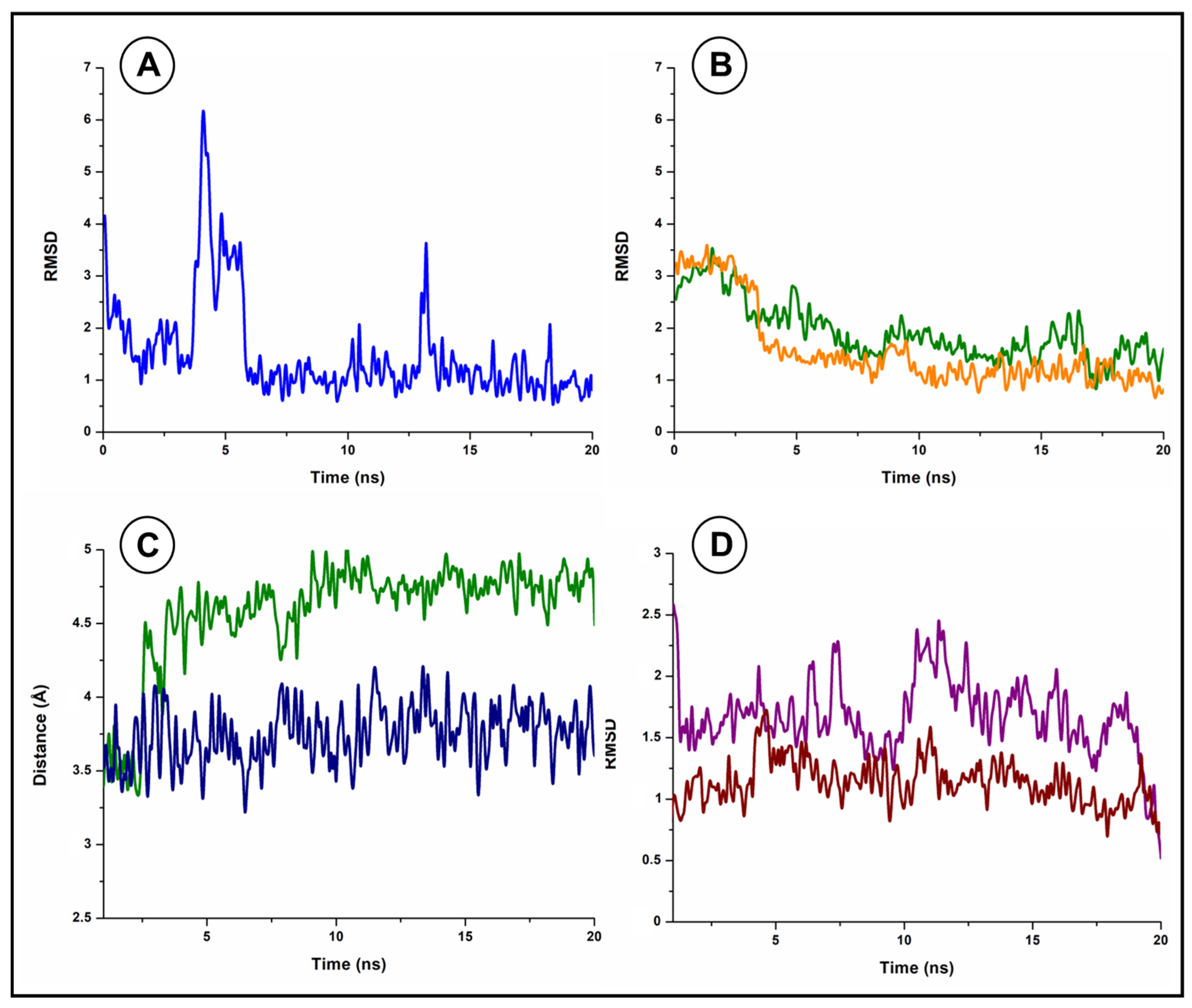
3.4. Molecular Docking Studies and Molecular Dynamics

The docking calculations for leu-enkephalin were validated through a redocking performed with the crystal structure of human neprilysin (NEP(h)) determined in the presence of the inhibitor omopatrilat (Omlat) (PDB ID 6SUK), which had a chemical structure similar to the analyzed peptide. The RMSD between the best calculated conformation for omopatrilat (Omlat) and its crystal structure was 0.7, confirming the reliability of AutoDock Vina for predicting binding conformations in similar systems.
The results obtained from the docking calculations with leu-enkephalin were evaluated based on a comparative analysis with the crystal complex between NEP(h) and Omlat, which was associated with the energy assessment of these results through the Prodigy webserver and led us to a promising conformation. This analysis suggests that leu-enkephalin interacts with NEP2(m) in a highly conservative manner when compared to the crystallized ligand (Figure 4A), forming direct contacts with the zinc ion and numerous surrounding amino acids, including several hydrophobic contacts with residues Phe50, Ala484, Phe485, Phe504, Val521, Trp634, and Val651, as well as polar contacts with His237, His258, Asn483, His652, and Arg658, among others, achieving a calculated binding energy of −6.42 Kcal/mol (Figure 5A and Table 1).
Additionally, the fact that leu-enkephalin is cleaved by NEP2(m) at the Gly3-Phe4 bond is in perfect accordance with the calculations since the OE2 group from the catalytic residue Glu525 is disposed 3.6 Å from the peptide bond mentioned above. A previous description involving the complex between NEP(h) and the inhibitor N-(3-Phenyl-2-sulfanylpropanoyl) phenylalanyl-alanine (PDB ID 1R1J) reported a distance of 3.3 Å for this bond, corroborating the calculated conformation. Nevertheless, since the interaction between the enzyme and the substrate was a dynamic process, MD simulations were performed with the aim of better understanding the behavior of leu-enkephalin at the catalytic site in the absence and presence of α-bisabolol.
We observed that leu-enkephalin exhibited a generally stable behavior at the catalytic site in the absence of α-bisabolol during the 20 ns of MD simulations, as observed by its RMSD (Figure 6A and Figure 7B). However, its terminal residues (Tyr1 and Leu5) underwent a significant rearrangement throughout the simulation, allowing leu-enkephalin to assume a folded conformation similar to another crystallized inhibitor [NEP(h)], sampatri-lat (Samlat), which had a more prolonged and more extended structure compared to Omlat (Figure 4D). Among the 11 polar contacts (including seven hydrogen bonds) predicted by docking calculations, only two were sustained along MD, established with residues Arg658 and His652. However, a new hydrogen bond was detected after the simulations between Tyr1 and Glu587 (Table 1 and Table 2). This rearrangement was associated with the observed flexibility of the segment Val481-Pro488, which directly interacted with the peptide (Figure 6C). Nevertheless, this adjusted conformation was quickly achieved and stabilized by the peptide (Figure 5B and Table 2), which presented a calculated binding energy of −6.59 Kcal/mol at the final frame after MD. Such a structural rearrangement possibly influenced the effectiveness and speed of the substrate cleavage as the precise alignment of chemical groups was crucial in the catalytic process.
To understand this effect, the length variation between the catalytic residue Glu525 and the peptide bond that linked residues Gly3 and Phe4 in leu-enkephalin was analyzed (Figure 6C). It was observed that the distance between these groups significantly increased after two ns of simulation. From 5 ns until the end of the simulation, it oscillated between 4.5 and 5 Å (Figure 7C). This was a far more considerable distance than observed in the crystal structures of neprilysin from different organisms in a complex with substrates with a peptide bond at this very position. For instance, in the case of NEP(h) complexed with the inhibitor N-(3-Phenyl-2-sulfanylpropanoyl) phenylalanyl-alanine (PDB ID 1R1J), the mentioned distance was 3.3 Å. Therefore, this computational evidence pointed toward a lower stability of longer peptides at the NEP2(m) catalytic site, which would have directly affected the rate of substrate processing.
Regarding the calculations with α-Bisabolol, blind docking primarily identified anchoring sites over the catalytic site of NEP2(m) and its vicinity, strengthening the hypothesis that this molecule can modulate neprilysin activity. As previously described, the reliability of AutoDock Vina for this purpose was validated through redocking calculations, this time involving the crystal structure of the complex between tobacco 5-epi-aristolochene synthase and the sesquiterpene germacrene A, which is a compound from the same group as α-bisabolol. The RMSD between the best calculated conformation for germacrene A and its crystal structure was 0.05, strongly confirming the reliability of AutoDock Vina.
Afterward, the search area for the docking calculations was delimited to the vicinities of the catalytic site, allowing the identification of a promising conformation in which α-Bisabolol interacted with NEP2(m) and leu-enkephalin. However, during MD simulations, an exciting accommodation of α-bisabolol was observed, as shown by its RMSD (Figure 7A), which quickly shifted its interacting site (initially over leu-enkephalin) to a small cavity located over segment Val481-Pro488, where it remained stable (Figure 6D). This cavity was not identified during the docking calculations due to its unavailability as the initial structure of NEP2(m) presented a closed conformation that opened and became accessible during MD simulations.
The α-bisabolol binding site presents a volume of 121.8 Å3 and a surface area of 223.7 Å2, composed mainly of residues Trp149, Ile476, Ile477, Asn483, Phe485, and Arg490, which anchor the ligand solely through hydrophobic interactions, achieving at its final frame after the MD-calculated binding energy of −7.16 Kcal/mol (Figure 8 and Table 3). This high hydrophobicity is likely the reason why the binding site remains closed in the absence of an appropriate ligand, a fact already described for different lipases that shift their conformations in the presence of detergents towards an open form, permitting hydrophobic ligands to access the active site through the interfacial activation of the enzyme.
The MD simulations demonstrate that the presence of α-bisabolol stabilizes the segment Val481-Pro488, as observed by its structural variation and RMSD (Figure 6D and Figure 7D). This segment stabilization is also reflected over leu-enkephalin since it establishes significant contact with this segment (Figure 5C and Figure 6B and Table 4). Consequently, the coordinates obtained for leu-enkephalin after MD in the presence of α-bisabolol closely resemble the result of the docking calculations, presenting a higher calculated binding energy of −6.99 Kcal/mol (Figure 4B,C). This sesquiterpene allowed the maintenance of 7 from the 11 polar contacts predicted by the docking calculations beyond establishing two new hydrogen bonds between Tyr1 and Glu587 (Table 1 and Table 4).
Although no direct contact between α-bisabolol and the peptide are observed, there are residues in NEP2(m) at the segment Val481-Pro488 that interact with both, playing an essential role in the stabilization of the peptide, such as Asn483 and Phe485. While Phe485 performs hydrophobic contact with α-bisabolol and the peptide, Asn483 interacts at a short range (2.8 Å) with the nitrogen group from the residue Phe4 in leu-enkephalin through a hydrogen bond, which is precisely the peptide bond cleaved by NEP2(m) (Figure 8). The distance variation between the catalytic residue Glu525 and the Gly3-Phe4 peptide bond in leu-enkephalin is considerably lower in the presence of α-bisabolol, remaining in the range between 3.5 and 4 Å throughout the entire simulation (Figure 7C). This fact can be associated with the described stabilizing effects of α-bisabolol, which corroborate its potential impact on the positive modulation of NEP2(m).

4. Discussion

Pockets and cavities found in receptors often correspond to the active sites responsible for biological processes. Therefore, comprehending their physicochemical properties is fundamental [79]. Thus, the predictions of binding hotspots were determined in the catalytic pocket of NEP2(m), a member of the M13 family of peptidases, which is a zinc-dependent type II integral membrane metallopeptidase [87]. For this purpose, small organic molecules were employed as probes. The integration of the results revealed the existence of only one hotspot in NEP2(m), which served as a region that could contribute to the ligand binding free energy (Gibbs free energy). The NEP2(m) is formed by 749 amino acid residues. Its structure consists of three main parts: (I) a short N-terminal cytoplasmic domain, (II) a single transmembrane helix, and (III) a C-terminal extracellular domain. The extracellular domain is bound to a zinc atom at its active site, serving as a cofactor for catalyzing substrates [16]. Among the substrates of NEP2(m) are hydrophobic residues found in peptides (size ≤ 3 kDa), with a preference for sites containing phenylalanine and leucine [88,89]. Additionally, the extracellular domain also features two helical structures that create a cleft housing the catalytic site of the enzyme [32].
Neprilysin is widely spread over human tissues (Figure 9) and is crucial in the processing and breakdown of various peptides, including vasoactive peptides involved in diuresis and natriuresis. The most notable peptides include natriuretic peptides (NPs), angiotensin I (Ang I), adrenomedullin (ADM), bradykinin (BK), neurokinin A, neuropeptide Y, substance P, and endothelin [90,91,92,93,94,95]. Additionally, neprilysin also antagonizes neurological processes, pain, inflammation, mitogenesis, angiogenesis, digestion, and other functions [96,97]. On the other hand, previous studies using α-bisabolol suggest that it has the potential to reduce beta-amyloid plaques, thus providing a possibility of treatment for Alzheimer’s disease [98,99]. These findings emphasize the neuroprotective role of α-bisabolol against Aβ-mediated neurotoxicity, attributed to its inhibition of Aβ fibrillization and a possible interaction with neprilysin.
Furthermore, the in silico prediction of the α-bisabolol ligand, based on Molinspiration virtual screening, indicated a low bioactivity score (−0.38) against multiple proteases, including metalloproteases such as NEP2(m). Bioactivity prediction performed by the Molinspiration virtual screening is based on the sum of the bioactivity scores of all individual fragments of the molecule investigated, typically ranging from −3 to 3. Corroborating with our predictions, a recent study indicated that α-bisabolol is capable of disrupting the proper functional processes of metalloproteases [56]. The weak inhibitory capacity of α-bisabolol on metalloproteases may be closely associated with the size of specific peptide substrates or the presence of poorly competitive or non-competitive ones. Thus, the presence of α-bisabolol in the catalytic site can prevent the adequate accommodation of larger peptide substrates and therefore induce weak inhibition as predicted for this class of proteases. However, the presence of α-bisabolol in the catalytic site of NEP2(m) appears to facilitate the better accommodation of small peptide substrates such as leu-enkephalin.
This was demonstrated by the molecular dynamics analysis of α-bisabolol, leu-enkephalin, and NEP2(m), as illustrated in Figure 6B,D. Our data suggest that α-bisabolol induces greater stability in the segment Val481-Pro488 of NEP2(m), creating a more favorable environment for interaction with the peptide leu-enkephalin. It is worth mentioning that NEP2(m) presents high affinity for hydrophobic peptides (≤3 kDa), especially those containing phenylalanine and leucine [100,101]. Many small molecules, including solvent molecules, substrate molecules, and cofactors, can play crucial roles in enzyme catalysis, influencing the speed and efficiency of specific biochemical reactions, which depend inherently on the dynamic nature of folded structures [102]. Generally, structures can give rise to an ensemble of interconverting conformations (sub-states) driven specifically by thermodynamic fluctuations [103,104,105], as illustrated in Figure 6A,C.
The physicochemical analysis of NEP2(m) based on the Ramachandran plot (omitted data) reveals that 99.8% of amino acid residues (φ/ψ pairs) are placed within the allowed regions, with 96% falling into the favored areas. These results suggest high-quality torsions and angles for each specific amino acid in the model generated for NEP2(m). It was initially proposed that more than 90% of the φ/ψ pairs should be located in the most favored regions of the plots [106]. However, these regions were later redefined, and the recent estimate suggests that over 98% of the angles should be found within them [73,74,75,76]. In this way, all thermodynamic fluctuations observed in the segment Val481-Pro488 of NEP2(m) are likely to occur when devoid of the α-bisabolol ligand. In addition, the induced fit enzymatic model suggests that binding with a specific ligand, such as α-bisabolol, can trigger structural and conformational changes compared to unbound structures. Therefore, the presence of α-bisabolol seems to make the enzymatic reaction of NEP2(m) more specifically about leu-enkephalin. On the other hand, the binding of leu-enkephalin without the presence of α-bisabolol seems to involve the exploration of the conformational space of NEP2(m) and results in a range of reactive possibilities over microsecond to millisecond time scales and longer.
Given the above, this study suggests that α-bisabolol can inhibit Aβ peptide aggregation in the cerebral cortex region of the brain, thus presenting a promising therapeutic strategy against Alzheimer’s disease. Not far from our findings, some studies on α-bisabolol have demonstrated that this compound could have advantageous therapeutic off-target effects [28,56,62,107]. This is in line with its common use in the cosmetic industry, where it is produced for various skin care products, personal hygiene products, and aromatherapy.

5. Conclusions

Here, we present computational evidence supporting the effect of α-bisabolol on the stabilization of leu-enkephalin at the catalytic site of NEP2(m), which can be linked to a positive modulation of the enzyme activity. This effect is achieved through the stabilization of the Val481-Pro488 segment in NEP2(m) by the binding of α-bisabolol, significantly reducing the distance between the catalytic residue Glu525 and the peptide bond Gly3-Phe4 in leu-enkephalin, likely enhancing enzyme processivity. As neprilysin has been shown to act as an Aβ-degrading agent, there have been increasing efforts to develop and discover new promising molecules to improve this capability. However, the findings here underscore the significance of the well-known compound as a promising drug for treating Alzheimer’s disease, validating historical knowledge and providing a new avenue for drug design.

Author Contributions

Conceptualization, J.E.R.M., F.S.L.V.-F. and D.d.S.d.A.; methodology, J.E.R.M., J.E.d.C.F., F.S.L.V.-F. and D.d.S.d.A.; validation, J.E.R.M., J.E.d.C.F., F.S.L.V.-F. and D.d.S.d.A.; formal analysis, V.M.C., J.E.d.C.F. and B.L.d.S.; investigation, J.E.R.M., F.S.L.V.-F., D.d.S.d.A. and V.M.C.; writing—original draft preparation, J.E.R.M., J.E.d.C.F., F.S.L.V.-F. and D.d.S.d.A.; writing—review and editing, V.M.C., J.E.d.C.F. and B.L.d.S.; visualization, V.M.C. and J.E.d.C.F.; supervision, B.L.d.S. All authors have read and agreed to the published version of the manuscript.

Funding

This research received no external funding.

Data Availability Statement

Data are contained within the article.

Acknowledgments

We acknowledge the Superior Institute of Biomedical Sciences—State University of Ceará, Fortaleza, CE, Brazil; the Dean of Culture, Federal University of Cariri, Juazeiro do Norte, Fortaleza, Ceará, Brazil; and the Faculty of Philosophy Dom Aureliano Matos, State University of Ceará, Limoeiro do Norte, Ceará, Brazil, for supporting this research, although not financially.

Conflicts of Interest

The authors declare no conflicts of interest.

References

  1. GBD 2019 Dementia Forecasting Collaborators. Estimation of the Global Prevalence of Dementia in 2019 and Forecasted Prevalence in 2050: An Analysis for the Global Burden of Disease Study 2019. Lancet Public Health 2022, 7, e105–e125. [Google Scholar] [CrossRef] [PubMed]
  2. Bovolenta, T.M.; Schumacher-Schuh, A.F.; Santos-Lobato, B.L.d.; Godeiro Júnior, C.d.O.; Silva, D.J.d.; Nicaretta, D.; Barbosa, E.R.; Cardoso, F.E.C.; Della Coletta, M.V.; Braga Neto, P.; et al. Average Annual Cost of Parkinson’s Disease in a Brazilian Multiethnic Population. Park. Relat. Disord. 2023, 117, 105897. [Google Scholar] [CrossRef] [PubMed]
  3. Bateman, R.J.; Xiong, C.; Benzinger, T.L.S.; Fagan, A.M.; Goate, A.; Fox, N.C.; Marcus, D.S.; Cairns, N.J.; Xie, X.; Blazey, T.M.; et al. Clinical and Biomarker Changes in Dominantly Inherited Alzheimer’s Disease. N. Engl. J. Med. 2012, 367, 795–804. [Google Scholar] [CrossRef] [PubMed]
  4. Foster, E.M.; Fernandes, M.; Dangla-Valls, A.; Hublitz, P.; Pangalos, M.; Lovestone, S.; Ribe, E.M.; Buckley, N.J. Glycosylated Clusterin Species Facilitate Aβ Toxicity in Human Neurons. Sci. Rep. 2022, 12, 18639. [Google Scholar] [CrossRef] [PubMed]
  5. Ghosh, S.; Ali, R.; Verma, S. Aβ-Oligomers: A Potential Therapeutic Target for Alzheimer’s Disease. Int. J. Biol. Macromol. 2023, 239, 124231. [Google Scholar] [CrossRef] [PubMed]
  6. Connor, J.P.; Quinn, S.D.; Schaefer, C. Sticker-and-Spacer Model for Amyloid Beta Condensation and Fibrillation. Front. Mol. Neurosci. 2022, 15, 962526. [Google Scholar] [CrossRef] [PubMed]
  7. Schraen-Maschke, S.; Duhamel, A.; Vidal, J.S.; Ramdane, N.; Vaudran, L.; Dussart, C.; Buée, L.; Sablonnière, B.; Delaby, C.; Allinquant, B.; et al. The Free Plasma Amyloid Aβ1–42/Aβ1–40 Ratio Predicts Conversion to Dementia for Subjects with Mild Cognitive Impairment with Performance Equivalent to That of the Total Plasma Aβ1–42/Aβ1–40 Ratio. The BALTAZAR Study. Neurobiol. Dis. 2024, 193, 106459. [Google Scholar] [CrossRef] [PubMed]
  8. Ju, Y.; Tam, K.Y. Pathological mechanisms and therapeutic strategies for Alzheimer’s disease. Neural Regen. Res. 2022, 17, 543. [Google Scholar] [CrossRef] [PubMed]
  9. Malfroy, B.; Kuang, W.-J.; Seeburg, P.H.; Mason, A.J.; Schofield, P.R. Molecular Cloning and Amino Acid Sequence of Human Enkephalinase (Neutral endopeptidase). FEBS Lett. 1988, 229, 206–210. [Google Scholar] [CrossRef]
  10. Malfroy, B.; Schofield, P.R.; Kuang, W.-J.; Seeburg, P.H.; Mason, A.J.; Henzel, W.J. Molecular Cloning and Amino Acid Sequence of Rat Enkephalinase. Biochem. Biophys. Res. Commun. 1987, 144, 59–66. [Google Scholar] [CrossRef]
  11. Devault, A.; Lazure, C.; Nault, C.; Le Moual, H.; Seidah, N.G.; Chrétien, M.; Kahn, P.; Powell, J.; Mallet, J.; Beaumont, A. Amino Acid Sequence of Rabbit Kidney Neutral Endopeptidase 24.11 (Enkephalinase) Deduced from a Complementary DNA. EMBO J. 1987, 6, 1317–1322. [Google Scholar] [CrossRef] [PubMed]
  12. Nakagawa, H.; Saito, Y. Roles of Natriuretic Peptides and the Significance of Neprilysin in Cardiovascular Diseases. Biology 2022, 11, 1017. [Google Scholar] [CrossRef]
  13. Nalivaeva, N.; Zhuravin, I.; Turner, A. Neprilysin Expression and Functions in Development, Ageing, and Disease. Mech. Ageing Dev. 2020, 192, 111363. [Google Scholar] [CrossRef] [PubMed]
  14. Zhi, J.; Yin, L.; Zhang, Z.; Lv, Y.; Wu, F.; Yang, Y.; Zhang, E.; Li, H.; Lu, N.; Zhou, M.; et al. Network Pharmacology-Based Analysis of Jin-Si-Wei on the Treatment of Alzheimer’s Disease. J. Ethnopharmacol. 2024, 319, 117291. [Google Scholar] [CrossRef] [PubMed]
  15. Xu, D.; Emoto, N.; Giaid, A.; Slaughter, C.; Kaw, S.; deWit, D.; Yanagisawa, M. ECE-1: A Membrane-Bound Metalloprotease That Catalyzes the Proteolytic Activation of Big Endothelin-1. Cell 1994, 78, 473–485. [Google Scholar] [CrossRef] [PubMed]
  16. Emoto, N.; Yanagisawa, M. Endothelin-Converting Enzyme-2 Is a Membrane-Bound, Phosphoramidon-Sensitive Metalloprotease with Acidic pH Optimum. J. Biol. Chem. 1995, 270, 15262–15268. [Google Scholar] [CrossRef] [PubMed]
  17. Pacheco-Quinto, J.; Clausen, D.; Pérez-González, R.; Peng, H.; Meszaros, A.; Eckman, C.B.; Levy, E.; Eckman, E.A. Intracellular Metalloprotease Activity Controls Intraneuronal Aβ Aggregation and Limits Secretion of Aβ via Exosomes. FASEB J. 2019, 33, 3758–3771. [Google Scholar] [CrossRef] [PubMed]
  18. Ullah, R.; Lee, E.J. Advances in Amyloid-β Clearance in the Brain and Periphery: Implications for Neurodegenerative Diseases. Exp. Neurobiol. 2023, 32, 216–246. [Google Scholar] [CrossRef]
  19. Bland, N.D.; Pinney, J.W.; Thomas, J.E.; Turner, A.J.; Isaac, R.E. Bioinformatic Analysis of the Neprilysin (M13) Family of Peptidases Reveals Complex Evolutionary and Functional Relationships. BMC Evol. Biol. 2008, 8, 16. [Google Scholar] [CrossRef]
  20. Turner, A.J.; Tanzawa, K. Mammalian Membrane Metallopeptidases: NEP, ECE, KELL, and PEX. FASEB J. 1997, 11, 355–364. [Google Scholar] [CrossRef]
  21. Lee, S.; Zambas, E.D.; Marsh, W.L.; Redman, C.M. Molecular Cloning and Primary Structure of Kell Blood Group Protein. Proc. Natl. Acad. Sci. USA 1991, 88, 6353–6357. [Google Scholar] [CrossRef] [PubMed]
  22. Campos, M.; Couture, C.; Hirata, I.Y.; Juliano, M.A.; Loisel, T.P.; Crine, P.; Juliano, L.; Boileau, G.; Carmona, A.K. Human Recombinant Endopeptidase PHEX Has a Strict S1’ Specificity for Acidic Residues and Cleaves Peptides Derived from Fibroblast Growth Factor-23 and Matrix Extracellular Phosphoglycoprotein. Biochem. J. 2003, 373, 271–279. [Google Scholar] [CrossRef] [PubMed]
  23. Du, L.; Desbarats, M.; Viel, J.; Glorieux, F.H.; Cawthorn, C.; Ecarot, B. cDNA Cloning of the Murine Pex Gene Implicated in X-Linked Hypophosphatemia and Evidence for Expression in Bone. Genomics 1996, 36, 22–28. [Google Scholar] [CrossRef] [PubMed]
  24. Ul-Haq, Z.; Iqbal, S.; Moin, S.T. Dynamic Changes in the Secondary Structure of ECE-1 and XCE Account for Their Different Substrate Specificities. BMC Bioinform. 2012, 13, 285. [Google Scholar] [CrossRef] [PubMed]
  25. Valdenaire, O.; Richards, J.G.; Faull, R.L.M.; Schweizer, A. XCE, a New Member of the Endothelin-Converting Enzyme and Neutral Endopeptidase Family, Is Preferentially Expressed in the CNS. Mol. Brain Res. 1999, 64, 211–221. [Google Scholar] [CrossRef] [PubMed]
  26. Ramirez, A.K.; Dankel, S.; Cai, W.; Sakaguchi, M.; Kasif, S.; Kahn, C.R. Membrane Metallo-Endopeptidase (Neprilysin) Regulates Inflammatory Response and Insulin Signaling in White Preadipocytes. Mol. Metab. 2019, 22, 21–36. [Google Scholar] [CrossRef] [PubMed]
  27. Pope, D.; Madura, J.D.; Cascio, M. β-Amyloid and Neprilysin Computational Studies Identify Critical Residues Implicated in Binding Specificity. J. Chem. Inf. Model. 2014, 54, 1157–1165. [Google Scholar] [CrossRef] [PubMed]
  28. Shanmuganathan, B.; Suryanarayanan, V.; Sathya, S.; Narenkumar, M.; Singh, S.K.; Ruckmani, K.; Pandima Devi, K. Anti-Amyloidogenic and Anti-Apoptotic Effect of α-Bisabolol against Aβ Induced Neurotoxicity in PC12 Cells. Eur. J. Med. Chem. 2018, 143, 1196–1207. [Google Scholar] [CrossRef] [PubMed]
  29. Ling, S.S.M.; Lilyanna, S.; Ng, J.Y.X.; Chong, J.P.C.; Lin, Q.; Yong, X.E.; Lim, T.K.; Lin, Q.; Richards, A.M.; Liew, O.W. Multiple Circulating Forms of Neprilysin Detected with Novel Epitope-Directed Monoclonal Antibodies. Cell. Mol. Life Sci. 2024, 81, 42. [Google Scholar] [CrossRef] [PubMed]
  30. Moss, S.; Subramanian, V.; Acharya, K.R. High-Resolution Crystal Structure of Substrate-Free Human Neprilysin. J. Struct. Biol. 2018, 204, 19–25. [Google Scholar] [CrossRef]
  31. Bayes-Genis, A.; Barallat, J.; Richards, A.M. A Test in Context: Neprilysin. J. Am. Coll. Cardiol. 2016, 68, 639–653. [Google Scholar] [CrossRef] [PubMed]
  32. Oefner, C.; D’Arcy, A.; Hennig, M.; Winkler, F.K.; Dale, G.E. Structure of Human Neutral Endopeptidase (Neprilysin) Complexed with Phosphoramidon. J. Mol. Biol. 2000, 296, 341–349. [Google Scholar] [CrossRef] [PubMed]
  33. Zhang, H.; Li, X.-J.; Martin, D.B.; Aebersold, R. Identification and Quantification of N-Linked Glycoproteins Using Hydrazide Chemistry, Stable Isotope Labeling and Mass Spectrometry. Nat. Biotechnol. 2003, 21, 660–666. [Google Scholar] [CrossRef] [PubMed]
  34. Sato, B.; Katagiri, Y.U.; Iijima, K.; Yamada, H.; Ito, S.; Kawasaki, N.; Okita, H.; Fujimoto, J.; Kiyokawa, N. The Human CD10 Lacking an N-Glycan at Asn628 Is Deficient in Surface Expression and Neutral Endopeptidase Activity. Biochim. Biophys. Acta (BBA) Gen. Subj. 2012, 1820, 1715–1723. [Google Scholar] [CrossRef] [PubMed]
  35. Deeb, S.J.; Cox, J.; Schmidt-Supprian, M.; Mann, M. N-Linked Glycosylation Enrichment for in-Depth Cell Surface Proteomics of Diffuse Large b-Cell Lymphoma Subtypes. Mol. Cell. Proteom. 2014, 13, 240–251. [Google Scholar] [CrossRef] [PubMed]
  36. Miners, J.S.; Van Helmond, Z.; Kehoe, P.G.; Love, S. Changes with Age in the Activities of β-Secretase and the Aβ-Degrading Enzymes Neprilysin, Insulin-Degrading Enzyme and Angiotensin-Converting Enzyme. Brain Pathol. 2010, 20, 794–802. [Google Scholar] [CrossRef] [PubMed]
  37. Shirotani, K.; Tsubuki, S.; Iwata, N.; Takaki, Y.; Harigaya, W.; Maruyama, K.; Kiryu-Seo, S.; Kiyama, H.; Iwata, H.; Tomita, T.; et al. Neprilysin Degrades Both Amyloid β Peptides 1–40 and 1–42 Most Rapidly and Efficiently among Thiorphan- and Phosphoramidon-Sensitive Endopeptidases. J. Biol. Chem. 2001, 276, 21895–21901. [Google Scholar] [CrossRef] [PubMed]
  38. Iwata, N.; Mizukami, H.; Shirotani, K.; Takaki, Y.; Muramatsu, S.; Lu, B.; Gerard, N.P.; Gerard, C.; Ozawa, K.; Saido, T.C. Presynaptic Localization of Neprilysin Contributes to Efficient Clearance of Amyloid-β Peptide in Mouse Brain. J. Neurosci. 2004, 24, 991–998. [Google Scholar] [CrossRef]
  39. Iwata, N.; Tsubuki, S.; Hama, E.; Takaki, Y.; Shirotani, K.; Saido, T.C. Reply to: “Clearance of Amyloid β-Peptide from Brain: Transport or Metabolism?". Nat. Med. 2000, 6, 718–719. [Google Scholar] [CrossRef]
  40. Iwata, N.; Tsubuki, S.; Takaki, Y.; Shirotani, K.; Lu, B.; Gerard, N.P.; Gerard, C.; Hama, E.; Lee, H.-J.; Saido, T.C. Metabolic Regulation of Brain Aβ by Neprilysin. Science 2001, 292, 1550–1552. [Google Scholar] [CrossRef]
  41. El-Amouri, S.S.; Zhu, H.; Yu, J.; Marr, R.; Verma, I.M.; Kindy, M.S. Neprilysin: An Enzyme Candidate to Slow the Progression of Alzheimer’s Disease. Am. J. Pathol. 2008, 172, 1342–1354. [Google Scholar] [CrossRef]
  42. Tampellini, D.; Rahman, N.; Lin, M.T.; Capetillo-Zarate, E.; Gouras, G.K. Impaired β-Amyloid Secretion in Alzheimer’s Disease Pathogenesis. J. Neurosci. 2011, 31, 15384–15390. [Google Scholar] [CrossRef] [PubMed]
  43. Li, Y.; Wang, J.; Liu, J.; Liu, F. A Novel System for in Vivo Neprilysin Gene Delivery Using a Syringe Electrode. J. Neurosci. Methods 2010, 193, 226–231. [Google Scholar] [CrossRef] [PubMed]
  44. Marr, R.A.; Guan, H.; Rockenstein, E.; Kindy, M.; Gage, F.H.; Verma, I.; Masliah, E.; Hersh, L.B. Neprilysin Regulates Amyloid β Peptide Levels. J. Mol. Neurosci. 2004, 22, 5–11. [Google Scholar] [CrossRef] [PubMed]
  45. Hemming, M.L.; Patterson, M.; Reske-Nielsen, C.; Lin, L.; Isacson, O.; Selkoe, D.J. Reducing Amyloid Plaque Burden via Ex Vivo Gene Delivery of an Aβ-Degrading Protease: A Novel Therapeutic Approach to Alzheimer Disease. PLoS Med. 2007, 4, e262. [Google Scholar] [CrossRef] [PubMed]
  46. Guan, H.; Liu, Y.; Daily, A.; Police, S.; Kim, M.-H.; Oddo, S.; LaFerla, F.M.; Pauly, J.R.; Murphy, M.P.; Hersh, L.B. Peripherally Expressed Neprilysin Reduces Brain Amyloid Burden: A Novel Approach for Treating Alzheimer’s Disease. J. Neurosci. Res. 2009, 87, 1462–1473. [Google Scholar] [CrossRef] [PubMed]
  47. Liu, Y.; Studzinski, C.; Beckett, T.; Guan, H.; Hersh, M.A.; Murphy, M.P.; Klein, R.; Hersh, L.B. Expression of Neprilysin in Skeletal Muscle Reduces Amyloid Burden in a Transgenic Mouse Model of Alzheimer Disease. Mol. Ther. 2009, 17, 1381–1386. [Google Scholar] [CrossRef] [PubMed]
  48. Liu, Y.; Studzinski, C.; Beckett, T.; Murphy, M.P.; Klein, R.L.; Hersh, L.B. Circulating Neprilysin Clears Brain Amyloid. Mol. Cell. Neurosci. 2010, 45, 101–107. [Google Scholar] [CrossRef] [PubMed]
  49. Spencer, B.; Marr, R.A.; Gindi, R.; Potkar, R.; Michael, S.; Adame, A.; Rockenstein, E.; Verma, I.M.; Masliah, E. Peripheral Delivery of a CNS Targeted, Metalo-Protease Reduces Aβ Toxicity in a Mouse Model of Alzheimer’s Disease. PLoS ONE 2011, 6, e16575. [Google Scholar] [CrossRef]
  50. Roques, B.P.; Noble, F.; Daugé, V.; Fournié-Zaluski, M.C.; Beaumont, A. Neutral Endopeptidase 24.11: Structure, Inhibition, and Experimental and Clinical Pharmacology. Pharmacol. Rev. 1993, 45, 87–146. [Google Scholar]
  51. Iijima-Ando, K.; Hearn, S.A.; Granger, L.; Shenton, C.; Gatt, A.; Chiang, H.-C.; Hakker, I.; Zhong, Y.; Iijima, K. Overexpression of Neprilysin Reduces Alzheimer Amyloid-Β42 (Aβ42)-Induced Neuron Loss and Intraneuronal Aβ42 Deposits but Causes a Reduction in Camp-Responsive Element-Binding Protein-Mediated Transcription, Age-Dependent Axon Pathology, and Premature Death in Drosophila. J. Biol. Chem. 2008, 283, 19066–19076. [Google Scholar] [CrossRef] [PubMed]
  52. Nagore, P.; Lokhande, P.; Mujawar, H. In-Vitro Bioevaluation, Pharmacokinetics and Molecular Docking Study of Unexplored Bisabolol-Rich Curcuma Inodora Blatt. Essential Oil from Konkan Region: A Biodiversity Hotspot. Plant Sci. Today 2023, 10, 223–231. [Google Scholar] [CrossRef]
  53. Nazarinia, D.; Moslehi, A.; Hashemi, P. (-)-α-Bisabolol Exerts Neuroprotective Effects against Pentylenetetrazole-Induced Seizures in Rats by Targeting Inflammation and Oxidative Stress. Physiol. Behav. 2023, 272, 114351. [Google Scholar] [CrossRef] [PubMed]
  54. Kim, S.; Yu, S.; Kim, J.; Khaliq, N.U.; Choi, W.I.; Kim, H.; Sung, D. Facile Fabrication of α-Bisabolol Nanoparticles with Improved Antioxidant and Antibacterial Effects. Antioxidants 2023, 12, 207. [Google Scholar] [CrossRef] [PubMed]
  55. Kamatou, G.P.P.; Viljoen, A.M. A Review of the Application and Pharmacological Properties of α-Bisabolol and α-Bisabolol-Rich Oils. J. Am. Oil Chem. Soc. 2010, 87, 1–7. [Google Scholar] [CrossRef]
  56. Eddin, L.B.; Jha, N.K.; Goyal, S.N.; Agrawal, Y.O.; Subramanya, S.B.; Bastaki, S.M.A.; Ojha, S. Health Benefits, Pharmacological Effects, Molecular Mechanisms, and Therapeutic Potential of α-Bisabolol. Nutrients 2022, 14, 1370. [Google Scholar] [CrossRef] [PubMed]
  57. Fernandes, M.Y.D.; Carmo, M.R.S.d.; Fonteles, A.A.; Neves, J.C.d.S.; Silva, A.T.A.d.; Pereira, J.F.; Ferreira, E.d.O.; Lima, N.M.R.d.; Neves, K.R.T.; Andrade, G.M.d. (-)-α-Bisabolol Prevents Neuronal Damage and Memory Deficits through Reduction of Proinflammatory Markers Induced by Permanent Focal Cerebral Ischemia in Mice. Eur. J. Pharmacol. 2019, 842, 270–280. [Google Scholar] [CrossRef] [PubMed]
  58. Braga, P.C.; Dal Sasso, M.; Fonti, E.; Culici, M. Antioxidant Activity of Bisabolol: Inhibitory Effects on Chemiluminescence of Human Neutrophil Bursts and Cell-Free Systems. Pharmacology 2009, 83, 110–115. [Google Scholar] [CrossRef] [PubMed]
  59. Zargaran, A.; Borhani Haghighi, A.; Faridi, P.; Daneshamouz, S.; Kordafshari, G.; Mohagheghzadeh, A. Potential Effect and Mechanism of Action of Topical Chamomile (Matricaria chamomila L.) Oil on Migraine Headache: A Medical Hypothesis. Med. Hypotheses 2014, 83, 566–569. [Google Scholar] [CrossRef]
  60. Nascimento, T.V.C. Evidence for the Involvement of TNF-α and IL-1β in the Antinociceptive and Anti-Inflammatory Activity of Stachys lavandulifolia Vahl. (Lamiaceae) Essential Oil and (-)-α-Bisabolol, Its Main Compound, in Mice. J. Ethnopharmacol. 2016, 191, 9–18. [Google Scholar]
  61. Sathya, S.; Shanmuganathan, B.; Devi, K.P. Deciphering the Anti-Apoptotic Potential of α-Bisabolol Loaded Solid Lipid Nanoparticles against Aβ Induced Neurotoxicity in Neuro-2a Cells. Colloids Surf. B Biointerfaces 2020, 190, 110948. [Google Scholar] [CrossRef] [PubMed]
  62. Jeyakumar, M.; Sathya, S.; Gandhi, S.; Tharra, P.; Suryanarayanan, V.; Singh, S.K.; Baire, B.; Pandima Devi, K. α-Bisabolol β-D-Fucopyranoside as a Potential Modulator of β-Amyloid Peptide Induced Neurotoxicity: An in vitro & in silico study. Bioorganic Chem. 2019, 88, 102935. [Google Scholar] [CrossRef] [PubMed]
  63. UniProt Consortium. UniProt: The Universal Protein Knowledgebase in 2023. Nucleic Acids Res. 2023, 51, D523–D531. [Google Scholar] [CrossRef] [PubMed]
  64. Sigrist, C.J.A.; de Castro, E.; Cerutti, L.; Cuche, B.A.; Hulo, N.; Bridge, A.; Bougueleret, L.; Xenarios, I. New and Continuing Developments at PROSITE. Nucleic Acids Res. 2013, 41, D344–D347. [Google Scholar] [CrossRef] [PubMed]
  65. Lu, S.; Wang, J.; Chitsaz, F.; Derbyshire, M.K.; Geer, R.C.; Gonzales, N.R.; Gwadz, M.; Hurwitz, D.I.; Marchler, G.H.; Song, J.S.; et al. CDD/SPARCLE: The Conserved Domain Database in 2020. Nucleic Acids Res. 2020, 48, D265–D268. [Google Scholar] [CrossRef] [PubMed]
  66. Blum, M.; Chang, H.-Y.; Chuguransky, S.; Grego, T.; Kandasaamy, S.; Mitchell, A.; Nuka, G.; Paysan-Lafosse, T.; Qureshi, M.; Raj, S.; et al. The InterPro Protein Families and Domains Database: 20 Years On. Nucleic Acids Res. 2021, 49, D344–D354. [Google Scholar] [CrossRef] [PubMed]
  67. Webb, B.; Sali, A. Comparative Protein Structure Modeling Using MODELLER. Curr. Protoc. Bioinform. 2016, 54, 5.6.1–5.6.37. [Google Scholar] [CrossRef] [PubMed]
  68. Camacho, C.; Boratyn, G.M.; Joukov, V.; Vera Alvarez, R.; Madden, T.L. ElasticBLAST: Accelerating Sequence Search via Cloud Computing. BMC Bioinform. 2023, 24, 117. [Google Scholar] [CrossRef] [PubMed]
  69. Altschul, S.F.; Madden, T.L.; Schäffer, A.A.; Zhang, J.; Zhang, Z.; Miller, W.; Lipman, D.J. Gapped BLAST and PSI-BLAST: A New Generation of Protein Database Search Programs. Nucleic Acids Res. 1997, 25, 3389–3402. [Google Scholar] [CrossRef]
  70. Burley, S.K.; Bhikadiya, C.; Bi, C.; Bittrich, S.; Chen, L.; Crichlow, G.V.; Christie, C.H.; Dalenberg, K.; Di Costanzo, L.; Duarte, J.M.; et al. RCSB Protein Data Bank: Powerful New Tools for Exploring 3D Structures of Biological Macromolecules for Basic and Applied Research and Education in Fundamental Biology, Biomedicine, Biotechnology, Bioengineering and Energy Sciences. Nucleic Acids Res. 2021, 49, D437–D451. [Google Scholar] [CrossRef]
  71. Moss, S.; Subramanian, V.; Acharya, K.R. Crystal Structure of Peptide-Bound Neprilysin Reveals Key Binding Interactions. FEBS Lett. 2020, 594, 327–336. [Google Scholar] [CrossRef]
  72. Shen, M.; Sali, A. Statistical Potential for Assessment and Prediction of Protein Structures. Protein Sci. 2006, 15, 2507–2524. [Google Scholar] [CrossRef]
  73. Chen, V.B.; Arendall, W.B.; Headd, J.J.; Keedy, D.A.; Immormino, R.M.; Kapral, G.J.; Murray, L.W.; Richardson, J.S.; Richardson, D.C. MolProbity: All-Atom Structure Validation for Macromolecular Crystallography. Acta Crystallogr. D Biol. Crystallogr. 2010, 66, 12–21. [Google Scholar] [CrossRef] [PubMed]
  74. Davis, I.W.; Leaver-Fay, A.; Chen, V.B.; Block, J.N.; Kapral, G.J.; Wang, X.; Murray, L.W.; Arendall, W.B.; Snoeyink, J.; Richardson, J.S.; et al. MolProbity: All-Atom Contacts and Structure Validation for Proteins and Nucleic Acids. Nucleic Acids Res. 2007, 35, W375–W383. [Google Scholar] [CrossRef]
  75. Prisant, M.G.; Williams, C.J.; Chen, V.B.; Richardson, J.S.; Richardson, D.C. New Tools in MolProbity Validation: CaBLAM for CryoEM Backbone, UnDowser to Rethink “Waters,” and NGL Viewer to Recapture Online 3D Graphics. Protein Sci. 2020, 29, 315–329. [Google Scholar] [CrossRef]
  76. Williams, C.J.; Headd, J.J.; Moriarty, N.W.; Prisant, M.G.; Videau, L.L.; Deis, L.N.; Verma, V.; Keedy, D.A.; Hintze, B.J.; Chen, V.B.; et al. MolProbity: More and Better Reference Data for Improved All-Atom Structure Validation. Protein Sci. 2018, 27, 293–315. [Google Scholar] [CrossRef]
  77. Volkamer, A.; Kuhn, D.; Rippmann, F.; Rarey, M. DoGSiteScorer: A Web Server for Automatic Binding Site Prediction, Analysis and Druggability Assessment. Bioinformatics 2012, 28, 2074–2075. [Google Scholar] [CrossRef] [PubMed]
  78. Tian, W.; Chen, C.; Lei, X.; Zhao, J.; Liang, J. CASTp 3.0: Computed Atlas of Surface Topography of Proteins. Nucleic Acids Res. 2018, 46, W363–W367. [Google Scholar] [CrossRef] [PubMed]
  79. da Cruz Freire, J.E.; Júnior, J.E.M.; Pinheiro, D.P.; da Cruz Paiva Lima, G.E.; do Amaral, C.L.; Veras, V.R.; Madeira, M.P.; Freire, E.B.L.; Ozório, R.G.; Fernandes, V.O.; et al. Evaluation of the Anti-Diabetic Drug Sitagliptin as a Novel Attenuate to SARS-CoV-2 Evidence-Based in Silico: Molecular Docking and Molecular Dynamics. 3 Biotech 2022, 12, 344. [Google Scholar] [CrossRef]
  80. Gonzatti, M.B.; Júnior, J.E.M.; Rocha, A.J.; de Oliveira, J.S.; Evangelista, A.J.d.J.; Fonseca, F.M.P.; Ceccatto, V.M.; de Oliveira, A.C.; da Cruz Freire, J.E. Mechanism of Molecular Interaction of Sitagliptin with Human DPP4 Enzyme—New Insights. Adv. Med. Sci. 2023, 68, 402–408. [Google Scholar] [CrossRef]
  81. Eberhardt, J.; Santos-Martins, D.; Tillack, A.F.; Forli, S. AutoDock Vina 1.2.0: New Docking Methods, Expanded Force Field, and Python Bindings. J. Chem. Inf. Model. 2021, 61, 3891–3898. [Google Scholar] [CrossRef]
  82. Humphrey, W.; Dalke, A.; Schulten, K. VMD: Visual Molecular Dynamics. J. Mol. Graph. 1996, 14, 33–38. [Google Scholar] [CrossRef] [PubMed]
  83. Abraham, M.J.; Murtola, T.; Schulz, R.; Páll, S.; Smith, J.C.; Hess, B.; Lindahl, E. GROMACS: High Performance Molecular Simulations through Multi-Level Parallelism from Laptops to Supercomputers. SoftwareX 2015, 1, 19–25. [Google Scholar] [CrossRef]
  84. Essmann, U.; Perera, L.; Berkowitz, M.L.; Darden, T.; Lee, H.; Pedersen, L.G. A Smooth Particle Mesh Ewald Method. J. Chem. Phys. 1995, 103, 8577–8593. [Google Scholar] [CrossRef]
  85. Berendsen, H.J.C.; Postma, J.P.M.; van Gunsteren, W.F.; DiNola, A.; Haak, J.R. Molecular Dynamics with Coupling to an External Bath. J. Chem. Phys. 1984, 81, 3684–3690. [Google Scholar] [CrossRef]
  86. Hess, B. P-LINCS:  A Parallel Linear Constraint Solver for Molecular Simulation. J. Chem. Theory Comput. 2008, 4, 116–122. [Google Scholar] [CrossRef] [PubMed]
  87. Schiering, N.; D’Arcy, A.; Villard, F.; Ramage, P.; Logel, C.; Cumin, F.; Ksander, G.M.; Wiesmann, C.; Karki, R.G.; Mogi, M. Structure of Neprilysin in Complex with the Active Metabolite of Sacubitril. Sci. Rep. 2016, 6, 27909. [Google Scholar] [CrossRef]
  88. Pankow, K.; Schwiebs, A.; Becker, M.; Siems, W.-E.; Krause, G.; Walther, T. Structural Substrate Conditions Required for Neutral Endopeptidase-Mediated Natriuretic Peptide Degradation. J. Mol. Biol. 2009, 393, 496–503. [Google Scholar] [CrossRef]
  89. Salazar, J.; Rojas-Quintero, J.; Cano, C.; Pérez, J.L.; Ramírez, P.; Carrasquero, R.; Torres, W.; Espinoza, C.; Chacín-González, M.; Bermúdez, V. Neprilysin: A Potential Therapeutic Target of Arterial Hypertension? Curr. Cardiol Rev. 2020, 16, 25–35. [Google Scholar] [CrossRef]
  90. Campbell, D.J.; Anastasopoulos, F.; Duncan, A.M.; James, G.M.; Kladis, A.; Briscoe, T.A. Effects of Neutral Endopeptidase Inhibition and Combined Angiotensin Converting Enzyme and Neutral Endopeptidase Inhibition on Angiotensin and Bradykinin Peptides in Rats. J. Pharmacol. Exp. Ther. 1998, 287, 567–577. [Google Scholar]
  91. Stephenson, S.L.; Kenny, A.J. The Hydrolysis of Alpha-Human Atrial Natriuretic Peptide by Pig Kidney Microvillar Membranes Is Initiated by Endopeptidase-24.11. Biochem J. 1987, 243, 183–187. [Google Scholar] [CrossRef]
  92. Jiang, W.; Jiang, H.-F.; Pan, C.-S.; Cai, D.-Y.; Qi, Y.-F.; Pang, Y.-Z.; Tang, C.-S. Relationship between the Contents of Adrenomedullin and Distributions of Neutral Endopeptidase in Blood and Tissues of Spontaneously Hypertensive Rats. Hypertens. Res. 2004, 27, 109–117. [Google Scholar] [CrossRef]
  93. Kokkonen, J.O.; Kuoppala, A.; Saarinen, J.; Lindstedt, K.A.; Kovanen, P.T. Kallidin- and Bradykinin-Degrading Pathways in Human Heart: Degradation of Kallidin by Aminopeptidase M-like Activity and Bradykinin by Neutral Endopeptidase. Circulation 1999, 99, 1984–1990. [Google Scholar] [CrossRef] [PubMed]
  94. Mangiafico, S.; Costello-Boerrigter, L.C.; Andersen, I.A.; Cataliotti, A.; Burnett, J.C. Neutral Endopeptidase Inhibition and the Natriuretic Peptide System: An Evolving Strategy in Cardiovascular Therapeutics. Eur. Heart J. 2013, 34, 886–893c. [Google Scholar] [CrossRef] [PubMed]
  95. Abassi, Z.; Golomb, E.; Keiser, H.R. Neutral Endopeptidase Inhibition Increases the Urinary Excretion and Plasma Levels of Endothelin. Metabolism 1992, 41, 683–685. [Google Scholar] [CrossRef]
  96. Jaffe, A.S.; Apple, F.S.; Mebazaa, A.; Vodovar, N. Unraveling N-Terminal pro-B-Type Natriuretic Peptide: Another Piece to a Very Complex Puzzle in Heart Failure Patients. Clin. Chem. 2015, 61, 1016–1018. [Google Scholar] [CrossRef] [PubMed]
  97. Riddell, E.; Vader, J.M. Potential Expanded Indications for Neprilysin Inhibitors. Curr. Heart Fail. Rep. 2017, 14, 134–145. [Google Scholar] [CrossRef] [PubMed]
  98. Laws III, J.S.; Shrestha, S.; Smid, S.D. Cannabis Terpenes Display Variable Protective and Anti-Aggregatory Actions against Neurotoxic β Amyloid in Vitro: Highlighting the Protective Bioactivity of α-Bisabolol in Motorneuronal-like NSC-34 Cells. Neuro Toxicol. 2022, 90, 81–87. [Google Scholar] [CrossRef] [PubMed]
  99. Sathya, S.; Shanmuganathan, B.; Manirathinam, G.; Ruckmani, K.; Devi, K.P. α-Bisabolol Loaded Solid Lipid Nanoparticles Attenuates Aβ Aggregation and Protects Neuro-2a Cells from Aβ Induced Neurotoxicity. J. Mol. Liq. 2018, 264, 431–441. [Google Scholar] [CrossRef]
  100. Khan, M.F.; Kundu, D.; Hazra, C.; Patra, S. A Strategic Approach of Enzyme Engineering by Attribute Ranking and Enzyme Immobilization on Zinc Oxide Nanoparticles to Attain Thermostability in Mesophilic Bacillus Subtilis Lipase for Detergent Formulation. Int. J. Biol. Macromol. 2019, 136, 66–82. [Google Scholar] [CrossRef]
  101. Sharma, U.; Cozier, G.E.; Sturrock, E.D.; Acharya, K.R. Molecular Basis for Omapatrilat and Sampatrilat Binding to Neprilysin-Implications for Dual Inhibitor Design with Angiotensin-Converting Enzyme. J. Med. Chem. 2020, 63, 5488–5500. [Google Scholar] [CrossRef]
  102. Verma, R.; Mitchell-Koch, K. In Silico Studies of Small Molecule Interactions with Enzymes Reveal Aspects of Catalytic Function. Catalysts 2017, 7, 212. [Google Scholar] [CrossRef]
  103. Agarwal, P.K.; Doucet, N.; Chennubhotla, C.; Ramanathan, A.; Narayanan, C. Conformational Sub-States and Populations in Enzyme Catalysis. Methods Enzym. 2016, 578, 273–297. [Google Scholar] [CrossRef]
  104. Bruice, T.C. Computational Approaches: Reaction Trajectories, Structures, and Atomic Motions. Enzyme Reactions and Proficiency. Chem. Rev. 2006, 106, 3119–3139. [Google Scholar] [CrossRef]
  105. Ramanathan, A.; Savol, A.; Burger, V.; Chennubhotla, C.S.; Agarwal, P.K. Protein Conformational Populations and Functionally Relevant Substates. Acc Chem. Res. 2014, 47, 149–156. [Google Scholar] [CrossRef]
  106. Laskowski, R.A.; MacArthur, M.W.; Moss, D.S.; Thornton, J.M. PROCHECK: A Program to Check the Stereochemical Quality of Protein Structures. J. Appl. Cryst. 1993, 26, 283–291. [Google Scholar] [CrossRef]
  107. Jeyakumar, M.; Sathya, S.; Gandhi, S.; Tharra, P.; Aarthy, M.; Balan, D.J.; Kiruthiga, C.; Baire, B.; Singh, S.K.; Devi, K.P. α-Bisabolol β-D-Fucopyranoside Inhibits β-Amyloid (Aβ)25–35 Induced Oxidative Stress in Neuro-2a Cells via Antioxidant Approaches. Process Biochem. 2022, 121, 493–503. [Google Scholar] [CrossRef]
Figure 1. General scheme of the degradation of β-amyloid (Aβ1–42) plaques by neprilysin (NEP). (A) Somatostatin-mediated signaling (SST) and induction of NEP expression in nerve cells. (B) Cleavage sites recognized by NEP in Aβ1–42 peptides. The amino acid numbers are according to the APP695 isoform, the most highly expressed isoform in neurons—transferrin receptor (TfR).
Processes 12 00885 g001
Figure 2. The general structure of leu-enkephalin in (A) and α-bisabolol in (B), along with their respective SMILES. The structures and SMILES were obtained from PubChem (https://pubchem.ncbi.nlm.nih.gov/, accessed on 18 April 2024) and the software MarvinSketch 6.2.2 (http://www.chemaxon.com, accessed on 18 April 2024), respectively.
Processes 12 00885 g002
Figure 3. The general structure of NEP2(m). (A) The predicted structure is presented as a light-purple cartoon covered by a transparent white surface. (B) Catalytic site of NEP2(m). The protein is depicted as light-purple lines and a cartoon, while the leu-enkephalin peptide is shown as pink sticks. The zinc ion is represented as a purple sphere in both panels.
Processes 12 00885 g003
Figure 4. Structural comparison of the binding modes of different ligands in the NEP(h) and NEP2(m) catalytic sites. The protein backbone and the zinc ion are represented as a light-purple transparent cartoon and a transparent purple sphere, respectively. (A) The best docking pose for leu-enkephalin is shown as orange sticks, while the omopatrilat crystal structure is presented as light-green sticks. (B) The best docking pose for leu-enkephalin and its final conformation after 20 ns MD without α-bisabolol are represented as orange and cyan sticks, respectively. (C) The best docking pose for leu-enkephalin and its final conformation after 20 ns MD in the presence of α-bisabolol are represented as orange and pink sticks, respectively. (D) The final conformation of leu-enkephalin after 20 ns MD in the absence of α-bisabolol and the crystal structure of sampatrilat are shown as cyan and dark-yellow sticks, respectively.
Processes 12 00885 g004
Figure 5. Leu-enkephalin binding analysis regarding NEP2(m). (A) The best docking pose for leu-enkephalin, represented as orange sticks. (B) Final conformation of leu-enkephalin after 20 ns MD in the absence of α-bisabolol, represented as cyan sticks. (C) Final conformation of leu-enkephalin after 20 ns MD in the presence of α-bisabolol, represented as pink sticks. The protein backbone and the zinc ion are represented as a light-purple transparent cartoon and a transparent purple sphere, respectively. NEP2(m) binding residues and molecular interactions are represented by light-purple lines and yellow dashes. For clarity, a few interactions and binding residues are omitted.
Processes 12 00885 g005
Figure 6. Molecular dynamics analysis of leu-enkephalin and NEP2(m). (A) Structural oscillation of leu-enkephalin along 20 ns MD without α-bisabolol. (B) Structural oscillation of leu-enkephalin along 20 ns MD in the presence of α-bisabolol. (C) Structural oscillation of segment Val481-Pro488 along 20 ns MD without α-bisabolol. (D) Structural oscillation of segment Val481-Pro488 along 20 ns MD in the presence of α-bisabolol. The highlighted structural oscillations are presented through time by color transition (red—initial conformation, white—conformation around 10 ns, blue—final conformation at 20 ns). The protein backbone is represented as a light-gray transparent cartoon, while leu-enkephalin and α-bisabolol are represented as colored sticks, and segment Val481-Pro488 is represented as a colored cartoon.
Processes 12 00885 g006
Figure 7. RMSD variation along molecular dynamics simulations. (A) α-bisabolol RMSD curve (blue). (B) Leu-enkephalin RMSD curves in the absence (orange) and presence (green) of α-bisabolol. (C) Bond distance variation between the atoms OE2 from the catalytic residue Glu525 and N from residue Phe4 (van der Waals) in leu-enkephalin in the absence (green) and presence (blue) of α-bisabolol. (D) Segment Val481-Pro488 RMSD curves in the absence (purple) and presence (wine) of α-bisabolol.
Processes 12 00885 g007
Figure 8. α-bisabolol binding site. Leu-enkephalin and α-bisabolol are represented as pink and orange sticks, respectively. The protein backbone and interacting residues from NEP2(m) are shown as a light-purple cartoon and lines, while the zinc ion is represented as a purple transparent sphere, respectively. Molecular interactions are presented as yellow dashes. For clarity, a few interactions and binding residues are omitted.
Processes 12 00885 g008
Figure 9. Human tissues and organs in which NEP2(m) mRNA are expressed. The right side of the image shows tissues and organs where the expression of mRNA coding for NEP2(m) occurs in men, while the left side shows tissues and organs where the expression of mRNA coding for NEP2(m) occurs in women. Data were extracted from the electronic source/address https://www.proteinatlas.org/, accessed on 5 April 2024.
Processes 12 00885 g009
Table 1. Docking calculations determine residues and bonds involved in leu-enkephalin coordination.
NeprilysinLeu-EnkephalinBond Properties
ResidueAtomResidueAtomType of InteractionDistance (Å)
His237NE2Tyr1OHH-bond3.3
His258ND1Tyr1OVan der Waals3.6
Ser538OTyr1OHH-bond3.0
Val651CG2Tyr1CD1Hydrophobic3.6
Glu525OE1Gly2OH-bond3.2
Phe485CD2Gly3CAHydrophobic3.8
Glu587OE2Gly3OH-bond3.5
ZNZNGly3OMetal coordination2.0
Phe50CZPhe4CE1Hydrophobic3.3
Asn483OD1Phe4NH-bond3.3
Ala484CBPhe4CBHydrophobic3.9
Phe504CE1Phe4CE1Hydrophobic3.5
Val521CG2Phe4CE2Hydrophobic3.7
Glu587OE2Phe4NVan der Waals3.6
Trp634CZ2Phe4CZHydrophobic3.6
His652NE2Phe4OH-bond3.2
Arg658NH1Phe4OH-bond3.0
Phe50CGLeu5CGHydrophobic3.6
Asn483OD1Leu5NH-bond3.0
Asn483ND2Leu5OXTHydrophobic2.9
His652ND1Leu5OVan der Waals3.6
Table 2. Docking calculations and molecular dynamics determine residues and bonds involved in leu-enkephalin coordination without α-bisabolol.
NeprilysinLeu-EnkephalinBond Properties
ResidueAtomResidueAtomType of InteractionDistance (Å)
Glu587CGTyr1CE1Hydrophobic3.6
Glu587OE2Tyr1OHH-bond2.8
His652CBTyr1CE1Hydrophobic3.8
ZNZNGly2OMetal coordination2.0
ZNZNGly3OMetal coordination2.1
Ala484CBPhe4CBHydrophobic3.8
Val521CG2Phe4CE1Hydrophobic3.7
His524CD2Phe4CD2Hydrophobic3.6
Glu587OE2Phe4OH-bond3.4
Arg658NH1Phe4OH-bond3.0
Phe50CE2Leu5CAHydrophobic3.6
Leu53CD2Leu5CD2Hydrophobic3.8
Val482CG1Leu5CD1Hydrophobic3.5
Ile499CDLeu5CD1Hydrophobic3.8
His652NE2Leu5OT2H-bond2.7
Table 3. Docking calculations and molecular dynamics determine residues and bonds involved in α-bisabolol coordination.
Neprilysinα-BisabololBond Properties
ResidueAtomAtomType of InteractionDistance (Å)
Trp149CH2C13Hydrophobic4.1
Ile476CG2C13Hydrophobic3.7
Ile476CBC13Hydrophobic3.9
Ile476OH14Van der Waals2.6
Ile477CBC12Hydrophobic4.0
Asn483ND2H11Van der Waals3.0
Phe485CentroidC7π-alkyl4.1
Arg490NH2H6Van der Waals3.0
Pro496CGC4Hydrophobic4.1
Table 4. Residues and bonds involved in leu-enkephalin coordination in the presence of α-bisabolol determined by docking calculations and molecular dynamics.
NeprilysinLeu-EnkephalinBond Properties
ResidueAtomResidueAtomType of InteractionDistance (Å)
His237ND1Tyr1OHH-bond3.0
Glu587OE2Tyr1NH-bond2.7
Glu587OE2Tyr1OH-bond3.1
Phe485CE1Gly2CAHydrophobic3.9
ZNZNGly2OMetal coordination2.1
ZNZNGly3OMetal coordination2.1
Asn483OD1Phe4NH-bond2.8
Glu525OE2Phe4NVan der Waals4.0
His524CE1Phe4CD2Hydrophobic3.9
Val521CG2Phe4CZHydrophobic3.9
Trp634CE2Phe4CZHydrophobic4.0
Val633CG2Phe4CZHydrophobic4.0
Phe50CZPhe4CE1Hydrophobic3.9
Arg654NH1Phe4OH-bond2.8
Asn483OD1Leu5NH-bond2.9
Asn483ND2Leu5OT2Van der Waals4.0
His652NE2Leu5OT1H-bond2.7
Val482CG1Leu5CD1Hydrophobic3.8
Phe50CGLeu5CD2Hydrophobic3.9
Disclaimer/Publisher’s Note: The statements, opinions and data contained in all publications are solely those of the individual author(s) and contributor(s) and not of MDPI and/or the editor(s). MDPI and/or the editor(s) disclaim responsibility for any injury to people or property resulting from any ideas, methods, instructions or products referred to in the content.

