Article

Effects of Rapid Palate Expansion Treatment in Growing Oral Respiratory Patients: Functional Assessment of the Upper Airway Using Active Anterior Rhinomanometry

1
Postgraduate School of Orthodontics, University of Ferrara, Via Luigi Borsari 46, 44121 Ferrara, Italy
2
Private Practice in Forlì-Cesena, Via Alfredo Oriani 1, 47122 Forlì, Italy
*
Author to whom correspondence should be addressed.
Submission received: 21 March 2024 / Revised: 24 April 2024 / Accepted: 24 April 2024 / Published: 27 April 2024

Abstract

:
This retrospective study aims to further investigate the effects of rapid maxillary expansion (RME) treatment on respiratory function in growing patients with contracted upper airways, by assessing nasal resistance using active anterior rhinomanometry (AAR). Methods: At T0 (pre-RME), 122 orthodontic patients aged 5 to 12 years, exhibiting oral breathing and/or snoring along with maxillary contraction, underwent AAR with a mask using the ATMOS Rhino 31 rhinomanometer. Additionally, the SRDB or PSQ-SRDB Questionnaire was utilized to validate the orthodontic history of obstructive sleep apnea syndrome (OSAS). If AAR results at T0 were clinically significant, they were repeated at T1, corresponding to a period of three months after screw fixation, to evaluate changes in nasal resistance values between pre- and post-treatment. Results: The study group comprised 42 oral respiratory patients with a mean age of 7.71 years, suspected of pediatric OSAS. Although the questionnaires did not yield statistically significant results, the therapy induced an overall reduction in airflow resistance values from both nostrils (−24.63% in the right nostril; −26.65% in the left nostril). Conclusions: This study demonstrates the beneficial effects of orthodontic treatment with RME on nasal function in terms of airflow resistance in cases of maxillary contraction.

1. Introduction

Mouth breathing is one of the most common deleterious oral habits among children and is often symptomatic of sleep-disordered breathing (SDB). Its prevalence ranges from 11 to 56% in children [1,2]. Mouth breathing typically occurs due to upper airway obstruction, which diminishes nasal airflow, compelling air to enter either partially or completely through the oral cavity. It is widely recognized that proper nasal respiratory function is essential for the harmonious and balanced growth of craniofacial structures, as supported by Moss’s functional matrix theory [3].
Mouth breathing can result from obstruction at any site in the upper airway. Since the upper airways lack support from hard tissue [4], they are directly influenced by the size, shape, and position of surrounding structures such as mucosa, tonsils, and adenoids. Pathological changes in these tissues can interfere with the passage of airflow [5,6].
The primary cause of mouth breathing in the pediatric population is adenotonsillar hypertrophy, which also leads to upper airway restriction during sleep. Adenoids grow actively between the ages of 2 and 6 years and begin to decrease in size after the age of 10, while tonsils typically undergo the most significant development between 2 and 5 years, gradually atrophying under physiological conditions and disappearing during adolescence [7].
Nasal airway impairment and nasal–oral breathing may lead to unfavorable facial growth and dental malocclusion. Airway impairment results in postural changes in the head and neck, changes in tongue placement and mandibular position, and abnormal oral and nasal pressures. Oral breathing and downward tongue posture are generally associated with transverse hypo-development of the upper maxilla and high palatal vault. Other craniofacial features associated with mouth breathing include adenoid facies, contracted upper jaw, elongated face, small and retruded chin and mandible, dental crowding in both arches, crossbite, increased overjet, increased mandibular angle, and lip incompetence [8,9].
Nasal airway obstruction, from anatomical, structural, and/or functional causes, can also lead to mouth breathing and is considered a significant risk factor for obstructive sleep apnea syndrome (OSAS), as it is frequently reported in affected patients [10]. In this regard, obstructive sleep apnea syndrome (OSAS) is a highly represented pathological condition in pediatric age and denotes one of the most severe forms of respiratory disorders in children. OSAS is defined as a breathing disorder characterized by prolonged partial obstruction of the upper airway and/or intermittent complete obstruction (obstructive apnea), which disrupts normal ventilation and physiological sleep architecture (with micro-awakenings or arousal), accompanied by signs or symptoms impacting the cardio-cerebrovascular system and quality of life at various levels [11]. During the night, there is a progressive reduction in pharyngeal reflexes. In particular, the pharynx becomes more easily deformed due to the physiological reduction in its own caliber, while airway resistance progressively increases.
Delayed diagnosis can lead to severe neurobehavioral alterations, cardiovascular, and metabolic complications that can adversely affect the child’s development [12]. Pediatric OSAS differs clinically from the adult form of the disease in physiological, developmental, and maturational factors related to breathing and sleep parameters [13]. In children, symptoms are not easily identifiable and a high diagnostic suspicion is often required to achieve screening to facilitate diagnosis.
Symptomatology is characterized by behavioral alterations, poor staturo-ponderal growth, neurocognitive deficits, fragmented and disturbed sleep, excessive sweating, oral breathing, apneas, wheezing or paradoxical breathing, and habitual snoring [11]. Snoring in the pediatric patient may be associated with micro-awakenings, sleep fragmentation, and be the cause of daytime symptoms such as irritability, attention deficit, hyperactivity, and drowsiness. More rarely, the patient with OSAS may manifest with hypertension, daytime sleepiness, and cardiac changes [14]. The prevalence of OSAS in development age is between 1% and 6% [13,14]. It can occur at any age although showing, according to current evidence, a maximum peak incidence between the second and sixth years of life, in conjunction with the rapid growth of lymphoid tissue. In pre-puberty, the disease manifests itself equally in both sexes; meanwhile, in adolescents, the data suggest a higher prevalence in favor of the male sex.
This condition is burdened by general misinformation and is often underdiagnosed. The gold standard test for OSAS is polysomnography (PSG) [15,16], although it has numerous limitations such as cost, accessibility, and requiring hospitalization. Alternative pediatric population screening tools are emerging including questionnaires (PSQ, PSQ-SRDB), which are effective in identifying mild OSAS, as well as being noninvasive, repeatable, and reproducible. Similarly, active anterior rhinomanometry (AAR) also allows for the highlighting of patients with clinically severe forms of the disease by assessment of nasal resistance opposed to airflow. Indeed, in the age of development, nasal resistance values correlate significantly with polysomnographic parameters [17].
With reference to the therapeutic approach of pediatric OSAS, the 2016 national Guidelines validate the use of orthodontic therapy, and in particular the benefit induced by orthopedic expansion of the upper jaw, in children with predominantly oral breathing and contracted upper jaw [16]. The rapid palatal expander (RPE) not only promotes a widening of the dental arches and hard palate but increases the diameter of the nasal cavity and nasopharynx [18,19], improving lingual posture and thus reducing the risk of obstruction [20], showing favorable effects on the growth of the maxillary complex [21]. RPE results in decreased nasal resistance values to airflow [22,23], improving nasal ventilation and oxygen saturation [24].
Since the association between adeno-tonsillar hypertrophy and OSAS in pediatric age is well-established in the literature [25,26], the present retrospective study aims to further investigate the effects of treatment with RPE in growing patients having contracted upper jaw, in whom there is or is not the concomitant presence of one or more sites of obstruction (adenoid and/or tonsillar hypertrophy and/or septal deviation), such as to promote oral breathing. This is to evaluate the effects on respiratory function, sleep quality, and related behavioral disorders generated by orthopedic expansion of the upper jaw.

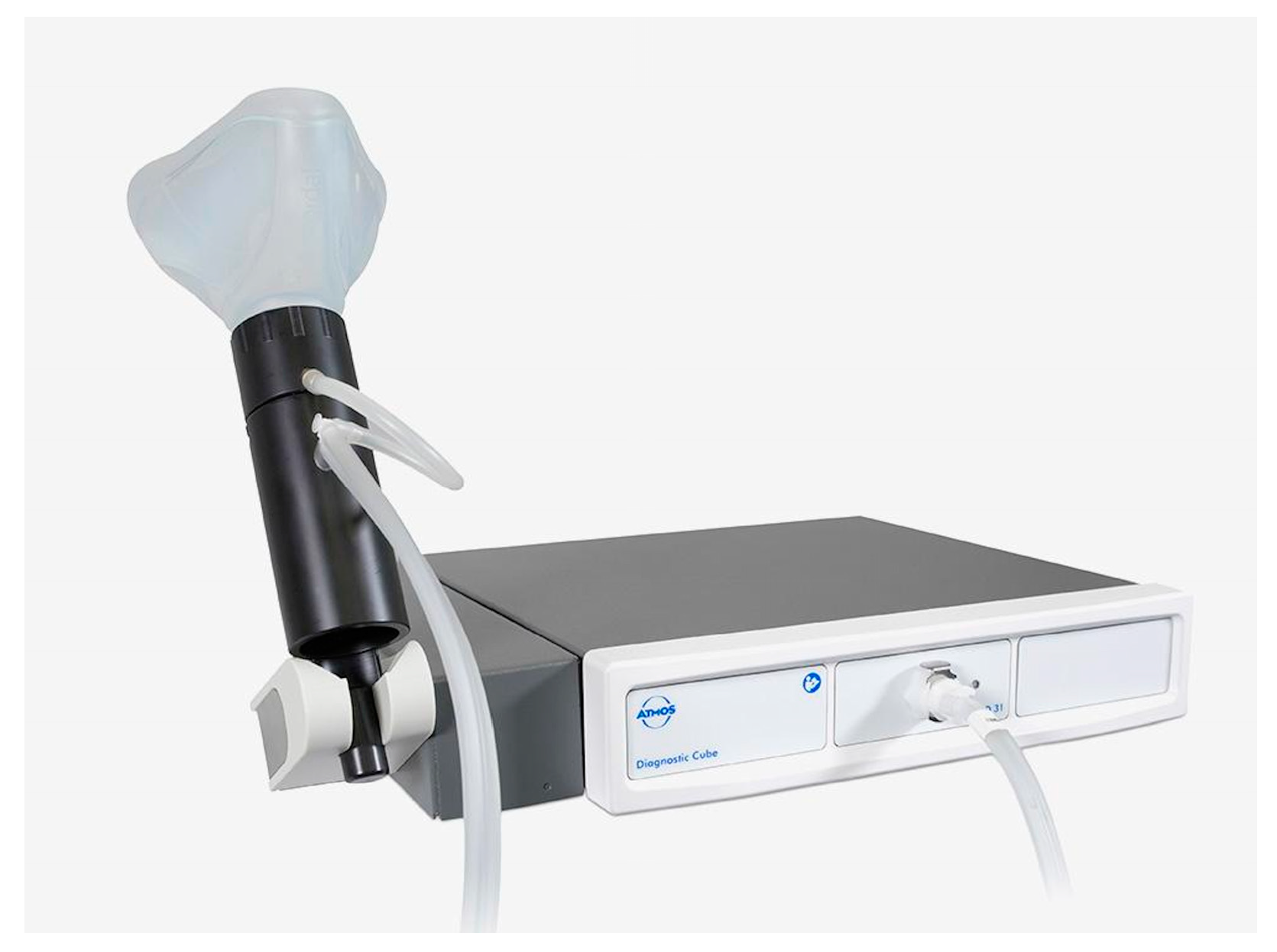
2. Materials and Methods

Study design: A retrospective study was performed on an orthodontic sample of pediatric patients with suspected OSAS and treated with RPE to further evaluate the functional effects on nasal resistance using active anterior rhinomanometry (AAR) with an ATMOS Rhino 31 rhinomanometer (Figure 1).
This study was conducted from an initial sample of 235 orthodontic patients, with a positive orthodontic history for oral breathing and/or snoring, provided by the same private practice (M.M). Specifically, the presence of the following: oral breathing related to obstructive cause (hypertrophy of adenoids and/or tonsils; hypertrophy of turbinate; septal deviation) or non-obstructive cause; disturbed sleep; and OSAS. All patients underwent a first orthodontic examination after otorhinic evaluation with a final diagnosis of mouth breathing.
Participants: From the initial sample, a first selection of patients was made by applying the following exclusion criteria: age less than 5 years or greater than 12 years, dento-cranial facial deformities in general, dental abnormalities, orthodontic treatment already performed or in progress; and the above inclusion criteria: contraction of the upper maxilla with a need to expand at least 5 mm, positive history of oral respiration.
Study size: The sample recruited consisted of 122 children, aged 5–12 years, 53 males and 69 females.
Data Collection: For each patient, at time T0 (pre-treatment), an active anterior rhinomanometry with mask (AAR) was conducted by the same operator (G.S.) to examine respiratory function. Specifically, the ATMOS Rhino 31 rhinomanometer was used in the present study to test nasal respiratory pressure and respiratory airflow. This device enables real-time monitoring of airflow trends by synchronous recording of pressure and flow difference.
In addition, each patient underwent the Scales for sleep-related breathing disorders Questionnaire (SRDB) or Pediatric Sleep Disorders Questionnaire (PSQ-SRDB), completed by the parent, in order to validate the orthodontic history of OSAS.
Moreover, intra- and extra-oral photos, imprints for study models, RX OPT, and TLL were performed for each subject. These records permitted confirmation of the diagnosis of upper jaw contraction, continuing with orthodontic treatment based on the application of a rapid palatal expander of the type Hyrax or Pendex on deciduous or permanent dentition. The protocol calls for the device to be activated 0.25 mm per day until, clinically, contact is detected between the palatal cusp of the upper first molar or upper deciduous second molar with the buccal cusp of the lower first molar or lower deciduous second molar. At this point, the expander is left in place for at least 4 months. If at T0 (pre-expansion) the AAR was clinically significant, the exam is repeated by the same operator (G.S.) at T1 (post-RPE), corresponding to a period of three months after screw fixation. Indeed, the purpose of the present study is to determine the magnitude of improvement on respiratory function by evaluation of nasal resistance values and the concomitant evolution in terms of sleep quality and OSAS. Because of this, the questionnaires, to be completed by the parent and mentioned above, were again administered at T1.
Changes between pre- and post-treatment in nasal resistance to airflow were assessed in order to examine any improvement induced by the application of RPE on respiratory function. Lastly, the final score that emerged from the questionnaires was compared between T0 and T1 with the aim of evaluating the subjective perception of the patient and parents regarding the potential improvement generated by the treatment on nasal breathing, sleep quality, and daytime behavior.
Statistical methods: The statistical analysis was conducted by analyzing the resistance to airflow opposed by the two nostrils, checking whether between post-treatment and pre-treatment the changes appeared similar between the right and left nasal side. By calculating the percentage difference between the values measured at T1 and those recorded at T0, subtracting the “pre” values from the “post” values, it was possible to infer for each patient belonging to the study group whether the treatment which they underwent generated a significant impact. Regarding the study group, the sample initially consisted of 44 subjects but 2 of them had out-of-scale resistance values (outliers) for one of the two nostrils. Because of this, they were excluded from the statistical analysis, reducing the final sample to 42 (Figure 2).
In addition, the null hypothesis H0 that RPE results in a mean change in nasal resistances of 0 was tested by the Student’s t-test on the variation variable. The use of this test instead of the similar Z-test was justified by not knowing the value of the variance in the population. Finally, the a posteriori formula was employed to calculate the margin of error and confidence level related to sample size and the Chi-square test for a single sample to verify the homogeneity of the sample by sex and age. In all the analyses mentioned, an alpha significance level of 0.05 was used. For the statistical analysis of data, the IBM SPSS Statistics software was employed, which was carried out using frequency tables for the qualitative variables, and then calculated in version 28.

3. Results

A total of 122 patients eligible for the study were evaluated. This initial group, at T0 (pre-RPE), underwent AAR and the SRDB or PSQ-SRDB questionnaire. At T0, the AAR was clinically significant in 42 subjects (study group), 27 females (64%) and 15 males (36%), with a mean age of 7.7 years (range 5–12 years; median 8 years) (Table 1). In this case, the AAR was repeated at T1 (post-RPE) in order to assess the extent of improvement on respiratory function and sleep quality at the end of orthopedic treatment. The population was homogeneous with respect to the sex variable (p = 0.217), while it was not homogeneous for the age variable (p = 0.000).
Among them, 37 were found to be oral respiratory from an obstructive cause while 5 manifested non-obstructive oral breathing. Compared with 37 oral breathing patients from an obstructive cause, 26 had adenoid hypertrophy (70%), 19 had tonsillar hypertrophy (51%); 21 showed hypertrophic turbinates (57%); and 19 were affected by septal deviation (51%). In addition, 15 patients were affected by snoring (41%) and 4 manifested apneas (11%) (Table 2). In contrast, of the five oral respiratory patients from a non-obstructive cause, four revealed the concomitant presence of snoring and apneas (80%) (Table 3).
Relative to the oral obstructive respirators, it was investigated whether the patients had previously undergone or were on the waiting list for adenotonsillectomy or adenoidectomy surgery. It was found that five of them had had prior surgery (four underwent adenotonsillectomy and one underwent adenoidectomy) while five subjects were on the waiting list for adenotonsillectomy.
Nasal resistance values (pre- and post-treatment) assessed with AAR: Figure 3a,b shows, using box plots, the distributions of the variables representing the change between T0 and T1 of the resistances divided by left and right nostril. In them, the dashed blue lines are placed at the mean value of these variables, indicative of the general effect of treatment for both nostrils on the subjects in the sample under study. From the analysis of these, it can be deduced that, on average, the therapy led to a general reduction in the values of resistance to airflow opposed by both nostrils: for the left cavity, a negative average pre-/post-treatment change of 24.63% is observed, while for the right nostril, an average decrease of 26.65% is observed between T0 and T1.
Using the Student’s t-test on the variable variation, with the null hypothesis that the mean variance is zero, it was analyzed whether the reduction in nasal resistance to airflow opposed by the two nostrils was statistically significant. The t-test, performed for each nostril, shows that in both nasal sides the null hypothesis was rejected (p = 0). From this, we infer that RPE treatment had a significant impact on nasal airflow resistance in both nostril sides. Specifically, the confidence intervals, calculated at 95% in the two nostrils, attest that in the sample, representative of the population examined, the variable variation takes on negative values between −11% and −37% (confidence interval 95%) (Table 4 and Table 5). As can be seen, in both tests the p-value has a value of 0, which suggests that the result of the t-test is very strong; thus, the variance on average was not zero. Therefore, despite the presence of zero or negative changes in resistance, it is still possible to show that treatment with RPE induces a statistically significant and positive improvement on nasal function, at least in terms of reduction in expressed nasal resistance.
  • SRDB and PSQ-SRDB Questionnaires (pre- and post-treatment): SRDB questionnaire was submitted to the parents of 36 patients in the study group. At pre-treatment, only in one case did it produce an outcome close (final score of 29) to that considered clinically significant for OSAS (greater than or equal to 33) (Figure 4). Evaluating the modification between T0 and T1 in the overall final score, completion of the above questionnaire was not statistically significant, if not confounding at times. In fact, although 49% of the subjects (17 patients) manifested a reduction in score following RME treatment, in contrast, 31% (11 cases) had an unchanged final score between pre- and post-treatment. Finally, in 20% of the cases (7 subjects) there was a worsening of the total score between T0 and T1.
    Similarly, the PSQ-SRDB questionnaire, administered to six subjects in the study group, in no case led to a final score of 0.33 or higher, a cut-off considered on the basis of the current literature to be significant for OSAS (Figure 5). Assessing the overall change in the score between pre- and post-treatment, a reduction in the score was observed in 67% of cases (four patients); in contrast, an increase is seen in 33% of cases (two subjects). So, once again the completion of the parental questionnaire was not found to be statistically significant.

4. Discussion

Rapid palatal expansion (RPE) is a validated and recommended therapeutic approach for treating a constricted maxillary upper jaw, particularly in growing patients. However, there has long been controversy surrounding the potential role of RPE in alleviating nasal obstruction and improving physiological nasal breathing. This controversy stems from the belief that restricted airways may contribute to the pathophysiology of obstructive sleep apnea syndrome (OSAS) in growing patients [27]. The scientific evidence shows that there is a direct correlation between nasal resistance, AHI, snoring time, and time spent with SpO2 below 90% in the pediatric population and a significant inverse correlation between total sleep time, sleep quality, and mean arterial oxygen saturation during sleep [17]. In order to assess any RPE-induced improvements on respiratory dynamics, it is necessary to quantify nasal aerodynamics, demonstrating changes between pre- and post-expansion. In recent times, rhinomanometry has made it possible to investigate nasal airway physiology with quantifiable and comparable parameters; in fact, numerous studies in the literature have evaluated the functional effects of RPE on nasal breathing with this instrumental method. Compadretti et al. [28] evaluated a sample of 27 children aged 5 to 13 years with maxillary contraction using AAR, acoustic rhinometry, and postero-anterior teleradiography before and 12 months after RPE, comparing it to an untreated control group. The AAR reports a remarkable reduction in nasal resistance after orthodontic treatment only under decongestion. De Filippe et al. [29] found a statistically significant reduction in NAR immediately after expansion (25.5%), stable over the following 9–12 months, similar to what was achieved by Monini et al. [30] and Halicioglu et al. [31]. Nasal airway impairment and the presence of obstruction are believed to be related to high nasal resistance and, in some cases, an oral breathing pattern. In order to determine the average nasal cross-sectional size and the effect of age on nasal breathing, Warren et al. [32] analyzed 102 patients aged 6 to 15 years under resting breathing conditions. The study showed that from 6 to 14 years, nasal airway size increases by an average of 0.032 cm2 per year: this explains the physiological reduction in nasal resistance. At the same time, the nasal breathing mode seems to increase with age: after the age of 8, most children have a nasal breathing mode. Thus, nasal breathing seems to be subject to physical and behavioral aspects in the context of an individual’s development. Beyond the known influence of nasal size on nasal breathing ability, there is evidence, even with adequate airways, of a certain percentage of children breathing orally.
The literature considers that RPE has a local effect on the upper airway, which is more evident at the bony level than at the mucosal level; it is probably related to compensatory post-expansion hypertrophy of the nasal mucosa and soft tissue adaptation [22,33]. Although most studies agree that the RPE induces a reduction in nasal resistance to airflow, the promoted improvements on nasal resistances show a weak correlation with dental expansion [34]. In addition, the influence of RPE on nasal resistances does not appear to be stable: a return to values close to baseline is expected about 30 months after treatment [22,23,35]. Therefore, it is not possible to demonstrate a long-lasting effect of RME on nasal respiratory function.
In most of the above studies, the study group was selected by placing the presence of obstructive diseases of the first airway as an exclusion criterion, performing the evaluation on an otolaryngologically healthy population. Only Monini et al. [30] and Langer et al. [35] included subjects with obstructive problems. The present retrospective study wanted to further investigate the possibility of obtaining beneficial effects on obstructive and non-obstructive pediatric oral respiratory patients with concomitant upper jaw contraction through the use of an RPE. The sample examined appeared to be mostly affected by obstructive respiratory disease, so as to favor oral breathing. Second, we aimed to explore the treatment potential available to the orthodontic specialist, who increasingly assesses patients with interdisciplinary problems.
Since the scientific evidence [36] shows that the skeletal and dento-alveolar effects of RPE are more significant when treatment is undertaken before the peak of pubertal growth, it was decided to include only growing patients aged 5 to 12 years. The current study group, consisting of 42 individuals with a mean age of 7.7 years, was not evenly distributed by age. The reason lies in the scientific evidence: depending on the age of the subject and the sutural activity present, there is a variable amount of orthopedic expansion. This variation is related to the fact that the resistance to maxillary expansion increases with sutural activity and thus the extent of expansion decreases as the degree of skeletal maturity increases [37,38]. For this reason, the literature agrees in emphasizing the importance of early treatment with RPE in order to maximize the magnitude of skeletal expansion in the presence of a perfectly adaptable palatine medial suture [39].
The results obtained from the AAR show a reduction in nasal resistance to airflow, confirming the findings achieved by the previously mentioned studies. In particular, the airflow varied in a statistically significant manner (p-value = 0.00) with an improvement that is similar for both nostrils, although showing slightly higher, by about two percentage points, on the right nasal side (Figure 3b).
Regarding the subjective perception of the patient and parents about the possible improvement in nasal breathing, quality of sleep, and daytime behavior, no statistically significant results emerged with the two questionnaires employed, SRDB and PSQ-SRDB. In fact, in no case did a subject in the study group score equal to or higher than that considered, based on the literature, to be significant for OSAS. On the other hand, evaluating the change in the overall score between T0 and T1, the PSQ-SRDB shows a reduction in the same in 67% of the cases, while in 33% the final score appears even increased (Figure 5). Relative to the SRDB scale, assessing the change in the final score between pre- and post-treatment, it appears reduced in 49% of cases, unchanged in 31%, and increased in 20% (Figure 4).
As is well known, obstructive sleep apnea syndrome (OSAS) in childhood is a severely underdiagnosed condition. Confirmatory PSG is held to be the gold standard [15,16] to diagnose this syndrome; however, it has numerous limitations. So, alternative tools are emerging, which include questionnaires. These simple, noninvasive, repeatable and reproducible devices can be the starting point for the correct diagnostic framing of the patient. Because they are filled out by the parent, they encourage more information about the disease, still burdened by general misinformation. In particular, the 22-item Pediatric Sleep Questionnaire shows the highest sensitivity for the identification of mild OSAS [40], as confirmed with the systematic review of Parenti et al. [41] and holds the best diagnostic accuracy. Another issue affecting this condition is the lack of a universally accepted classification for estimating the severity of pediatric OSAS. Many studies employ the apnea and hypopnea index (AHI) to classify the disease as mild (1 < AHI > 4.9), moderate (5 < AHI > 9.9), or severe (AHI > 10). In this context, active anterior rhinomanometry (AAR) with mask, the gold standard for accuracy and precision in detection, can emerge as a valuable tool to identify pediatric patients with clinically severe OSAS, helping in the management of long waiting lists so as to prioritize access to confirmatory PSG [17,42].
Limits: The limitations of this study include selection bias (based on one center), lack of diversity considering one person conducted all measurements, the lack of a control group to which to relate the results obtained, and the small sample size of the study group. It would be appropriate to implement the cohort of subjects with pure oral breathing, an expression of a flawed habit inherent in them, not related to any impediment or obstacle to physiological nasal breathing. In this way, it would be possible to compare the extent of improvement on nasal airflow resistance and to assess under which condition, whether in the presence of one or more obstructive sites or whether in the absence of obstruction, a more significant improvement is seen. It might be useful to also include in the study protocol the performance of AAR under nasal decongestion, in order to eliminate the possible influence that the turbinate variable might exert on the results obtained. Finally, it is necessary to carefully examine the long-term stability of the results obtained, taking into account the role of remaining craniofacial growth and the rate of relapse in nasal airflow resistance post-maxillary expansion.

5. Conclusions

Based on the results emerged from AAR, taking into account the small sample size examined, we can conclude as follows:
  • In cases of maxillary contraction, the orthodontic treatment with RPE has positive effects on nasal respiratory function, in terms of resistance to airflow.
  • In the orthodontic patient requiring upper jaw expansion, with concomitant presence of obstructive pathology of the first airway (adenotonsillar hypertrophy), rapid palate expander treatment should be evaluated as a priority.
  • Only at a later stage should patients be re-evaluated by the ENT specialist in order to re-investigate the status of their nasal function post-expansion, to assess the actual need for the surgical procedure.
  • Given the positive effects attested in the current literature on sleep disorders, it is possible that, following orthodontic treatment, the evolution of apneic/snore peak to full-blown adult OSAS will be interrupted.
  • With a view to greater interdisciplinarity and for the benefit of the young patient, it is desirable that ENT specialists and pediatricians be aware of the potential of this orthodontic method, less invasive and burdened with fewer risks than the surgical approach.

Author Contributions

Conceptualization, L.L. and F.C.; methodology, F.C.; software, M.M.; validation, M.M. and L.L.; formal analysis, M.F.; investigation, G.S.; resources, M.M.; data curation, M.F.; writing—original draft preparation, M.F. and C.P.; writing—review and editing, F.C. and C.P.; visualization, F.C.; supervision, F.C.; project administration, M.M.; All authors have read and agreed to the published version of the manuscript.

Funding

This research received no external funding.

Institutional Review Board Statement

This study was conducted in accordance with the Declaration of Helsinki, and approved by the Institutional Ethics Committee of University of Ferrara (protocol code 10/2022: 10/2/2022).

Informed Consent Statement

Informed consent was obtained from all subjects involved in the study.

Data Availability Statement

All authors assured that all data and materials as well as software application or custom code support their published claims and comply with field standards. The raw data supporting the conclusions of this article will be made available by the corresponding author on request.

Conflicts of Interest

The authors declare no conflicts of interest.

References

  1. Dhull, K.; Verma, T.; Dutta, B. Prevalence of deleterious oral habits among 3- to 5-year-old preschool children in Bhubaneswar, Odisha, India. Int. J. Clin. Pediatr. Dent. 2018, 11, 3. [Google Scholar]
  2. Majorana, A.; Bardellini, E.; Amadori, F.; Conti, G.; Polimeni, A. Timetable for oral prevention in childhood–developing dentition and oral habits: A current opinion. Prog. Orthod. 2015, 16, 39. [Google Scholar] [CrossRef] [PubMed]
  3. Moss, M.; Salentijn, L. The primary role of functional matrices in facial growth. Am. J. Orthod. 1969, 55, 77. [Google Scholar] [CrossRef] [PubMed]
  4. Kamínková, J.; Fornůsek, L.; Vĕtvicka, V.; Ríhová, B.; Kaspárek, L.; Vránová, M.; Pekárek, J. The effect of transfer factor on phagocytosis and humoral immunity in children with recurrent middle ear inflammations. Cas. Lek. Cesk. 1983, 122, 9. [Google Scholar]
  5. Iwasaki, T.; Sugiyama, T.; Yanagisawa-Minami, A.; Oku, Y.; Yokura, A.; Yamasaki, Y. Effect of adenoids and tonsil tissue on pediatric obstructive sleep apnea severity determined by computational fluid dynamics. J. Clin. Sleep. Med. 2020, 16, 8. [Google Scholar] [CrossRef] [PubMed]
  6. Zhao, T.; He, H. Pediatric mouth breathing and malocclusion. Chin. J. Orthod. 2019, 26, 8. [Google Scholar]
  7. Vogler, R.; Ii, F.; Pilgram, T. Age-specific size of the normal adenoid pad on magnetic resonance imaging. Clin. Otolaryngol. Allied Sci. 2000, 25, 392–395. [Google Scholar] [CrossRef] [PubMed]
  8. Pacheco, M.; Fiorott, B.; Finck, N.; Araújo, M. Craniofacial changes and symptoms of sleep-disordered breathing in healthy children. Dental Press. J. Orthod. 2015, 20, 7. [Google Scholar] [CrossRef] [PubMed]
  9. Katz, E.; D’Ambrosio, C. Pediatric obstructive sleep apnea syndrome. Clin. Chest Med. 2010, 31, 34. [Google Scholar] [CrossRef]
  10. Magliulo, G.; Iannella, G.; Ciofalo, A.; Polimeni, A.; De Vincentiis, M.; Pasquariello, B.; Montevecchi, F.; Vicini, C. Nasal pathologies in patients with obstructive sleep apnoea. Acta Otorhinolaryngol. Ital. 2019, 39, 250–256. [Google Scholar] [CrossRef]
  11. American Thoracic Society. Standards and indications for cardiopulmonary sleep studies in children. American Thoracic Society. Am. J. Respir. Crit. Care Med. 1996, 153, 866–878. [Google Scholar] [CrossRef] [PubMed]
  12. Sateia, M.J. International classification of sleep disorders-third edition: Highlights and modifications. Chest 2014, 146, 1387–1394. [Google Scholar] [CrossRef] [PubMed]
  13. Alsubie, H.; BaHammam, A. Obstructive Sleep Apnoea: Children are not little Adults. Paediatr. Respir. Rev. 2017, 21, 72–79. [Google Scholar] [CrossRef] [PubMed]
  14. Marcus, C.; Brooks, L.; Draper, K.; Gozal, D.; Halbower, A.; Jones, J.; Schechter, M.; Sheldon, S.; Spruyt, K.; Ward, S.; et al. Diagnosis and management of childhood obstructive sleep apnea syndrome. Am. Acad. Pediatr. 2012, 130, 576–584. [Google Scholar]
  15. Muzumdar, H.; Arens, R. Diagnostic issues in pediatric obstructive sleep apnea. Proc. Am. Thorac. Soc. 2008, 5, 263–273. [Google Scholar] [CrossRef] [PubMed]
  16. M. D. S. S. G. U. I. e. DCOM Linee Guida Nazionali per la Prevenzione ed il Trattamento Odontoiatrico del Russamento e Della Sindrome Delle Apnee Ostruttive nel Sonno in età Evolutiva. 2016. Available online: https://www.salute.gov.it/imgs/C_17_pubblicazioni_2484_allegato.pdf (accessed on 16 March 2016).
  17. Clement, P.; Gordts, F. Clement PA, Gordts F.; Standardisation Committee on Objective Assessment of the Nasal Airway, IRS, and ERS. Consensus report on acoustic rhinometry and rhinomanometry. Rhinology 2005, 43, 169–179. [Google Scholar] [PubMed]
  18. Lagravere, M.; Major, P.; Flores-Mir, C. Long-term skeletal changes with rapid maxillary expansion: A systematic review. Angle Orthod. 2005, 75, 1046–1052. [Google Scholar]
  19. Izuka, E.; Feres, M.; Pignatari, S. Immediate impact of rapid maxillary expansion on upper airway dimensions and on the quality of life of mouth breathers. Dental Press. J. Orthod. 2015, 20, 43–49. [Google Scholar] [CrossRef]
  20. Ozbek, M.; Memikoglu, U.; Altug-Atac, A.; Lowe, A. Stability of maxillary expansion and tongue posture. Angle Orthod. 2009, 79, 214–220. [Google Scholar] [CrossRef]
  21. McNamara, J.J.; Lione, R.; Franchi, L.; Angelieri, F.; Cevidanes, L.; Darendeliler, M.; Cozza, P. The role of rapid maxillary expansion in the promotion of oral and general health. Prog. Orthod. 2015, 16, 33. [Google Scholar] [CrossRef]
  22. Matsumoto, M.; Itikawa, C.; Valera, F.; Faria, G.; Anselmo-Lima, W. Long-term effects of rapid maxillary expansion on nasal area and nasal airway resistance. Am. J. Rhinol. Allergy 2010, 24, 161–165. [Google Scholar] [CrossRef] [PubMed]
  23. Itikawa, C.; Valera, F.; Matsumoto, M.; Lima, W. Effect of rapid maxillary expansion on the dimension of the nasal cavity and on facial morphology assessed by acoustic rhinometry and rhinomanometry. Dental Press. J. Orthod. 2012, 17, 129–133. [Google Scholar] [CrossRef]
  24. Fastuca, R.; Perinetti, G.; Zecca, P.; Nucera, R.; Caprioglio, A. Airway compartments volume and oxygen saturation changes after rapid maxillary expansion: A longitudinal correlation study. Angle Orthod. 2015, 85, 955–961. [Google Scholar] [CrossRef] [PubMed]
  25. Zeng, B.; Ng, A.; Qian, J.; Petocz, P.; Darendeliler, M.; Cistulli, P. Influence of nasal resistance on oral appliance treatment outcome in obstructive sleep apnea. Sleep 2008, 31, 543–547. [Google Scholar] [CrossRef] [PubMed]
  26. Suratt, P.; Turner, B.; Wilhoit, S. Effect of intranasal obstruction on breathing during sleep. Chest 1986, 90, 324–329. [Google Scholar] [CrossRef] [PubMed]
  27. Alexander, N.S.; Schroeder, J.W. Pediatric obstructive sleep apnea syndrome. Pediatr. Clin. N. Am. 2013, 60, 827–840. [Google Scholar] [CrossRef] [PubMed]
  28. Compadretti, G.; Tasca, I.; Bonetti, G. Nasal airway measurements in children treated by rapid maxillary expansion. Am. J. Rhinol. 2006, 20, 385–393. [Google Scholar] [CrossRef]
  29. De Felippe, N.O.; Da Silveira, A.; Viana, G.; Kusnoto, B.; Smith, B.; Evans, C. Relationship between rapid maxillary expansion and nasal cavity size and airway resistance: Short- and long-term effects. Am. J. Orthod. Dentofac. Orthop. 2008, 134, 370–382. [Google Scholar] [CrossRef]
  30. Monini, S.; Malagola, C.; Villa, M.; Tripodi, C.; Tarentini, S.; Malagnino, I.; Marrone, V.; Lazzarino, A.; Barbara, M. Rapid maxillary expansion for the treatment of nasal obstruction in children younger than 12 years. Arch. Otolaryngol. Head. Neck Surg. 2009, 135, 2. [Google Scholar] [CrossRef]
  31. Halicioğlu, K.; Kiliç, N.; Yavuz, İ.; Aktan, B. Effects of rapid maxillary expansion with a memory palatal split screw on the morphology of the maxillary dental arch and nasal airway resistance. Eur. J. Orthod. 2010, 32, 716–720. [Google Scholar] [CrossRef]
  32. Warren, D.W.; Hairfield, W.; Dalston, E. Effect of age on nasal cross-sectional area and respiratory mode in children. Laryngoscope 1990, 100, 89–93. [Google Scholar] [CrossRef] [PubMed]
  33. Enoki, C.; Valera, F.; Lessa, F.; Elias, A.; Matsumoto, M.; Anselmo-Lima, W. Effect of rapid maxillary expansion on the dimension of the nasal cavity and on nasal air resistance. Int. J. Pediatr. Otorhinolaryngol. 2006, 70, 1225–1230. [Google Scholar] [CrossRef] [PubMed]
  34. Timms, D. The effect of rapid maxillary expansion on nasal airway resistance. Br. J. Orthod. 1986, 13, 221–228. [Google Scholar] [CrossRef] [PubMed]
  35. Langer, M.; Itikawa, C.; Valera, F.; Matsumoto, M.; Anselmo-Lima, W. Does rapid maxillary expansion increase nasopharyngeal space and improve nasal airway resistance? Int. J. Pediatr. Otorhinolaryngol. 2011, 75, 122–125. [Google Scholar] [CrossRef] [PubMed]
  36. Baccetti, T.; Franchi, L.; Cameron, C.; McNamara, J.J. Treatment timing for rapid maxillary expansion. Angle Orthod. 2001, 71, 343–350. [Google Scholar] [PubMed]
  37. Wertz, R. Skeletal and dental changes accompanying rapid midpalatal suture opening. Am. J. Orthod. 1970, 58, 41–66. [Google Scholar] [CrossRef]
  38. Melsen, B.; Melsen, F. The postnatal development of the palatomaxillary region studied on human autopsy material. Am. J. Orthod. 1982, 82, 329–342. [Google Scholar] [CrossRef] [PubMed]
  39. Bala, A.; Campbell, P.; Tadlock, L.; Schneiderman, E.; Buschang, P. Short-term skeletal and dentoalveolar effects of overexpansion. Angle Orthod. 2022, 92, 55–63. [Google Scholar] [CrossRef]
  40. Chervin, R.; Hedger, K.; Dillon, J.; Pituch, K. Pediatric sleep questionnaire (PSQ): Validity and reliability of scales for sleep-disordered breathing, snoring, sleepiness, and behavioral problems. Sleep Med. 2000, 1, 21–32. [Google Scholar] [CrossRef]
  41. Parenti, S.I.; Fiordelli, A.; Bartolucci, M.; Martina, S.; D’Antò, V.; Alessandri-Bonetti, G. Diagnostic accuracy of screening questionnaires for obstructive sleep apnea in children: A systematic review and meta-analysis. Sleep Med. Rev. 2021, 57, 101464. [Google Scholar] [CrossRef]
  42. Klaus, V.; Gregor, B.-H.; Andreas, L.; Alina, N.; Franz, P.; Klaus-Dieter, W. The new agreement of the international RIGA consensus conference on nasal airway function tests. Rhinology 56 2018, 2, 133–143. [Google Scholar]
Figure 1. ATMOS Rhino 31. The rhinomanometer is equipped with operational buttons, electromechanical pressure transducers, amplifiers, and analog-to-digital transducers. It features a display for real-time monitoring and visualization of results, as well as an integrated printer. Additionally, it consists of a measuring probe with olives or a mask for recording airflow; a pressure probe, a flexible tube, or a tube with nasal adapter to record pressure differences; and a handle for gripping.
Applsci 14 03721 g001
Figure 2. Flow chart diagram.
Applsci 14 03721 g002
Figure 3. Distribution of pre-/post-treatment airflow change in the left (a) and right nostrils (b). (a) % variation in pressure pre-/post-treatment—left nostril; (b) % variation in pressure pre-/post-treatment—right nostril.
Applsci 14 03721 g003
Figure 4. Questionnaire scales for sleep-related breathing disorders (SRBDs).
Applsci 14 03721 g004
Figure 5. Pediatric Sleep Questionnaire (PSQ) in the reduced 22-item form (PSQ-SRDB), translated and validated in Italian.
Applsci 14 03721 g005
Table 1. Demographic characteristics of the study group categorized by sex and age variables.
SexN ObservedN ExpectedResidualMean AgeMedian Age
F2521.04.0
M172.0−4.0
Total42 7.718
Table 2. Descriptive analysis of the frequency of obstructive variables within the obstructive oral breather population (N = 37) in the study group.
TimeN TotAdenoid
Hypertrophy
Tonsillar HypertrophyHypertrophied TurbinatesSeptal DeviationSnoringApnea
Pre (T0)3726192119154
Pre (T0)3770%51%57%51%41%11%
Table 3. Descriptive analysis of the variables of apnea and snoring investigated in non-obstructive oral breathers (N = 5) belonging to the study group.
TimeN TotSnoringApnea
Pre (T0)544
Pre (T0)580%80%
Table 4. Descriptive statistics of the variation variable.
Right NostrilNMeanStandard DeviationMean Standard Error
% Variation42−26.1375%27.57237%4.25451%
Left nostrilNMeanStandard deviationMean standard error
% Variation42−24.63007%40.688442%6.278363%
Table 5. Results of Student’s t-test.
Test Value = 0
tglTwo-Sided p-ValueDifference of Mean95% Confidence Interval of Difference
LowerUpper
Left nostril% Variation−3.923410.000−24.630071%−37.30948%−11.95066%
Right nostril% Variation−6.143410.000−26.13747%−34.7296%−17.5453%
Disclaimer/Publisher’s Note: The statements, opinions and data contained in all publications are solely those of the individual author(s) and contributor(s) and not of MDPI and/or the editor(s). MDPI and/or the editor(s) disclaim responsibility for any injury to people or property resulting from any ideas, methods, instructions or products referred to in the content.

