Article

Polydopamine-Coated Copper-Doped Co3O4 Nanosheets Rich in Oxygen Vacancy on Titanium and Multimodal Synergistic Antibacterial Study

1
College of Chemistry, Chemical Engineering and Resource Utilization, Northeast Forestry University, Harbin 150040, China
2
Key Laboratory of Microsystems and Microstructures Manufacturing, School of Medicine and Health, Harbin Institute of Technology, Harbin 150080, China
3
School of Life Science and Technology, Harbin Institute of Technology, Harbin 150001, China
*
Authors to whom correspondence should be addressed.
Submission received: 2 April 2024 / Revised: 18 April 2024 / Accepted: 20 April 2024 / Published: 26 April 2024
(This article belongs to the Special Issue Nanocomposite Based Materials for Various Applications)

Abstract

:
Medical titanium-based (Ti-based) implants in the human body are prone to infection by pathogenic bacteria, leading to implantation failure. Constructing antibacterial nanocoatings on Ti-based implants is one of the most effective strategies to solve bacterial contamination. However, single antibacterial function was not sufficient to efficiently kill bacteria, and it is necessary to develop multifunctional antibacterial methods. This study modifies medical Ti foils with Cu-doped Co3O4 rich in oxygen vacancies, and improves their biocompatibility by polydopamine (PDA/Cu-Ov-Co3O4). Under near-infrared (NIR) irradiation, nanocoatings can generate •OH and 1O2 due to Cu+ Fenton-like activity and a photodynamic effect of Cu-Ov-Co3O4, and the total reactive oxygen species (ROS) content inside bacteria significantly increases, causing oxidative stress of bacteria. Further experiments prove that the photothermal process enhances the bacterial membrane permeability, allowing the invasion of ROS and metal ions, as well as the protein leakage. Moreover, PDA/Cu-Ov-Co3O4 can downregulate ATP levels and further reduce bacterial metabolic activity after irradiation. This coating exhibits sterilization ability against both Escherichia coli and Staphylococcus aureus with an antibacterial rate of ca. 100%, significantly higher than that of bare medical Ti foils (ca. 0%). Therefore, multifunctional synergistic antibacterial nanocoating will be a promising strategy for preventing bacterial contamination on medical Ti-based implants.

1. Introduction

Bacterial infection has become a critical threat to global human health, which needs to be urgently addressed. The hazard of bacterial infections from implantable medical devices during clinical treatment is quite difficult to avoid [1], and can even be directly described as a fatal threat to patients. Medical titanium (Ti) and its alloys have been widely recognized for the clinical application of implants in orthopedics and dentistry due to their considerable chemical inertness, favorable mechanical properties, and excellent biocompatibility [2,3,4], but the problem of bacterial infections associated with their pre- and post-operative periods threatens their successful operation to a great extent, and the infections frequently manifest themselves in the localized pain and postponed healing [5], which can cause additional disturbances and economic losses to the patients and their families. In the past, the utilization of antibiotics for the treatment of bacterial infections arising from implants has obtained satisfactory results, but the abuse of antibiotics has also caused a variety of negative effects, such as the emergence of bacterial drug resistance and rapid bacterial evolution and mutation, etc., which have significantly diminished the efficacy of antibiotics [6,7]. Therefore, it is necessary to develop and innovate multifunctional nanomaterial platforms with higher efficiency and controllability to compensate for antibiotics.
In this regard, multifunctional nanomaterials’ platform has drawn tremendous attention in recent years to defend against pathogenic bacterial infections, owing to their distinctive physicochemical properties including a quantum size effect and high specific surface area, superior antibacterial efficiency, lower bacterial drug resistance, and biocompatibility [2,8]. The nanomaterials’ modification of Ti implants showed promising potential for improving the antibacterial performance. For instance, polydopamine (PDA) was able to form a stable and thin nanofilm on Ti implants, which could further coordinate with metal ions or organic compounds to prepare a multifunctional antibacterial layer [9,10]. Yiwen Li recently summarized progress of PDA-based antibacterial composites, indicating their convenient process and efficient activity for fabricating PDA composite antibacterial interfaces [11].
It is worth noting that Co-based nanomaterials have proven to be promising candidates for sterilization. For example, Li et al. found that a Co3O4 nanowire electrode was capable of discharging low voltage electricity to effectively prevent bacterial skin infections [12]. Tian et al. developed a hierarchical macro/mesoporous Co3O4-SiO2, which could eradicate the antibiotic-resistant bacteria [13]. The porous Co3O4 nanoplates possessed synergetic photothermal/photodynamic therapy through a DNA damage route [14]. Thus, applying Co3O4-based composites on Ti-based implants will be an alternative way to suppress bacterial infection. In addition, Cu is a perspective inorganic antibacterial agent and Cu-based nanocoatings can potentially combat bacterial adhesion as well as prevent the formation of biofilms on implantable medical devices [15]. For instance, the minimum bactericidal concentration (MBC) of Cu2+ against S. aureus was 7.04 μg/mL [16]. Ning et al. found that the MBC of Cu2+ against S. aureus was only 1.6 μg/mL [17]. Zhao et al. found that Cu2O-TiO2/Ti2O3/TiO exhibited the contact sterilization property [18]. Recently, Wang’s work proved that Cu2+ sterilization and contact sterilization were involved when 0.034 μg/mL of Cu2+ was released [19]. Inspired by these findings, Cu-doped Co3O4 is assumed to display enhanced Fenton-like activity due to the synergetic Co−Cu electronic interaction [20]. However, few papers on Cu-doped Co3O4-based antibacterial nanocoating on Ti-based implants were reported.
In this work, PDA/Cu-Ov-Co3O4 was designed and synthesized by electrochemical deposition, alkaline oxidation polymerization, and plasma-enhanced chemical vapor deposition (PECVD) methods. The presence of PDA coating enhanced its biocompatibility and photothermal property. Herein, PECVD was an effective strategy to modulate the micro-/nanostructures and composition of Cu-Ov-Co3O4, such as pores, oxygen defects, etc., which was regarded to be a potential factor for modulating the physicochemical properties. Co3O4, Ov-Co3O4, and Cu-Ov-Co3O4 were also prepared by similar methods. The antibacterial property of these materials to Gram-negative Escherichia coli (E. coli) and the Gram-positive Staphylococcus aureus (S. aureus) was examined under 808 nm of irradiation and compared. It was found that PDA/Cu-Ov-Co3O4 showed synergistic effects for sterilization including a metal ion release, Fenton-like reaction, and photothermal and photodynamic effect compared to these control groups. A systematic study disclosed that the ATP and protein levels in bacteria were downregulated, and the membrane permeability was increased after treatment by PDA/Cu-Ov-Co3O4.

2. Materials and Methods

2.1. Materials

Medical Ti foils (0.1 mm thickness, 10 mm in length and width, 99.9%) were purchased from Haiyuan Research Metals (Dongwan, China). Chloride hexahydrate (CoCl2·6H2O, 99.0%), potassium nitrate (KNO3, 99.0%), ammonium chloride (NH4Cl, 99.5%), sodium phosphate monobasic dihydrate (NaH2PO4·2H2O, 99.0%), ethanol absolute (CH3CH2OH, 99.7%), and copper chloride (CuCl2·2H2O, 99.0%) were bought from Chemical Reagent Co., Ltd. (Tianjin, China). Dopamine hydrochloride (C8H11NO2·HCl, 98.0%) and dimethyl sulfoxide (DMSO, 99.7%) were acquired from Macklin Biochemical Co., Ltd. (Shanghai, China). Tris base (C4H11NO3, 99.9%) was obtained from Gentihold Biotechnology Co., Ltd. (Beijing, China). In addition, a phosphate buffer solution (PBS) was purchased from Bio-Channel Biotechnology Co., Ltd. (Nanjing, China). Nutrient broth (NB) and nutrient agar (NA) were purchased from Hope Bio-Technology Co., Ltd. (Qingdao, China). Alternatively, the as-prepared NA plates could be stored at 4 °C for further use.

2.2. Characterization

The scanning electron microscope (SEM) with Quanta 200FEG (Thermo Fisher Scientific Inc., Waltham, MA, USA) and field emission transmission electron microscope (FETEM) with JEM-2100F (JEOL, Tokyo, Japan) were employed to analyze the surface morphology and compositions in the as-prepared samples, respectively. The high voltage was ranging from 10 to 20 kV. And the working distance was between 10 and 20 mm. The physical phases were recognized by X-ray diffraction (XRD, X’Pert PRO, Almelo, The Netherlands) with slow scanning modes at 3°/min in the range of 10 to 90°. The chemical element components were detected by X-ray photoelectron spectroscopy (XPS, ESCALAB 250Xi, Thermo Fisher Scientific Inc., Waltham, MA, USA). The electron paramagnetic resonance spectra (EPR) were conducted on a Bruker A300 spectrometer (A300-10/12, Ettlingen, Germany) with the X-band at a frequency of 9.853 GHz. The Raman spectra were acquired by a confocal Raman spectrometer (inVia-Reflex, New Mills, UK) with excitation wavelengths of 532, 633, or 785 nm. The chemical structure was determined with a Fourier transform infrared spectrometer (FTIR, Nicolet is50, Green Bay, WI, USA) using KBr blanks as controls in the range of 4000 to 400 cm−1. The optical properties of the samples were measured with an ultraviolet-visible-near-infrared (UV-Vis-NIR) spectrophotometer (U-4100, Tokyo, Japan) at a scanning rate of 600 nm/min. The fluorescence spectra were performed on a fluorescence spectrometer (Fluoromax-4, Irvine, CA, USA) with an excitation wavelength at 315 nm. The zeta potential was measured by Zetasizer Nano-ZS90 (Malvern Instruments, Malvern, UK). An atomic force microscope (AFM) was used on Agilent 5500 (Santa Clara, CA, USA). The topography images were used to measure the root mean square roughness (RMS) by Gwyddion 2.65 software.

2.3. Synthesis of Co3O4 Nanosheets on Ti foils

Medical Ti foils (10 mm × 20 mm × 0.1 mm) were polished sequentially with silicon carbide (SiC) sandpaper (1000, 2000, and 3000#) and cleaned by ultrasonication with anhydrous ethanol and deionized water for 10 min, respectively. Cleaned Ti foils were oven-dried at 50 °C and stored at room temperature for further use. Typically, Co3O4 precursors were prepared by the electrochemical deposition method in a three-electrode system including Ti foil as the working electrode, Pt foil as the counter electrode, and Ag/AgCl as the reference electrode. Herein, the electrolyte solution contained CoCl2 (0.02 mol/L), KNO3 (0.04 mol/L), and NH4Cl (0.2 mol/L). Also, the chronopotentiometry method was used to deposit the Co3O4 precursors at a current of 12.5 mA. After deposition, the Co3O4-precursor-modified Ti foils were placed in a quartz tube furnace and then calcined at 450 °C for 2 h under an N2 atmosphere to obtain porous Co3O4 nanosheets.

2.4. Fabrication of Ov-Co3O4 and Cu-Ov-Co3O4 Nanocoatings

The porous Co3O4 nanosheets were immersed in a CuCl2 (4 mmol/L) aqueous solution at room temperature for 24 h, which were denoted as Cu-Co3O4. Subsequently, the Co3O4 and Cu-Co3O4 nanosheets were treated by H2 plasma (3 sccm) at 30 W for 10 min under a vacuum of 30 Pa (named as Ov-Co3O4 and Cu-Ov-Co3O4, respectively).

2.5. Preparation of PDA/Cu-Ov-Co3O4 Nanocoatings

The as-prepared Cu-Ov-Co3O4 nanosheets were immersed in a dopamine hydrochloride solution (0.75 mg/mL) containing 1.5 mg/mL Tris (pH 8.5) for 24 h to form PDA films, which were named as PDA/Cu-Ov-Co3O4.

2.6. Photothermal Experiment

Different samples (10 mm × 10 mm) were placed into a 24-well plate with 500 μL of PBS to determine the photothermal property, and each well was irradiated by an 808 nm NIR laser (1.5 W/cm2, 10 min). Subsequently, the photostability curves were also conducted under the NIR irradiation (1.5 W/cm2, 10 min) with five cycles of a heating-cooling test. What’s more, photothermal tests with different power densities (0.5, 1.0, 1.5, and 2.0 W/cm2) were also documented separately. Notably, all the thermal mapping was monitored via a thermal imager (FLIR i7, Washington, DC, USA) together with a cell phone to steadily record the actual temperature change on surfaces every 30 s during the process.

2.7. Antibacterial Activity Assessment

E. coli (ATCC 25922, China) from Shanghai Bioresource Collection Center (SHBCC) (Shanghai, China) and S. aureus (CMCC(B) 26003, China) from Shanghai Yingxin laboratory equipment Co., Ltd. (Shanghai, China) were selected as assay strains whose antibacterial activity was validated using the plate-counting method. The two thawed strains were firstly incubated in a sterilized NB medium in a 37 °C shaker at 120 rpm and then gathered until the concentration of bacterial suspension reached ~108 colony-forming units per milliliter (CFUs/mL). In the meantime, five groups of different types of samples were placed in a 24-well plate, respectively, and sterilized together via ultraviolet light for an hour. Afterward, 500 μL of an as-prepared bacterial solution was dropped onto the surface of each sample, respectively, and co-incubated in a 37 °C incubator for 6 h. To maintain humidity and prevent the evaporation of the solution, between the wells of placed samples were full of sterile deionized water. Then, the samples were handled with the NIR laser (808 nm, 1.5 W/cm2) for 10 min, while the thermal imager was exposed to recording the temperature difference.
Furthermore, each sample was transferred to the containers with 2 mL of PBS before ultrasonic detachment (150 W, 50 Hz), and adherent bacteria were released from substrates after 5 min. After sequential dilutions, 40 μL of the bacterial liquids was dropped onto standard NA plates and spread well, then incubated at 37 °C overnight. Ultimately, bacterial colonies on the plates were captured in photos and counted. The relevant antibacterial rate was calculated utilizing the following formula: Antibacterial rate R = C E / E × 100 % , where C is the colony amount of the control group (Ti), and E is the colony amount of the modified samples.
In order to study the morphology of the bacteria attached to the sample surface, the bacteria on samples were firstly fixed with a 4% paraformaldehyde (Biosharp, Beijing, China) solution at 4 °C for 4 h, and then gradually dehydrated utilizing a gradient ethanol solution (10, 30, 50, 70, 80, 90, and 100%) for 15 min. Finally, all samples were dried overnight and covered with gold for further SEM observation.
The bacteria live/dead staining was performed with SYTO9 Green Fluorescent Nucleic Acid Stain (Mao Kang, Shanghai, China) and Propidium Iodide (PI) Red Fluorescent Nucleic Acid Stain (Beyotime, Shanghai, China) to visualize the antibacterial ability on different samples. The specific methods for dye staining were in accordance with the instructions. After a series of operations, the live/dead bacteria were recorded utilizing a confocal laser scanning microscope (CLSM, Zeiss LSM880, Jena, Germany).

2.8. Analysis of Antibacterial Mechanism

2.8.1. Bacterial Membrane Permeability Assay

To determine the integrity of the bacterial membrane, 8-anilino-1-naphthalenesulfonic acid (ANS, 96%, Aladdin, Shanghai, China) was devoted to evaluating the change in outer membrane permeability towards E. coli, due to the outer membrane unique to the Gram-negative bacteria. In contrast, o-Nitrophenyl β-D-galactopyranoside (ONPG, 98%, Yuanye, Shanghai, China) was employed to measure bacterial inner membrane change in the above two species of bacteria. Briefly, after the antibacterial process, each bacterial solution was disposed of a 500 μL ONPG solution (0.75 mol/L in NaH2PO4 buffer, pH 7.0). Finally, the yellow supernatant was removed and detected at the absorption of 420 nm (OD420) with a SPARK multifunctional microplate reader (Tecan, Austria GmbH, Kärnten, Austria).

2.8.2. Bacterial Total ROS Level Detection

The generation of total ROS levels within the bacteria was determined by a 2′, 7′-dichlorodihydrofluorescein diacetate (H2DCFDA) probe and a ROS Assay Kit (Cat#BL714A, Biosharp, Beijing, China). After irradiation for 10 min, the bacterial suspension was co-cultured with H2DCFDA (10 μmol/L in PBS) at 37 °C for 30 min, standing in the dark. After that, 150 μL of bacterial solutions was seeded onto a 96-well plate in order and analyzed by a SPARK multifunctional microplate reader (Tecan, Austria GmbH, Kärnten, Austria) at the excitation/emission wavelength of 488/525 nm.

2.8.3. Singlet Oxygen Measurement

The generation of singlet oxygen (1O2) from different sample surfaces was related to 1, 3-Diphenylisobenzofuran (DPBF). Firstly, 500 μL of DPBF (5 mmol/L in DMSO) was co-incubated with different samples in darkness for 10 min. After 808 nm of irradiation, the supernatant was collected and measured at the absorption of 420 nm (OD420) with a SPARK multifunctional microplate reader (Tecan, Austria GmbH, Kärnten, Austria).

2.8.4. Hydroxyl Radical Evaluation

The production of the hydroxyl radical (•OH) was measured with disodium terephthalate (DST). Different samples were immersed with 5mL of DST (0.5 mmol/L in deionized water) for 10 min in advance. After, with/without 808 nm of irradiation, the ability to generate •OH was detected with the fluorescence spectrometer with the excitation/emission wavelength of 315 nm/425 nm.

2.8.5. Bacterial Protein Leakage Assay

The protein assay kit (Cat#PC0020, Solarbio, Beijing, China) based on the bicinchoninic acid (BCA) method was carried out to study the bacterial protein leakage after irradiation, which was detected with absorbance values at 562 nm (OD562) by a SPARK multifunctional microplate reader (Tecan, Austria GmbH). In brief, after being irradiated for 10 min, respectively, 20 μL of as-treated bacterial solutions was removed and added into a 48-well plate with 200 μL of a solution, placing it in a 37 °C incubator for 30 min. Finally, withdraw the supernatant immediately and analyze the corresponding bacterial protein leakage by a SPARK multifunctional microplate reader (Tecan, Austria GmbH, Kärnten, Austria) at 562 nm.

2.8.6. Bacterial ATP Level Test

The enhanced ATP assay kit (Cat#S0026, Beyotime, Shanghai, China) was applied to research the bacterial metabolic activity. After irradiation, the already treated bacteria solution was collected and co-cultured with 100 μL of the lysate before being sonicated for 3 min to fully lyse. Lastly, suck out 20 μL of the supernatant and 100 μL of the working solution, inject them into a 96-well black plate together, and then measure the ATP levels through the program “chemiluminescence” via a SPARK multifunctional microplate reader (Tecan, Austria GmbH, Kärnten, Austria).

2.9. Metal Ion Release Test

The metal ion release behavior was determined by inductively coupled plasma optical emission spectrometry (ICP-OES, iCAP 7400, Thermo Fisher Scientific Inc., Waltham, MA, USA). The sample (10 mm × 10 mm) was placed into centrifuge tubes containing 10 mL of PBS (pH 7.4) and simultaneously immersed in a 37 °C water bath for 3, 6, 9, and 12 h, where the untreated PBS was used as a control group. At each time, the sample was removed and the corresponding solutions were transferred to a −80 °C refrigerator for freezing. After that, all sample tubes were freeze-dried for 24 h. Finally, 10 mL of HNO3 (2%) was added to each tube and diluted 3-fold for further ICP detection.

2.10. Cytotoxicity Evaluation

Mouse fibroblasts (L-929, Beijing, China) were used to study the biocompatibility of different samples [21]. The L-929 cells were cultured in a 37 °C incubator with 5% CO2 for 24 h and the cell medium was changed every other day. Subsequently, the cytotoxicity towards L-929 fibroblasts was evaluated by methyl thiazolyl tetrazolium (MTT) assays.
Briefly, 500 μL (3.0 × 10⁴ cells/mL) of the L-929 suspension was added onto the surface of various sterilized samples in a 24-well plate, respectively, and co-incubated for 1, 2, and 3 days, ensuring the sample surface was completely immersed by the cell medium. At each time point, the cell medium was removed before 50 μL of MTT (0.5 mg/mL) was added into the well and returned to the incubator for 4 h in darkness. The supernatant was carefully sucked out twice and then dissolved in 150 μL of DMSO, also being shaken, and vibrated for another 15 min. Finally, the supernatant was evaluated at the absorption of 490 nm (OD490) on a SPARK multifunctional microplate reader (Tecan, Austria GmbH, Kärnten, Austria) while the cell medium was applied as the control.

3. Results and Discussions

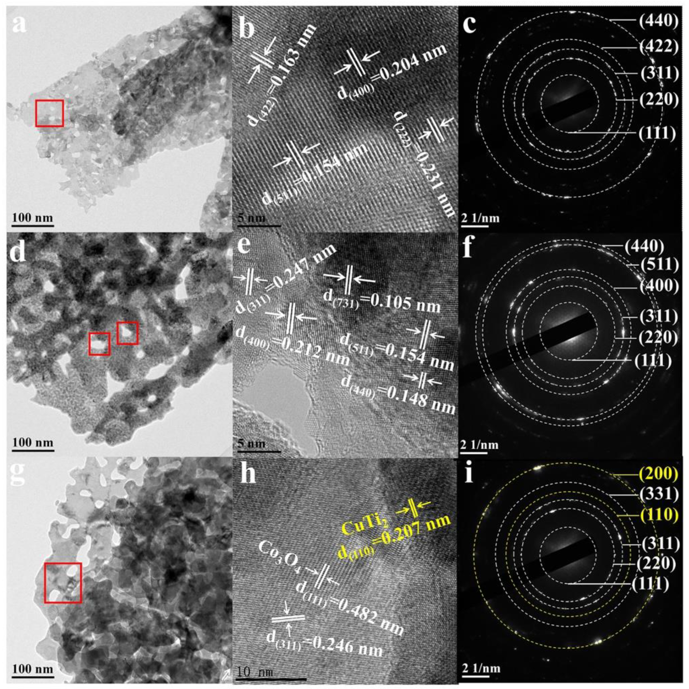
3.1. Synthesis and Physicochemical Properties

As illustrated in Figure 1a, the electrodeposition and H2 plasma annealing process were applied to synthesize Co3O4 ultrathin nanosheets on as-polished Ti foils’ surface. After modifying with PDA in Figure S1a–e, it could be found that the color on the PDA/Cu-Ov-Co3O4 surface obviously varied from brown to black. The morphology and structure features on different samples were observed by the SEM. As demonstrated in Figure 1b and Figure S1f, the surface of bare Ti foils was smooth. In contrast, after electrodeposition (Step 1 in Figure 1a), Co3O4 (Figure S1g and Figure 1c) ultrathin nanosheets were successfully prepared on the Ti surface and its morphology (Figure S1h and Figure 1d) did not change after calcination treatment, with an average pore size of approximately 1.5 μm. Interestingly, the surface of the Ov-Co3O4 (Figure S1h) and Cu-Ov-Co3O4 sample (Figure S1i and Figure 1d) displayed nanopores (Step 2 in Figure 1a), which were caused by high-energy H2 plasma. It is assumed that Cu doping and plasma treatment were able to regulate Ov concentration on the Co3O4 surface. Engineering of constructive surface defects allows us to optimize the energy band structure productively, thereby enhancing the optical absorption capacity and the photothermal conversion efficiency [22,23]. SEM images in Figure S1j and Figure 1e showed that a layer of polymer nanofilm was covered on the surface of nanosheets (Step 3 in Figure 1a), resulting from the polymerization of dopamine.
XRD was used to confirm the phase patterns related to as-prepared nanosheets. As revealed in Figure 1f, diffraction peaks at 40.17, 53.00, and 70.66° corresponded to (101), (102), and (103) crystal facets, which were matched with Ti (PDF#44-1294). The diffraction peaks for Co3O4@Ti and Ov-Co3O4@Ti were observed at 19.00, 31.27, 36.85, 38.53, and 65.25°, which were associated with the (111), (220), (311), (222), and (440) facets of Co3O4 (PDF#43-1003) [24,25]. In addition, for Cu-doped Ov-Co3O4 (named as Cu-Ov-Co3O4), the (002), (004), (110), and (006) peaks were found, in which the presence of Co3O4 and CuTi2 were observed assuming that Cu doping destroyed the long-range order of Ov-Co3O4. The mixture was mainly caused by high-energy H2 plasma treatment.
FTIR and Raman spectra were used to verify the presence of PDA. As presented in Figure 1g, the three bands at 1543, 1616, and 3315 cm−1 were related to the vibration mode of C−O, C=O, and O−H/N−H groups in PDA, respectively [26,27]. In contrast, no other bands for PDA were found in FTIR spectra of Cu-Ov-Co3O4@Ti, indicating the successful preparation of PDA coating. Furthermore, Raman spectra were carried out to compare various samples before and after H2 plasma treatment and Cu doping. As seen in Figure S2, two bands from 456 to 756 cm−1 appeared compared with PDA powder besides the stretching and deformation of PDA in the range of 1350 to 1650 cm−1 [4,10]. These emerging bands at 193, 467, 511, 606, and 678 cm−1 were ascribed to F12g, E2g, F22g, F32g, and A1g symmetry modes of Co3O4, respectively [28,29]. It is worth noting that A1g modes in Ov-Co3O4 and Cu-Ov-Co3O4 were red-shifted by 9 cm−1, probably owing to the introduction of Ov.
To verify the existence of Ov, EPR was conducted to explore the Ov quantity contained in Figure 1i as it is well known that a stronger signal represents more Ov sites. The EPR signal at g = 2.003 was caught in both Ov-Co3O4@Ti and Cu-Ov-Co3O4@Ti, the intensity of which was around 8.5 and 19.9 times that of Co3O4@Ti, respectively. Such improvements further proved that massive Ov was successfully introduced in the Ov-Co3O4 sample, which was further increased in Cu-Ov-Co3O4 due to Cu doping and H2 plasma treatment. Ov had been demonstrated to substantially decelerate the photogenerated carriers’ recombination rates, which resulted in the generation of more ROS, and then the photocatalytic antibacterial performance would be enhanced [22,30,31].
A TEM was further used to investigate the morphology and crystalline boundary. As seen in Figure 2a,d,g, the introduction of Ov led to a large amount of nanopores on nanosheets, as indicated by red squares. These pores would favor the exposure of active sites for the generation of ROS. HRTEM in Figure 2b,e shows the crystalline facets of (222), (400), (422), (511), (311), and (440), ascribed to Co3O4 [8]. It is seen that the main crystalline phase in Ov-Co3O4@Ti was still Co3O4 in Figure 2f. Besides the crystalline facets of Co3O4, the interlayer distance of 0.207 nm was found in Cu-Ov-Co3O4@Ti in Figure 2h, corresponding to the crystalline CuTi2 (110) facet. Eventually, SAED patterns in Figure 2i disclosed the coexistence of Co3O4 and CuTi2, consistent with XRD results.
The XPS analysis was carried out to evaluate the chemical component and changes in element status on the sample surface. As observed in Figure S3, XPS spectra revealed that the PDA/Cu-Ov-Co3O4 sample consisted of C, O, Co, N, and Cu elements. The fine deconvolution of Cu 2p in Figure 3a shows two peaks at a binding energy of 932.74 and 952.68 eV, respectively, consistent with the spin-orbital doublet peaks of Cu+ 2p3/2 and Cu+ 2p1/2, indicating that Cu+ ions were generated during H2 plasma treatment [32,33]. N 1s XPS spectra in Figure 3b display three peaks at 398.61, 400.14, and 401.53 eV, respectively, belonging to tertiary/aromatic (=N−R), secondary (R−NH−R), and primary (R−NH2) amine, respectively, further determining the successful attachment of PDA [34]. Co 2p XPS spectra of Co3O4 and Ov-Co3O4 in Figure 3c were deconvoluted into four peaks, indicating the coexistence of Co2+ and Co3+ on the sample surface [8,35,36]. Interestingly, Co0 was found in Cu-Ov-Co3O4, which might be caused by the reduction in high-energy H2 plasma. O 1s XPS spectra in Figure 3d indicate that the peaks for the Co−O−Co bond in Cu-Ov-Co3O4@Ti decreased greatly compared with Co3O4@Ti and Ov-Co3O4@Ti [7,9,36]. In addition, the peaks related to Ov increased from 24.3 to 43.4% after introducing Ov by H2 plasma, and finally increased to 70.9%, demonstrating that a large amount of Ov sites were generated in Cu-Ov-Co3O4@Ti.
Moreover, water contact angle measurement was conducted to verify the wettability of different samples in Figure S4. The Ti substrate exhibited hydrophilicity and the contact angle was 73.73°. However, Co3O4 and Ov-Co3O4 nanosheets became hydrophobic (149.44 and 153.78°), which might be caused by the nanostructured surface [37,38]. After the introduction of Cu, the contact angle decreased to 79.96° in the Cu-Ov-Co3O4@Ti, and that of PDA/Cu-Ov-Co3O4@Ti further decreased to 34.41° after PDA coating. It was assumed that the hydrophilic surface of PDA/Cu-Ov-Co3O4 could facilitate the infiltration of the electrolyte solution and subsequent attachment of bacteria, then triggering the next sterilization.

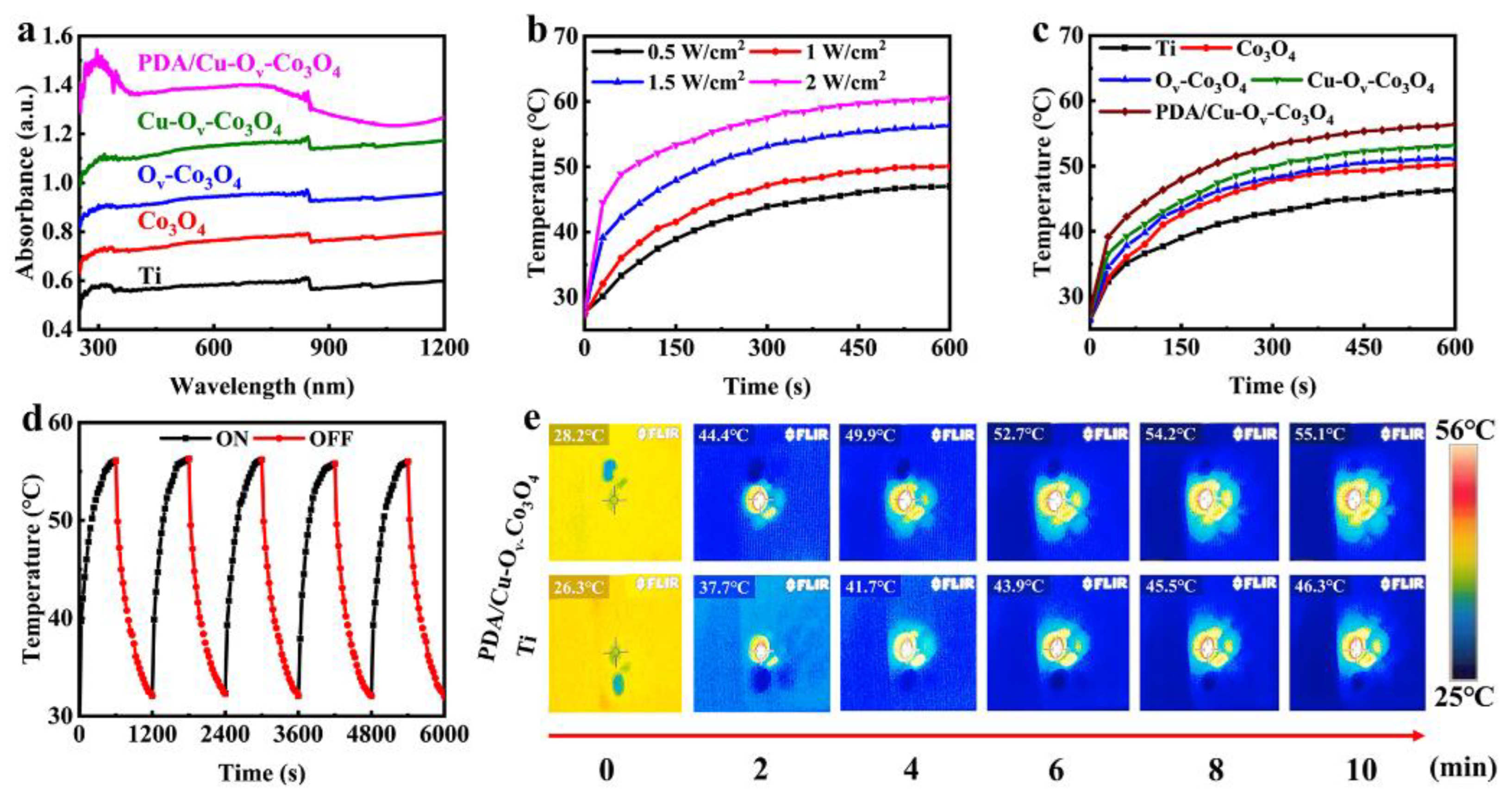
3.2. Photothermal Performance

The NIR absorption properties were investigated by the UV-Vis-DRS in Figure 4a. The absorbance intensity at 808 nm increased by 11.5, 24.4, and 48.3%, respectively, after the modification of Co3O4, Ov-Co3O4, and Cu-Ov-Co3O4 on Ti, which was caused by the decreased band gap due to the presence of Ov. According to solid physics, Ov could generate defective energy around the Fermi level, thus resulting in a smaller band gap [31,39]. The absorbance of PDA/Cu-Ov-Co3O4 further increased by 77.0% compared to bare Ti foils after coating a thin layer of PDA nanofilm. When irradiated by an 808 nm laser in Figure 4b, the surface temperature for PDA/Cu-Ov-Co3O4 in PBS was dependent on laser power densities of 0.5, 1.0, 1.5, and 2.0 W/cm2, respectively. To protect normal cell tissue, all thermal tests were performed at 1.5 W/cm2 for 10 min in Figure 4c. In the case of PDA/Cu-Ov-Co3O4@Ti, the temperature finally increased to 55.1 °C within 10 min, higher than that of control groups consisting of Ti (46.3 °C), Co3O4@Ti (50.2 °C), Ov-Co3O4@Ti (51.2 °C), and Cu-Ov-Co3O4@Ti (53.1 °C), respectively. The thermal stability was measured in Figure 4d. The highest temperature at the fifth cycle was almost unchanged compared to that in the first cycle, indicating good thermal stability in the aqueous solution. The real-time thermal mapping in Figure 4e reflects the photothermal region of PDA/Cu-Ov-Co3O4@Ti and bare Ti foils, and the real-time thermal mapping of the other three samples is shown in Figure S5. It is seen that the surface temperature was even for all cases. However, the PDA/Cu-Ov-Co3O4@Ti substrate clearly exhibited higher temperature than the medical bare Ti foils.

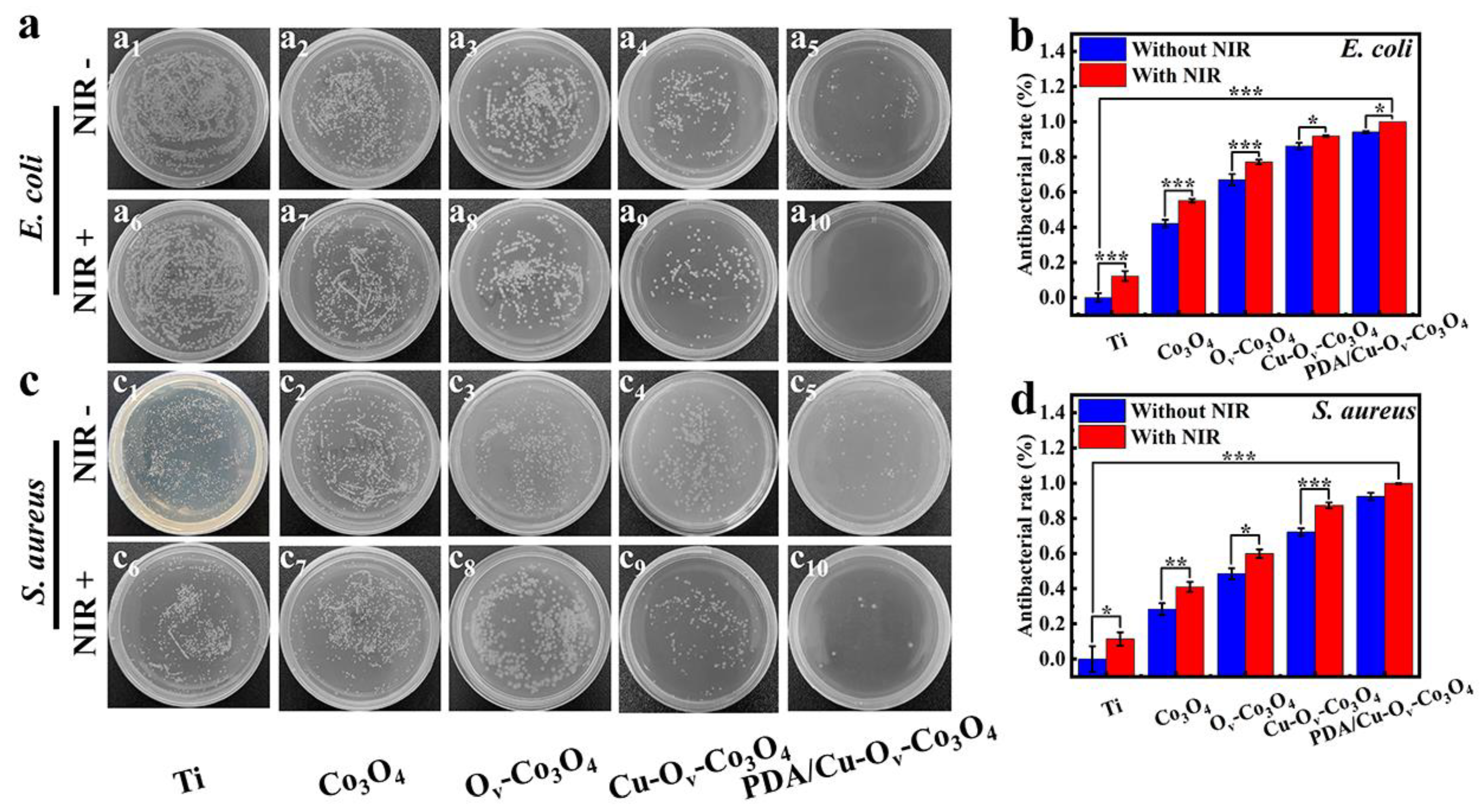
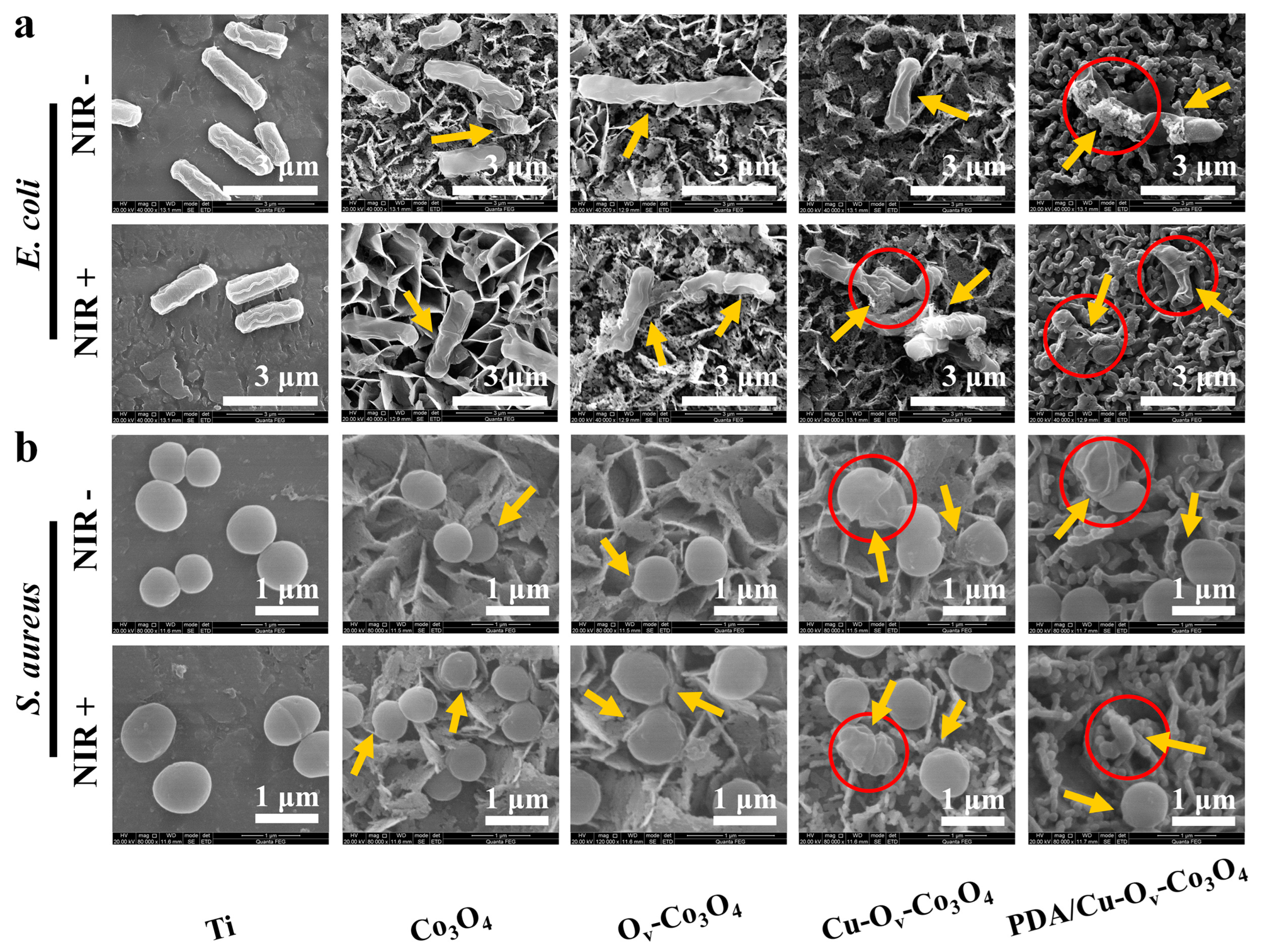
3.3. Antibacterial Activity

Next, the antibacterial performance was evaluated using different substrates. Based on the above characterization, the bacteria were synergistically sterilized by the photothermal effect, Fenton-like activity of Cu+ ions, and photodynamic effect of the semiconductor. The standard plate-counting experiment was conducted to study the antibacterial efficacy. In our study, E. coli (Gram-negative) and S. aureus (Gram-positive) were utilized as the strain types of investigation. Firstly, the dynamic growth curves of E. coli and S. aureus were measured on a 24-well plate by optical density at 600 nm (OD600) methods and the results are displayed in Figure S6, and bacteria in the logarithmic phase of microbial growth were selected for the following study.
After co-incubation with various samples and with or without 808 nm of irradiation, the amounts of E. coli and S. aureus on agar plates are shown in Figure 5a,c. The antibacterial efficacy results are shown in Figure 5b,d. In the dark condition, the Co3O4 nanosheets expressed weak toxicity against E. coli (44.22%) and S. aureus (22.27%) in comparison with bare Ti foils, whereas after the introduction of Ov, the antibacterial capability had an increase on E. coli (67.05%) and S. aureus (47.27%), respectively. In addition, the inhibition rate towards E. coli and S. aureus for Cu-Ov-Co3O4@Ti increased to around 86.14 and 72.69%, and further increased to 94.17 and 94.93% for PDA/Cu-Ov-Co3O4@Ti. Under NIR (808 nm, 1.5 W/cm2) conditions, the antibacterial activity for bare Ti foils was improved against E. coli (12.28%) and S. aureus (11.85%), whereas the inhibition rate for Co3O4@Ti increased to 55.14 and 37.58%, respectively. Similarly, the Ov-Co3O4 sample showed a higher inhibition rate to E. coli (77.08%) and S. aureus (53.43%) as well. Moreover, after the introduction of Cu, the Cu-Ov-Co3O4 sample killed 93.27% of E. coli and 91.85% of S. aureus. Most importantly, the PDA/Cu-Ov-Co3O4 sample demonstrated the highest inhibition rate of ca. 100% against both bacteria. Compared to the reported Ti-PDA/BP/ZnO, TiO2/MoS2/PDA/RGD, CpTi-SiO2-3Cu, and Ti-Co15 coatings, the good antibacterial result of PDA/Cu-Ov-Co3O4 was speculated to be attributed to the ROS, localized hyperthermia, and metal ion release effects [8,40,41,42].
Basically, in order to kill E. coli, the direct heating temperature should not be lower than 50 °C; otherwise, there is no substantial threat to the viability of E. coli. On the other hand, only at an irradiation temperature of up to 88.8 °C and a duration of 15 min for the NIR laser could S. aureus be completely killed [43]. However, higher localized temperature will cause serious damage to normal cells. Thus, as stated above, we would like to kill bacteria by utilizing a synergistic strategy at relatively lower temperature in our study.
The SEM was capitalized on to observe two bacterial morphologies and integrities on the sample surface. As seen in Figure 6a,b, the bacteria were detected on bare Ti foils after co-culturing for 6 h, exhibiting smooth and complete rod-shaped E. coli and spherical-shaped S. aureus without or with NIR laser irradiation. However, a slightly wrinkled and distorted structure change was observed in the Co3O4-based group (yellow arrows), respectively, ascribed to its physical interaction between bacterial membranes and nanosheets. In sharp contrast, under irradiation, Co3O4+NIR, Ov-Co3O4+NIR, Cu-Ov-Co3O4+NIR, and PDA/Cu-Ov-Co3O4+NIR groups caused the deformation of the bacterial membrane. It was noticed that the most severe disruptive effects containing lesions, holes, and shrinkage were observed in the PDA/Cu-Ov-Co3O4 group, which were related to its multifunctional antibacterial activity.
The antibacterial effect was further evaluated through a live/dead fluorescence staining assay. The live bacteria could be stained to green by STYO 9, and the dead bacteria showed a red color after PI staining. When both staining reagents are present, the fluorescence intensity originating from PI will be more intense. Figure S7 shows CLSM images on the sample surface after co-culturing for 6 h. It is shown that E. coli and S. aureus on the surface of Ti foils were stained with green with or without NIR irradiation. As for PDA/Cu-Ov-Co3O4@Ti, after exposure to NIR irradiation, dead bacteria with red fluorescence were seen, indicating that E. coli and S. aureus were substantially killed.
In addition, the bacterial membrane permeability was verified by ANS and ONPG analyses, respectively. ANS was utilized to determine the outer membrane permeability towards E. coli. Due to the difference in bacteria structure, the outer membrane was solely present in Gram-negative bacteria [44]. ANS was able to reinforce the fluorescence imaging via integrating with the hydrophobic district of the external membrane [45,46]. As displayed in Figure 7a, after being co-cultured with ANS, the Ti+NIR group was non-fluorescent, whereas blue fluorescence could be detected in the other four photothermal groups. In particular, as shown in Figure 7b, the PDA/Cu-Ov-Co3O4+NIR group exhibited higher fluorescence intensity than the Ti+NIR group, Co3O4+NIR group, Ov-Co3O4+NIR group, and Cu-Ov-Co3O4+NIR group. It is assumed that the photothermal effect played a significant role in severely disrupting the outer membrane structure of E. coli, thus accelerating the bacterial death.
The changing permeability of the bacterial internal membrane was also evaluated by ONPG hydrolysis assays. The ONPG could be hydrolyzed by intracellular β-galactosidase (β-Gal) to produce galactose and yellow ο-nitrophenol (ONP) detected with a strong absorption peak at 420 nm [47,48,49]. As indicated in Figure 7c,d, the OD420 values for bare Ti were lower than 0.2 for both E. coli and S. aureus. However, the OD420 value for PDA/Cu-Ov-Co3O4 increased by 55 and 57% for both E. coli and S. aureus compared with bare Ti in the dark, indicating enhanced inner membrane permeability. And upon NIR irradiation, the OD420 value further increased in both cases. The above results illustrated that the physical impedance of nanosheets and the photothermal effect synergistically enhanced the bacterial membrane permeability. Previous work also found that high temperature (around 50 °C) was able to enhance membrane permeability in the bacteria [40]. As a result, more external toxic substances such as ROS or metal ions could enter the cell interior and lead to its death [50].
Besides the enhanced membrane permeability, we next investigated the ROS levels in bacteria. After NIR irradiation, the ROS levels were detected by the H2DCFDA analysis. ROS in bacteria will oxidize DCFH to produce the fluorescein, named 2′, 7′-dichlorofluorescein (DCF), and the corresponding fluorescence intensity directly reflecting the intracellular ROS levels within the bacteria [13,51]. Figure 8a,b show that PDA/Cu-Ov-Co3O4@Ti promoted the generation of DCF in the dark compared to that of bare Ti, and the intensity increased by 51.5% for E. coli and 41.5% for S. aureus upon NIR irradiation. This result indicated that bacteria underwent severe oxidative stress on the PDA/Cu-Ov-Co3O4 substrate, thus inducing bacteria to produce more ROS. Hence, the antibacterial effect was partially attributed to internal damage induced by a considerable ROS attack within the bacteria, thus resulting in their oxidative stress.
Moreover, it was found in Figure S8 that the zeta potentials of pure E. coli and S. aureus suspensions were −34.46 and −27.56 mV. The zeta potentials of Co3O4@Ti, Ov-Co3O4@Ti, Cu-Ov-Co3O4@Ti, and PDA/Cu-Ov-Co3O4@Ti were −16.60, −8.84, −6.38, and −10.97 mV, respectively. It is seen that the introduction of Ov and Cu increased the zeta potentials. PDA coating slightly decreased the zeta potential, which was attributed to the negatively charged •OH groups in PDA. In brief, the electrostatic repulsion between bacteria and PDA/Cu-Ov-Co3O4@Ti was reduced compared with Co3O4@Ti, being conducive to capturing the bacteria [52].
As stated above, cell permeability was improved, which might lead to the protein leakage. Thus, the protein leakage assay was used to evaluate the protein levels in bacterial solutions. According to standard curves in Figure S9, the protein concentration was related to OD562 values. In general, higher OD562 values represent more protein released, which indicates that the severe disruption of the bacterial membrane or increase in membrane permeability to some extent occurred in the presence of antibacterial substrates. As shown in Figure 9a,b, PDA/Cu-Ov-Co3O4@Ti after irradiation exhibited the highest protein leakage for both E. coli and S. aureus, suggesting that the bacterial cell membrane was severely damaged by the photothermal effect [53,54]. As a result, more protein leakage led to the metabolic imbalance and ultimate death of bacteria.
The presence of hyperthermia, ROS, and metal ions resulted in the imbalanced bacterial metabolism; thus, ATP levels might be downregulated. It could be said that the inactivation of bacteria cells was associated with the ATP deficiency [55,56]. Figure 9c,d display that a significant decrease in ATP concentration was observed for PDA/Cu-Ov-Co3O4@Ti in the dark compared with bare Ti, and it kept decreasing under NIR irradiation. The downregulated ATP was sufficient to provide essential energy for bacterial proliferation during the cellular respiration and metabolism. Thus, the bacteria-respiratory process was severely disturbed upon NIR irradiation, thus leading to cell death.
ROS was of great importance to cell death. Thus, the identification of various ROS was conducive to disclosing the antibacterial mechanism. The DST fluorescent probe was used as the trap to detect the production capability of •OH [57,58]. As shown in Figure 10a, the characteristic fluorescence peak of DST alone was negligible in the dark, indicating that DST itself did not generate •OH. In the case of bare Ti foils, the fluorescence intensity was almost unchanged. However, the fluorescence intensity for the PDA/Cu-Ov-Co3O4 sample increased by 21.38% after incubating for 10 min in the dark, which was related to the self-supplied H2O2 due to the oxidation of PDA in the presence of solubilized O2. And this intensity further increased by 35.44% upon NIR irradiation, indicating that the photothermal effect could promote the generation of •OH. Cu-doped Ov-Co3O4 as a photosensitizer might possess a photodynamic effect during irradiation. 1O2 was measured by monitoring the DPBF absorbance at 420 nm [59,60]. As seen in Figure 10b, the fluorescence intensity of pure DPBF and in the presence of bare Ti foils was almost unchanged when placed in the dark for 10 min. However, an obvious intensity decrease was observed when PDA/Cu-Ov-Co3O4@Ti was irradiated by NIR irradiation for 10 min. It is assumed that an energy transfer occurred from O2 to 1O2 during NIR irradiation in semiconductive Cu-Ov-Co3O4.
EPR was applied to solidly validate the presence of •OH and 1O2. Under dark conditions, neither the Cu-Ov-Co3O4 nor PDA/Cu-Ov-Co3O4 sample could present any EPR signals corresponding to •OH or 1O2. However, the EPR signals of the Cu-Ov-Co3O4@Ti sample were significantly enhanced upon NIR irradiation. As seen in Figure 10c, the intensities show 1:2:2:1, which was ascribed to the typical •OH peaks. The 1:1:1 profile in Figure 10d means that the presence of 1O2 was due to photodynamic effects. Importantly, both signal intensities obviously increased towards PDA/Cu-Ov-Co3O4@Ti after irradiation for 10 min, indicating the generation of larger amounts of either •OH or 1O2 than that of the Cu-Ov-Co3O4@Ti sample.

3.4. Cell Compatibility

To further support the future application, the cytotoxicity of bare Ti foils and PDA/Cu-Ov-Co3O4@Ti was measured towards L-929 cells by MTT assays for 3 days. In Figure 11, the cell viability of the Ti group is about 92.5% at 3 days, indicating its negligible cytotoxicity towards L-929 cells. In the whole culturing process, the cell viability of the PDA/Cu-Ov-Co3O4@Ti sample was about 90.5% after culturing for 3 days. Consequently, PDA/Cu-Ov-Co3O4@Ti could be favorable to the normal cell propagation and growth, showing higher biocompatibility. In addition, Co and Cu ions were slowly released into the PBS over 12 h and the concentrations were determined by ICP tests. As seen in Figure S10, the concentrations of Co and Cu ions for PDA/Cu-Ov-Co3O4@Ti were 0.201 and 0.179 μg/mL after 6 h, followed by 0.533 and 0.290 μg/mL after 12 h, respectively. Combined with the spread plate results, it was demonstrated that the released metal ions in solutions might trigger the bacterial cytotoxicity along with the contact sterilization.

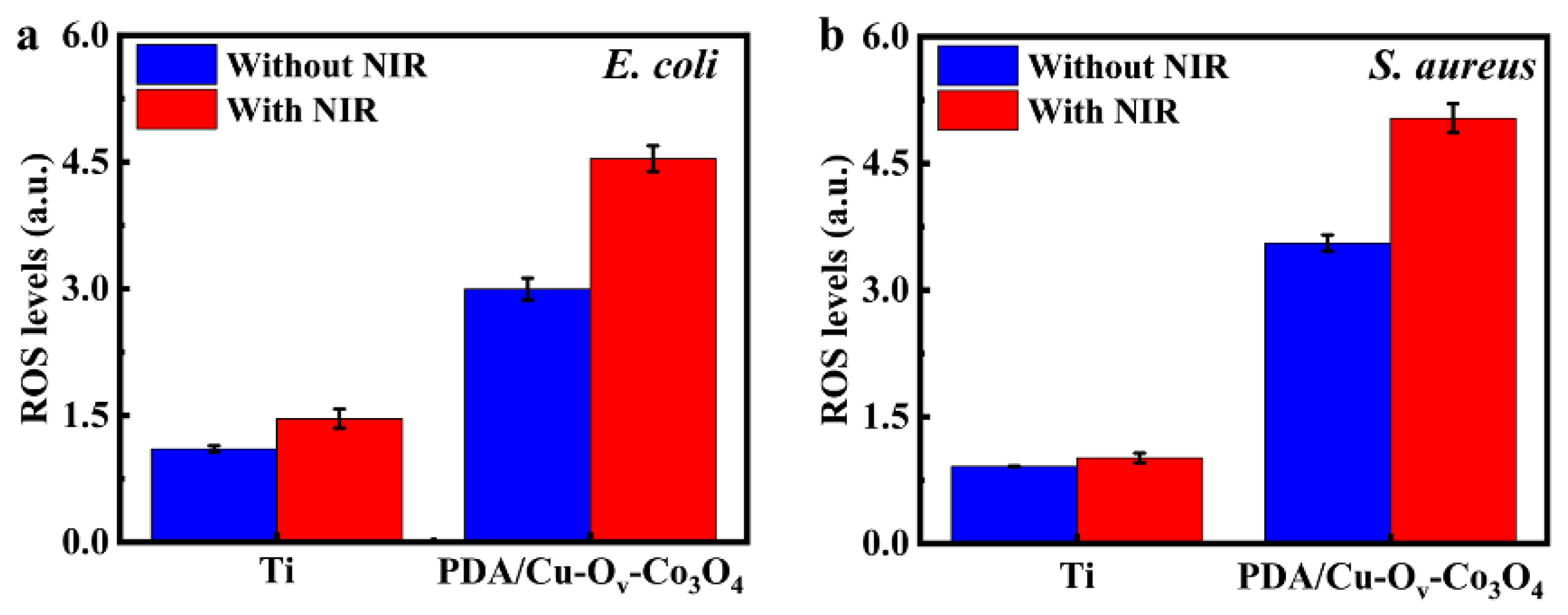
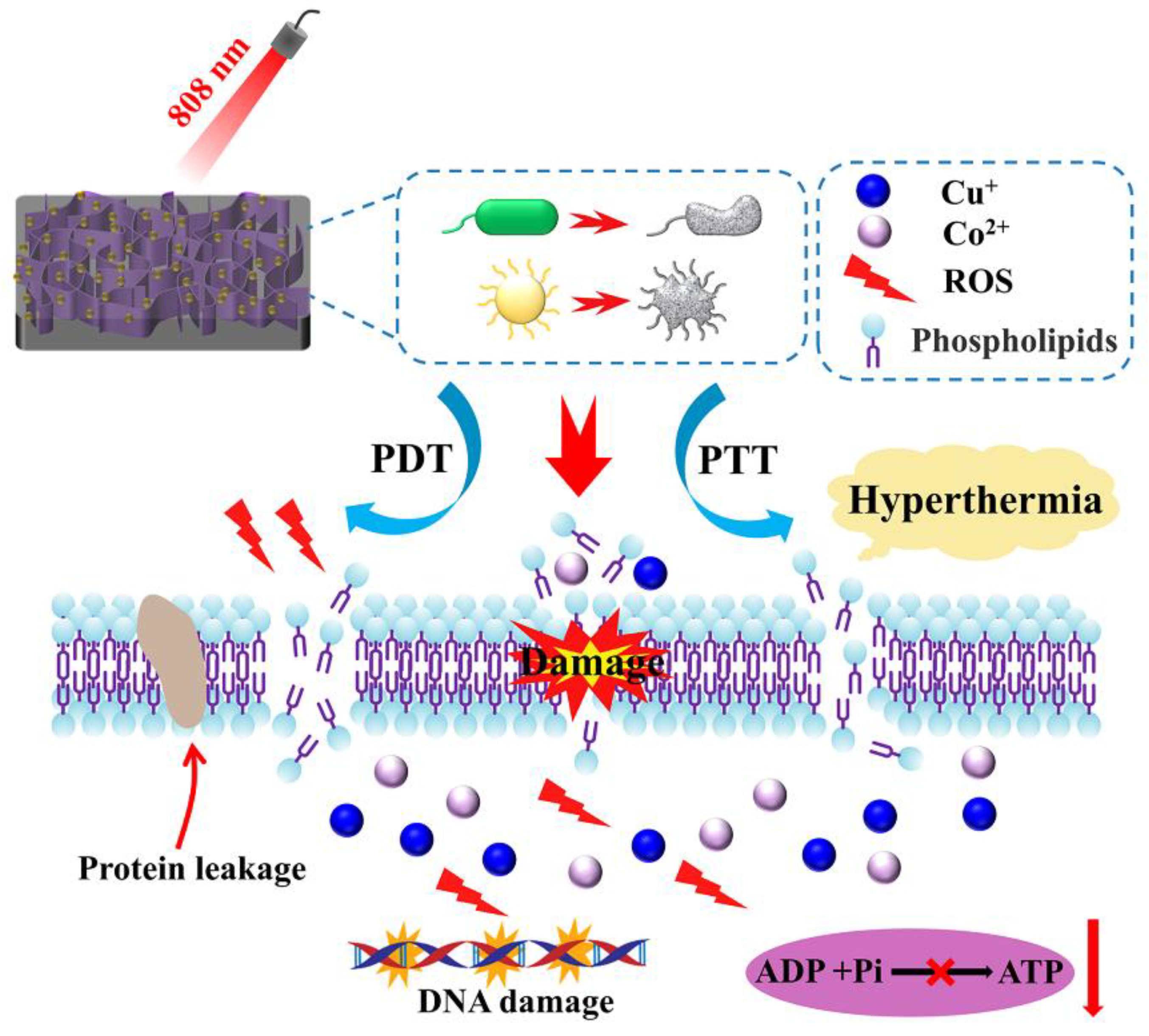
3.5. Synergistic Antibacterial Mechanism

According to the above results, a possible mechanism is proposed for PDA/Cu-Ov-Co3O4@Ti in Figure 12. AFM topography images of bare Ti and PDA/Cu-Ov-Co3O4@Ti are shown in Figure S11. The bare Ti shows a relatively smooth surface with an RMS value of 14.5 nm. In contrast, the RMS value of PDA/Cu-Ov-Co3O4@Ti increased to 92.4 nm. It is seen that the smooth surface of bare Ti became rough after the modification of these interconnected nanosheets. In general, the rough surface was conducive to the bacterial adhesion and subsequent killing. Based on the above characterization, released metal ions, the photothermal effect, and the photodynamic effect will be involved in our antibacterial study.
Firstly, the released metal ions from substrates could lead to cytotoxicity towards bacteria. Moreover, the catechol structure in PDA could spontaneously undergo an oxidation reaction with O2 to produce the o-benzoquinone and H2O2 [61]. As proven by XPS, Cu+ ions existed on PDA/Cu-Ov-Co3O4@Ti, which possessed Fenton-like activity with H2O2 for the generation of •OH. In particular, the Fenton-like activity could be further improved by the synergistic Cu−Co electronic coupling, which caused the upshift of the d-band center towards the Fermi level in Cu-Ov-Co3O4; then, the dissociation of H2O2 into •OH was boosted due to the favorable electron donation to H2O2 [20]. The high electrochemical oxidation potential of •OH could be used effectively to result in both DNA damage and protein denaturation.
Secondly, the photothermal effect could cause physical damage to bacteria, inducing intracellular oxidative stress accompanied with the physical impedance with ultrathin nanosheets. In addition, the photothermal effect led to the enhanced membrane permeability for the invasion of metal ions or ROS, and the release of protein. In addition, such a mechano-bactericidal mechanism has been proven in a nanostructured surface by mimicking insect wings [62]. Meanwhile, the photothermal effect could promote the generation of •OH and 1O2, as confirmed by DST and DPBF probe tests, as well as EPR characterization upon NIR irradiation. All these factors cause severe oxidative stress on bacteria and then the metabolism was imbalanced, as confirmed by the downregulated ATP levels.
Thirdly, electron transition occurred from VB to CB in PDA/Cu-Ov-Co3O4@Ti when excited by an 808 nm laser, resulting in the generation of electron-hole pairs. The high-energy electrons would reduce the o-benzoquinone structure during the generation of the H2O2 process, thus restoring the catechol structure. Meanwhile, type II energy transfer in photodynamic therapy could convert the dissolved O2 to 1O2, which has been proven by TEMP-trapped EPR tests. It is well concluded that the two types of ROS would efficiently inactivate the bacteria.

4. Conclusions

In summary, a multifunctional and highly biocompatible antibacterial nanocoating with medical Ti foils has been designed through H2 plasma treatment and PDA coating. By introducing Ov- and Cu-doped atoms, the light absorption of the coating in the NIR region can be improved by 77.0%, resulting in an enhanced photothermal effect. The hyperthermia generated by NIR irradiation greatly improves the permeability of the bacterial outer and inner membranes, and the released Cu and Co ions and exogenous ROS can smoothly penetrate the bacterial membrane. At the same time, combined with the Fenton-like effect of Cu+, it can convert H2O2, supplied by the self-oxidation of PDA, into •OH. The antibacterial efficiency was about 100% for both Gram-negative and Gram-positive bacteria. Hyperthermia and physical impedance further stimulate bacteria to produce more endogenous ROS, disrupting their metabolic balance. Under the synergistic action of various functions mentioned above, the bacterial membrane ruptures, leading to protein leakage and a decrease in ATP levels, and resulting in its death. In addition, MTT results indicate that the viability of the L-929 cell is higher than 90%, indicating that PDA nanocoating has cell compatibility. This work proposes a promising route for multifunctional synergistic antibacterial medical Ti-based coatings to address issues such as post-implantation infections in the future.

Supplementary Materials

The following supporting information can be downloaded at: https://0-www-mdpi-com.brum.beds.ac.uk/article/10.3390/ma17092019/s1, Figure S1: Digital and SEM images of (a,f) bare Ti foils, (b,g) Co3O4@Ti, (c,h) Ov-Co3O4@Ti, (d,i) Cu-Ov-Co3O4@Ti, and (e,j) PDA/Cu-Ov-Co3O4@Ti; Figure S2: Raman spectra of PDA powder and PDA/Cu-Ov-Co3O4@Ti; Figure S3: XPS survey scan of different samples including Co3O4@Ti, Ov-Co3O4@Ti, Cu-Ov-Co3O4@Ti, and PDA/Cu-Ov-Co3O4@Ti; Figure S4: Water contact angles of bare Ti, Co3O4@Ti, Ov-Co3O4@Ti, Cu-Ov-Co3O4@Ti, and PDA/Cu-Ov-Co3O4@Ti; Figure S5: Real-time thermal mapping corresponding to Co3O4@Ti, Ov-Co3O4@Ti and Cu-Ov-Co3O4@Ti during NIR irradiation for 10 min (808 nm, 1.5 W/cm2); Figure S6: Colony pictures corresponding to (a) E. coli and (b) S. aureus suspension in logarithmic phase stepwise dilutions during proliferation. Dynamic growth curves of (c) E. coli at 10 h and (d) S. aureus at 24 h in a 37 °C incubator; Figure S7: CLSM images of (a) E. coli and (b) S. aureus. Live bacteria were stained green with SYTO 9 and dead bacteria were stained red with PI; Figure S8: Zeta potentials of E. coli, S. aureus, Co3O4@Ti, Ov-Co3O4@Ti, Cu-Ov-Co3O4@Ti, and PDA/Cu-Ov-Co3O4@Ti; Figure S9: Standard curves for protein leakage and ATP assay on bacteria; Figure S10: ICP tests corresponding to Cu and Co ions under different time; Figure S11: AFM topography images of (a) bare Ti and (b) PDA/Cu-Ov-Co3O4@Ti.

Author Contributions

Conceptualization, Y.Q., T.S. and S.L.; Data curation, Y.Q. and Z.M.; Formal analysis and Methodology, J.Q., M.Y., Y.L., J.Z., X.L. and Z.M.; Investigation, J.Q. and Y.Q.; Writing—original draft and Writing—review and editing, J.Q., Y.Q., T.S. and S.L. All authors have read and agreed to the published version of the manuscript.

Funding

This work is supported by National Natural Science Foundation of China (No. 52272271), the National Science Fund for Distinguished Young Scholars (grant no. 51825202), and the Foundation for Heilongjiang Touyan Innovation Team (grant no. HITTY-20190036).

Institutional Review Board Statement

Not applicable.

Informed Consent Statement

Not applicable.

Data Availability Statement

Data are contained within the article and Supplementary Materials.

Conflicts of Interest

The authors declare no conflicts of interest.

References

  1. Yuan, Z.; Tao, B.; He, Y.; Mu, C.; Liu, G.; Zhang, J.; Liao, Q.; Liu, P.; Cai, K. Remote eradication of biofilm on titanium implant via near-infrared light triggered photothermal/photodynamic therapy strategy. Biomaterials 2019, 223, 119479. [Google Scholar] [CrossRef] [PubMed]
  2. Abd-Elaziem, W.; Darwish, M.A.; Hamada, A.; Daoush, W.M. Titanium-Based alloys and composites for orthopedic implants Applications: A comprehensive review. Mater. Des. 2024, 241, 112850. [Google Scholar] [CrossRef]
  3. Kelly, C.N.; Wang, T.; Crowley, J.; Wills, D.; Pelletier, M.H.; Westrick, E.R.; Adams, S.B.; Gall, K.; Walsh, W.R. High-strength, porous additively manufactured implants with optimized mechanical osseointegration. Biomaterials 2021, 279, 121206. [Google Scholar] [CrossRef] [PubMed]
  4. Lin, M.-H.; Wang, Y.-H.; Kuo, C.-H.; Ou, S.-F.; Huang, P.-Z.; Song, T.-Y.; Chen, Y.-C.; Chen, S.-T.; Wu, C.-H.; Hsueh, Y.-H.; et al. Hybrid ZnO/chitosan antimicrobial coatings with enhanced mechanical and bioactive properties for titanium implants. Carbohydr. Polym. 2021, 257, 117639. [Google Scholar] [CrossRef] [PubMed]
  5. Xie, X.; Mao, C.; Liu, X.; Zhang, Y.; Cui, Z.; Yang, X.; Yeung, K.W.K.; Pan, H.; Chu, P.K.; Wu, S. Synergistic Bacteria Killing through Photodynamic and Physical Actions of Graphene Oxide/Ag/Collagen Coating. ACS Appl. Mater. Interfaces 2017, 9, 26417–26428. [Google Scholar] [CrossRef] [PubMed]
  6. Blair, J.M.A.; Webber, M.A.; Baylay, A.J.; Ogbolu, D.O.; Piddock, L.J.V. Molecular mechanisms of antibiotic resistance. Nat. Rev. Microbiol. 2015, 13, 42–51. [Google Scholar] [CrossRef] [PubMed]
  7. Hughes, D.; Andersson, D.I. Evolutionary Trajectories to Antibiotic Resistance. Annu. Rev. Microbiol. 2017, 71, 579–596. [Google Scholar] [CrossRef] [PubMed]
  8. Fang, J.; Wan, Y.; Sun, Y.; Sun, X.; Qi, M.; Cheng, S.; Li, C.; Zhou, Y.; Xu, L.; Dong, B.; et al. Near-infrared-activated nanohybrid coating with black phosphorus/zinc oxide for efficient biofilm eradication against implant-associated infections. Chem. Eng. J. 2022, 435, 134935. [Google Scholar] [CrossRef]
  9. Avcu, E.; Bastan, F.E.; Guney, M.; Yildiran Avcu, Y.; Ur Rehman, M.A.; Boccaccini, A.R. Biodegradable Polymer Matrix Composites Containing Graphene-Related Materials for Antibacterial Applications: A Critical Review. Acta Biomater. 2022, 151, 1–44. [Google Scholar] [CrossRef] [PubMed]
  10. Yeroslavsky, G.; Lavi, R.; Alishaev, A.; Rahimipour, S. Sonochemically-Produced Metal-Containing Polydopamine Nanoparticles and Their Antibacterial and Antibiofilm Activity. Langmuir 2016, 32, 5201–5212. [Google Scholar] [CrossRef]
  11. Fu, Y.; Yang, L.; Zhang, J.; Hu, J.; Duan, G.; Liu, X.; Li, Y.; Gu, Z. Polydopamine antibacterial materials. Mater. Horiz. 2021, 8, 1618–1633. [Google Scholar] [CrossRef] [PubMed]
  12. Li, C.; Li, Z.; Zeng, Y.; Cao, X.; Zhao, H.; Yang, Y.Y.; Yuan, P.; Lu, X.; Ding, X. Co3O4 Nanowires Capable of Discharging Low Voltage Electricity Showing Potent Antibacterial Activity for Treatment of Bacterial Skin Infection. Adv. Healthc. Mater. 2022, 11, 2102044. [Google Scholar] [CrossRef]
  13. Tian, Y.; Yao, S.; Zhou, L.; Hu, Y.; Lei, J.; Wang, L.; Zhang, J.; Liu, Y.; Cui, C. Efficient removal of antibiotic-resistant bacteria and intracellular antibiotic resistance genes by heterogeneous activation of peroxymonosulfate on hierarchical macro-mesoporous Co3O4-SiO2 with enhanced photogenerated charges. J. Hazard. Mater. 2022, 430, 127414. [Google Scholar] [CrossRef]
  14. Yuan, M.; Xu, S.; Zhang, Q.; Zhao, B.; Feng, B.; Ji, K.; Yu, L.; Chen, W.; Hou, M.; Xu, Y.; et al. Bicompatible porous Co3O4 nanoplates with intrinsic tumor metastasis inhibition for multimodal imaging and DNA damage–mediated tumor synergetic photothermal/photodynamic therapy. Chem. Eng. J. 2020, 394, 124874. [Google Scholar] [CrossRef]
  15. Shahed, C.A.; Ahmad, F.; Günister, E.; Foudzi, F.M.; Ali, S.; Malik, K.; Harun, W.S.W. Antibacterial mechanism with consequent cytotoxicity of different reinforcements in biodegradable magnesium and zinc alloys: A review. J. Magnes. Alloys 2023, 11, 3038–3058. [Google Scholar] [CrossRef]
  16. Li, K.Q.; Xia, C.; Qiao, Y.Q.; Liu, X.Y. Dose-response relationships between copper and its biocompatibility/antibacterial activities. J. Trace Elem. Med. Biol. 2019, 55, 127–135. [Google Scholar] [CrossRef]
  17. Ning, C.; Wang, X.; Li, L.; Zhu, Y.; Li, M.; Yu, P.; Zhou, L.; Zhou, Z.; Chen, J.; Tan, G.; et al. Concentration Ranges of Antibacterial Cations for Showing the Highest Antibacterial Efficacy but the Least Cytotoxicity against Mammalian Cells: Implications for a New Antibacterial Mechanism. Chem. Res. Toxicol. 2015, 28, 1815–1822. [Google Scholar] [CrossRef] [PubMed]
  18. Zhao, X.; Cai, D.; Hu, J.; Nie, J.; Chen, D.; Qin, G.; Zhang, E. A high-hydrophilic Cu2O-TiO2/Ti2O3/TiO coating on Ti-5Cu alloy: Perfect antibacterial property and rapid endothelialization potential. Biomater. Adv. 2022, 140, 213044. [Google Scholar] [CrossRef]
  19. Wang, J.; Liang, M.F.; Pan, Y.; Sun, S.; Shen, T.; Wei, X.; Zhu, Y.; Liu, J.; Huang, Q. Control of surface composition and microstructure of nano super-hydrophilic TiO2-CuOy coatings through reactive sputtering to improve antibacterial ability, corrosion resistance, and biocompatibility. Appl. Surf. Sci. 2022, 578, 151893. [Google Scholar] [CrossRef]
  20. Guo, X.; Hu, B.; Wang, K.; Wang, H.; Li, B.; Guo, M.; Tian, Y.; Zhang, R.; Shi, S.; Han, Y. Cu embedded Co oxides and its fenton-like activity for metronidazole degradation over a wide pH range: Active sites of Cu doped Co3O4 with {112} exposed facet. Chem. Eng. J. 2022, 435, 132910. [Google Scholar] [CrossRef]
  21. Bandyopadhyay, A.; Mitra, I.; Goodman, S.B.; Kumar, M.; Bose, S. Improving biocompatibility for next generation of metallic implants. Prog. Mater Sci. 2023, 133, 101053. [Google Scholar] [CrossRef] [PubMed]
  22. Dias, L.F.G.; Abou-Hassan, A. Different applications, same story: Inspiring nanomedicine from photothermal catalysis to modulate the photothermal activity of nanomaterials through defects engineering. Coord. Chem. Rev. 2024, 507, 215751. [Google Scholar] [CrossRef]
  23. Zhang, J.; Chen, H.; Duan, X.; Sun, H.; Wang, S. Photothermal catalysis: From fundamentals to practical applications. Mater. Today 2023, 68, 234–253. [Google Scholar] [CrossRef]
  24. Yu, Q.; Liu, C.; Li, X.; Wang, C.; Wang, X.; Cao, H.; Zhao, M.; Wu, G.; Su, W.; Ma, T.; et al. N-doping activated defective Co3O4 as an efficient catalyst for low-temperature methane oxidation. Appl. Catal. B 2020, 269, 118757. [Google Scholar] [CrossRef]
  25. Zhou, J.; Yang, S.; Wan, W.; Chen, L.; Chen, J. Synergistic catalysis of mesoporous Cu/Co3O4 and surface oxygen vacancy for CO2 fixation to carbamates. J. Catal. 2023, 418, 178–189. [Google Scholar] [CrossRef]
  26. Jayaseelan, R.; Thennarasu, S.; Rajaji, P.; Nethaji, P.; Revathi, P.; Ramalingam, R.J.; Arokiyaraj, S. Long-life stability and high energy density storage MnCoPB-PDA/NF electrode material in hybrid supercapacitors. J. Energy Storage 2023, 72, 108303. [Google Scholar] [CrossRef]
  27. Dreyer, D.R.; Miller, D.J.; Freeman, B.D.; Paul, D.R.; Bielawski, C.W. Elucidating the Structure of Poly(dopamine). Langmuir 2012, 28, 6428–6435. [Google Scholar] [CrossRef] [PubMed]
  28. Li, Y.; Chen, T.; Zhao, S.; Wu, P.; Chong, Y.; Li, A.; Zhao, Y.; Chen, G.; Jin, X.; Qiu, Y.; et al. Engineering Cobalt Oxide with Coexisting Cobalt Defects and Oxygen Vacancies for Enhanced Catalytic Oxidation of Toluene. ACS Catal. 2022, 12, 4906–4917. [Google Scholar] [CrossRef]
  29. Wang, X.; Li, X.; Mu, J.; Fan, S.; Chen, X.; Wang, L.; Yin, Z.; Tadé, M.; Liu, S. Oxygen Vacancy-rich Porous Co3O4 Nanosheets toward Boosted NO Reduction by CO and CO Oxidation: Insights into the Structure–Activity Relationship and Performance Enhancement Mechanism. ACS Appl. Mater. Interfaces 2019, 11, 41988–41999. [Google Scholar] [CrossRef]
  30. Zhang, D.; Wang, M.; Wei, G.; Li, R.; Wang, N.; Yang, X.; Li, Z.; Zhang, Y.; Peng, Y. High visible light responsive ZnIn2S4/TiO2−x induced by oxygen defects to boost photocatalytic hydrogen evolution. Appl. Surf. Sci. 2023, 622, 156839. [Google Scholar] [CrossRef]
  31. Li, G.; Blake, G.R.; Palstra, T.T.M. Vacancies in functional materials for clean energy storage and harvesting: The perfect imperfection. Chem. Soc. Rev. 2017, 46, 1693–1706. [Google Scholar] [CrossRef] [PubMed]
  32. Singh, H.; Kumar, S.; Sharma, P.K. Tunable exciton-plasmon coupled resonances with Cu2+/Cu+ substitution in self-assembled CuS nanostructured films. Appl. Surf. Sci. 2023, 612, 155831. [Google Scholar] [CrossRef]
  33. Fornero, E.L.; Murgida, G.E.; Bosco, M.V.; Hernández Garrido, J.C.; Aguirre, A.; Calaza, F.C.; Stacchiola, D.; Verónica Ganduglia-Pirovano, M.; Bonivardi, A.L. CuGaO2 delafossite as a high-surface area model catalyst for Cu+-activated reactions. J. Catal. 2023, 427, 115107. [Google Scholar] [CrossRef]
  34. Zangmeister, R.A.; Morris, T.A.; Tarlov, M.J. Characterization of Polydopamine Thin Films Deposited at Short Times by Autoxidation of Dopamine. Langmuir 2013, 29, 8619–8628. [Google Scholar] [CrossRef] [PubMed]
  35. Liu, S.; Kang, L.; Hu, J.; Jung, E.; Zhang, J.; Jun, S.C.; Yamauchi, Y. Unlocking the Potential of Oxygen-Deficient Copper-Doped Co3O4 Nanocrystals Confined in Carbon as an Advanced Electrode for Flexible Solid-State Supercapacitors. ACS Energy Lett. 2021, 6, 3011–3019. [Google Scholar] [CrossRef]
  36. Sun, D.; Pang, X.; Cheng, Y.; Ming, J.; Xiang, S.; Zhang, C.; Lv, P.; Chu, C.; Chen, X.; Liu, G.; et al. Ultrasound-Switchable Nanozyme Augments Sonodynamic Therapy against Multidrug-Resistant Bacterial Infection. ACS Nano 2020, 14, 2063–2076. [Google Scholar] [CrossRef] [PubMed]
  37. Zhang, C.; Cao, M.; Ma, H.; Yu, C.; Li, K.; Yu, C.; Jiang, L. Morphology-Control Strategy of the Superhydrophobic Poly(Methyl Methacrylate) Surface for Efficient Bubble Adhesion and Wastewater Remediation. Adv. Funct. Mater. 2017, 27, 1702020. [Google Scholar] [CrossRef]
  38. Fan, H.; Wang, J.; Wu, P.; Zheng, L.; Xiang, J.; Liu, H.; Han, B.; Jiang, L. Hydrophobic ionic liquid tuning hydrophobic carbon to superamphiphilicity for reducing diffusion resistance in liquid-liquid catalysis systems. Chem 2021, 7, 1852–1869. [Google Scholar] [CrossRef]
  39. Chen, C.; Jiang, T.; Hou, J.; Zhang, T.; Zhang, G.; Zhang, Y.; Wang, X. Oxygen vacancies induced narrow band gap of BiOCl for efficient visible-light catalytic performance from double radicals. J. Mater. Sci. Technol. 2022, 114, 240–248. [Google Scholar] [CrossRef]
  40. Zhang, G.; Zhang, X.; Yang, Y.; Chi, R.; Shi, J.; Hang, R.; Huang, X.; Yao, X.; Chu, P.K.; Zhang, X. Dual light-induced in situ antibacterial activities of biocompatibleTiO2/MoS2/PDA/RGD nanorod arrays on titanium. Biomater. Sci. 2020, 8, 391–404. [Google Scholar] [CrossRef]
  41. Ciliveri, S.; Bandyopadhyay, A. Additively Manufactured SiO2 and Cu-Added Ti Implants for Synergistic Enhancement of Bone Formation and Antibacterial Efficacy. ACS Appl. Mater. Interfaces 2024, 16, 3106–3115. [Google Scholar] [CrossRef]
  42. Madiwal, V.; Khairnar, B.; Rajwade, J. Enhanced antibacterial activity and superior biocompatibility of cobalt-deposited titanium discs for possible use in implant dentistry. iScience 2024, 27, 108827. [Google Scholar] [CrossRef]
  43. Lei, W.; Ren, K.; Chen, T.; Chen, X.; Li, B.; Chang, H.; Ji, J. Polydopamine Nanocoating for Effective Photothermal Killing of Bacteria and Fungus upon Near-Infrared Irradiation. Adv. Mater. Interfaces 2016, 3, 1600767. [Google Scholar] [CrossRef]
  44. Rojas, E.R.; Billings, G.; Odermatt, P.D.; Auer, G.K.; Zhu, L.; Miguel, A.; Chang, F.; Weibel, D.B.; Theriot, J.A.; Huang, K.C. The outer membrane is an essential load-bearing element in Gram-negative bacteria. Nature 2018, 559, 617–621. [Google Scholar] [CrossRef] [PubMed]
  45. Lisboa, J.; Pereira, C.; Pinto, R.D.; Rodrigues, I.S.; Pereira, L.M.G.; Pinheiro, B.; Oliveira, P.; Pereira, P.J.B.; Azevedo, J.E.; Durand, D.; et al. Unconventional structure and mechanisms for membrane interaction and translocation of the NF-κB-targeting toxin AIP56. Nat. Commun. 2023, 14, 7431. [Google Scholar] [CrossRef]
  46. Schnaider, L.; Brahmachari, S.; Schmidt, N.W.; Mensa, B.; Shaham-Niv, S.; Bychenko, D.; Adler-Abramovich, L.; Shimon, L.J.W.; Kolusheva, S.; DeGrado, W.F.; et al. Self-assembling dipeptide antibacterial nanostructures with membrane disrupting activity. Nat. Commun. 2017, 8, 1365. [Google Scholar] [CrossRef] [PubMed]
  47. Acurio Cerda, K.; Kathol, M.; Purohit, G.; Zamani, E.; Morton, M.D.; Khalimonchuk, O.; Saha, R.; Dishari, S.K. Cationic Lignin as an Efficient and Biorenewable Antimicrobial Material. ACS Sustain. Chem. Eng. 2023, 11, 10364–10379. [Google Scholar] [CrossRef]
  48. Galbadage, T.; Liu, D.; Alemany, L.B.; Pal, R.; Tour, J.M.; Gunasekera, R.S.; Cirillo, J.D. Molecular Nanomachines Disrupt Bacterial Cell Wall, Increasing Sensitivity of Extensively Drug-Resistant Klebsiella pneumoniae to Meropenem. ACS Nano 2019, 13, 14377–14387. [Google Scholar] [CrossRef] [PubMed]
  49. Lee, H.; Lim, S.I.; Shin, S.-H.; Lim, Y.; Koh, J.W.; Yang, S. Conjugation of Cell-Penetrating Peptides to Antimicrobial Peptides Enhances Antibacterial Activity. ACS Omega 2019, 4, 15694–15701. [Google Scholar] [CrossRef]
  50. Li, Y.; Liu, X.; Tan, L.; Cui, Z.; Yang, X.; Zheng, Y.; Yeung, K.W.K.; Chu, P.K.; Wu, S. Rapid Sterilization and Accelerated Wound Healing Using Zn2+ and Graphene Oxide Modified g-C3N4 under Dual Light Irradiation. Adv. Funct. Mater. 2018, 28, 1800299. [Google Scholar] [CrossRef]
  51. Reiniers, M.J.; van Golen, R.F.; Bonnet, S.; Broekgaarden, M.; van Gulik, T.M.; Egmond, M.R.; Heger, M. Preparation and Practical Applications of 2′,7′-Dichlorodihydrofluorescein in Redox Assays. Anal. Chem. 2017, 89, 3853–3857. [Google Scholar] [CrossRef]
  52. Arakha, M.; Saleem, M.; Mallick, B.C.; Jha, S. The effects of interfacial potential on antimicrobial propensity of ZnO nanoparticle. Sci. Rep. 2015, 5, 9578. [Google Scholar] [CrossRef] [PubMed]
  53. Li, J.; Wu, X.; Shi, Q.; Li, C.; Chen, X. Effects of hydroxybutyl chitosan on improving immunocompetence and antibacterial activities. Mater. Sci. Eng. C 2019, 105, 110086. [Google Scholar] [CrossRef] [PubMed]
  54. Dediu, V.; Ghitman, J.; Gradisteanu Pircalabioru, G.; Chan, K.H.; Iliescu, F.S.; Iliescu, C. Trends in Photothermal Nanostructures for Antimicrobial Applications. Int. J. Mol. Sci. 2023, 24, 9375. [Google Scholar] [CrossRef]
  55. Yin, L.; Ma, H.; Fones Elizabeth, M.; Morris David, R.; Harwood Caroline, S. ATP Is a Major Determinant of Phototrophic Bacterial Longevity in Growth Arrest. mBio 2023, 14, e03609-22. [Google Scholar] [CrossRef] [PubMed]
  56. Bekale, L.A.; Sharma, D.; Bacacao, B.; Chen, J.; Maria, P.L.S. Eradication of bacterial persister cells by leveraging their low metabolic activity using adenosine triphosphate coated gold nanoclusters. Nano Today 2023, 51, 101895. [Google Scholar] [CrossRef]
  57. Bellanger, X.; Schneider, R.; Dezanet, C.; Arroua, B.; Balan, L.; Billard, P.; Merlin, C. Zn2+ leakage and photo-induced reactive oxidative species do not explain the full toxicity of ZnO core Quantum Dots. J. Hazard. Mater. 2020, 396, 122616. [Google Scholar] [CrossRef]
  58. Dissanayake, D.; Achola, L.A.; Kerns, P.; Rathnayake, D.; He, J.; Macharia, J.; Suib, S.L. Aerobic oxidative coupling of amines to imines by mesoporous copper aluminum mixed metal oxides via generation of Reactive Oxygen Species (ROS). Appl. Catal. B 2019, 249, 32–41. [Google Scholar] [CrossRef]
  59. Singh, N.; Sen Gupta, R.; Bose, S. A comprehensive review on singlet oxygen generation in nanomaterials and conjugated polymers for photodynamic therapy in the treatment of cancer. Nanoscale 2024, 16, 3243–3268. [Google Scholar] [CrossRef]
  60. Korupalli, C.; Kuo, C.-C.; Getachew, G.; Dirersa, W.B.; Wibrianto, A.; Rasal, A.S.; Chang, J.-Y. Multifunctional manganese oxide-based nanocomposite theranostic agent with glucose/light-responsive singlet oxygen generation and dual-modal imaging for cancer treatment. J. Colloid Interface Sci. 2023, 643, 373–384. [Google Scholar] [CrossRef]
  61. Liu, H.; Qu, X.; Tan, H.; Song, J.; Lei, M.; Kim, E.; Payne, G.F.; Liu, C. Role of polydopamine’s redox-activity on its pro-oxidant, radical-scavenging, and antimicrobial activities. Acta Biomater. 2019, 88, 181–196. [Google Scholar] [CrossRef] [PubMed]
  62. Jenkins, J.; Mantell, J.; Neal, C.; Gholinia, A.; Verkade, P.; Nobbs, A.H.; Su, B. Antibacterial effects of nanopillar surfaces are mediated by cell impedance, penetration and induction of oxidative stress. Nat. Commun. 2020, 11, 1626. [Google Scholar] [CrossRef] [PubMed]
Figure 1. Characterization of PDA/Cu-Ov-Co3O4@Ti. (a) Schematic procedure of antibacterial nanocoating. SEM images of (b) bare Ti, (c) Co3O4@Ti, (d) Cu-Ov-Co3O4@Ti, and (e) PDA/Cu-Ov-Co3O4@Ti. (f) Corresponding XRD patterns, (g) FTIR spectra, (h) Raman spectra, and (i) EPR spectra.
Materials 17 02019 g001
Figure 2. TEM characterization of the synthesized nanosheets. (a,d,g) TEM, (b,e,h) HRTEM, and (c,f,i) SAED images of Co3O4@Ti, Ov-Co3O4@Ti, and Cu-Ov-Co3O4@Ti, respectively. Red squares represent nanopores.
Materials 17 02019 g002
Figure 3. High-resolution XPS spectra for (a) Cu 2p, (b) N 1s, (c) Co 2p, and (d) O 1s. Different color lines represent elements with different valence states.
Materials 17 02019 g003
Figure 4. Evaluation of photothermal properties. (a) Ultraviolet–visible–Diffuse-Reflectance Spectra (UV-Vis-DRS) of all samples. (b) The heating curves of PDA/Cu-Ov-Co3O4@Ti treated with different power densities (0.5, 1.0, 1.5, and 2.0 W/cm2) for 10 min (808 nm, 1.5 W/cm2). (c) The heating curves of different samples upon NIR irradiation for 10 min (808 nm, 1.5 W/cm2). (d) The five cycle curves irradiated with NIR irradiation (808 nm, 1.5 W/cm2). (e) Real-time thermal mapping corresponding to bare Ti foils and PDA/Cu-Ov-Co3O4@Ti.
Materials 17 02019 g004
Figure 5. Antibacterial activity. Spread plate images against (a) E. coli and (c) S. aureus. The corresponding antibacterial rates against (b) E. coli and (d) S. aureus. *, ** and *** represent p < 0.05, p < 0.01 and p < 0.001, respectively.
Materials 17 02019 g005
Figure 6. Bacterial morphology visualization. SEM images of (a) E. coli and (b) S. aureus with and without NIR irradiation by various substrates. Red circles indicated bacterial deformation or rupture.
Materials 17 02019 g006
Figure 7. Evaluation of bacterial membrane permeability. (a) Fluorescence images of substrates covered with E. coli, which were tested by ANS probe with excitation length of 380 nm. (b) Corresponding fluorescence intensities. Bacterial inner membrane permeability of (c) E. coli and (d) S. aureus monitored by ONPG assay.
Materials 17 02019 g007
Figure 8. The relative intracellular ROS levels of (a) E. coli and (b) S. aureus.
Materials 17 02019 g008
Figure 9. Protein levels released by (a) E. coli and (b) S. aureus and reduced ATP levels in (c) E. coli and (d) S. aureus.
Materials 17 02019 g009
Figure 10. Photodynamic assessments. Identification of (a) •OH by fluorescence probe of DST and (b) 1O2 by DPBF. EPR tests of different samples trapped with (c) DMPO for •OH and (d) TEMP for 1O2, respectively (laser power density of 1.5 W/cm2, irradiation distance of 10 cm, and irradiation time of 10 min).
Materials 17 02019 g010
Figure 11. Cell viability of L-929 on bare Ti and PDA/Cu-Ov-Co3O4 samples.
Materials 17 02019 g011
Figure 12. Schematic diagram for synergistic antibacterial mechanism.
Materials 17 02019 g012
Disclaimer/Publisher’s Note: The statements, opinions and data contained in all publications are solely those of the individual author(s) and contributor(s) and not of MDPI and/or the editor(s). MDPI and/or the editor(s) disclaim responsibility for any injury to people or property resulting from any ideas, methods, instructions or products referred to in the content.

