Article

Design and Synthesis of Dimethylaminomethyl-Substituted Curcumin Derivatives: Potent Anti-Inflammatory, Anti-Oxidant, and Radioprotection Activity, Improved Aqueous Solubility Compared with Curcumin

1
School of Pharmacy, Henan University, Kaifeng 475001, China
2
Department of Pharmaceutical Science, Beijing Institute of Radiation Medicine, Beijing 100850, China
3
School of Pharmacy, Guangdong Pharmaceutical University, Guangzhou 510006, China
*
Authors to whom correspondence should be addressed.
These authors contributed equally to this work.
Submission received: 1 April 2024 / Revised: 24 April 2024 / Accepted: 25 April 2024 / Published: 26 April 2024
(This article belongs to the Special Issue Design, Synthesis, and Analysis of Potential Drugs, 3rd Edition)

Abstract

:
Although the wide variety of bioactivities of curcumin has been reported by researchers, the clinical application of curcumin is still limited due to its poor aqueous solubility. In view of this, a series of dimethylaminomethyl-substituted curcumin derivatives were designed and synthesized (compounds 115). Acetate of these derivatives were prepared (compounds 1a15a). The Mannich reaction and aldol condensation reaction are the main reactions involved in this study. Compounds 6, 10, 12, 3a, 5a, 6a, 7a, 8a, 10a, 11a, 12a, 13a, 14a, and 15a exhibited better in vitro anti-inflammatory activity compared to curcumin in the RAW264.7 cell line. Compounds 5, 1a, 5a, 8a, and 12a exhibited better in vitro antioxidant activity compared to curcumin in the PC 12 cell line. Compounds 11, 13, 5a, 7a, and 13a exhibited better in vitro radiation protection compared to curcumin in the PC 12 cell line. The aqueous solubilities of all the curcumin derivative acetates were greatly improved compared to curcumin.

1. Introduction

Curcumin is a natural product isolated from the plant Curcuma Longa L. in the ginger family. Although a wide variety of bioactivities of curcumin have been reported by researchers, including anti-inflammation [1], anti-oxidation [2], anti-radiation [3,4,5], etc., the clinical application of curcumin is still limited due to its poor aqueous solubility (<0.1 mg/mL) [6]. Poor aqueous solubility of drugs can lead to many problems, such as making it difficult for drugs to be made into oral formulations, affecting the metabolism of drugs in the body, reducing the exposure of compounds, and affecting the efficacy of drugs [7,8]. Common methods to improve drug aqueous solubility include salification [9] and the development of prodrugs [10] or drug delivery systems [9,11,12]. Compared to other methods, salt formation is a direct and effective strategy to improve the aqueous solubility of drugs. As for curcumin, the weak acidity of curcumin and its instability in alkaline environments make it difficult for curcumin to form a salt [13]. Dimethylaminomethyl is a common group for structural modification of curcumin. The function of curcumin derivatives containing quaternary amine groups in inducing DNA strand cross-linking and late apoptosis of tumor cells has been the concern of many researchers [14,15,16,17]. Moreover, the basic nitrogen atom of dimethylaminomethyl endows the target compounds with the potential for salt formation, and researchers have designed and synthesized many dimethylaminomethyl-substituted curcumin derivatives to facilitate salt formation. At present, dimethylaminomethyl-substituted curcumin derivatives can be mainly divided into two categories. One is the derivatives in which the methoxy group of curcumin are substituted by dimethylaminomethyl, such as the curcumin derivatives designed and synthesized by Fang X. et al. [14] and Lei F. et al. [18] The aqueous solubility of curcumin derivative salts synthesized by Fang X. et al. was much improved, reaching 367.88 mg/mL. The dimethylaminomethyl-substituted curcumin derivatives synthesized by Lei F. et al. also belong to this category, and showed better in vitro free-radical scavenging ability (DPPH, IC50 1.5–29.9 μM) and aqueous solubility (16.7 mg/mL) compared to curcumin. Another class of dimethylaminomethyl-substituted curcumin derivatives have their hydroxyl ortho hydrogen substituted by dimethylaminomethyl. The curcumin derivatives synthesized by Kurnia A. et al. [15], Liu B. et al. [16], and Dong J. et al. [17] belong to this category. The curcumin derivatives synthesized by Kurnia A. et al. exhibited a potent cytotoxic agent (IC50 = 5.70, 5.55, and 2.97 μM) against WiDr (colorectal carcinoma) cells lines. Among the series of curcumin derivatives synthesized by Liu B. et al., curcumin derivatives containing dimethylaminomethyl showed certain anti-tumor activity (IC50 = 27.07 μM and 27.96 μM against Hela and MCF-7 cell lines, respectively). The dimethylaminomethyl-substituted curcumin derivatives synthesized by Dong J. et al. exhibited a potent cytotoxic agent (IC50 = 1.70 μM) against human chronic myeloid leukemia cells.
These studies indicated that the aqueous solubility of dimethylaminomethyl-substituted curcumin derivatives could be greatly increased through the formation of salt. Meanwhile, these derivatives exhibited excellent anti-oxidant and anti-tumor activities. However, the anti-inflammatory and anti-radiation activities of dimethylaminomethyl-substituted curcumin derivatives were not involved in previous studies, and the number of derivatives synthesized was relatively small. In this study, a series of unsymmetric curcumin derivatives, which belongs to the second category of dimethylaminomethyl substituted curcumin derivatives were designed and synthesized. As shown in Figure 1, firstly, the hydrogen atom adjacent to the phenolic hydroxyl group on one aromatic ring of curcumin was replaced by the dimethylaminomethyl group. Then, a series of dimethylaminomethyl-substituted curcumin derivatives (compounds 115) were designed by diversifying the substituents on the other aromatic ring. The acetates (compounds 1a15a) of these derivatives were prepared. Subsequently, various in vitro physiological activities of curcumin derivatives and their acetates were detected and compared with curcumin, including anti-inflammatory activity, anti-oxidant activity, anti-radiation activity, and cytotoxic activity. Finally, aqueous solubilities of these curcumin derivative acetates were measured.

2. Results

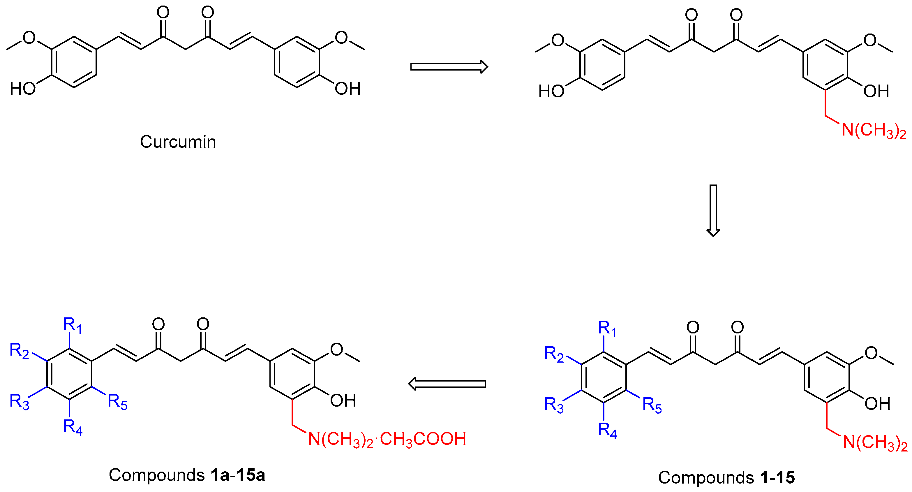
2.1. Synthesis of Dimethylaminomethyl Substituted Curcumin Derivatives and Their Acetates

The synthesis route of the target compounds is shown in Figure 2. Intermediate a was synthesized using the method in reference [19]. Vanillin is a commonly used raw material for the synthesis of curcumin and curcumin derivatives. Owing to p-π conjugation effect, the hydrogen atom on ortho-position to hydroxyl group in vanillin is very active, so that it is suitable for performing a Mannich reaction to give compound a. Meanwhile, according to the method in reference [20], various substituted benzaldehydes were subjected to condensation reaction with 2, 4-pentanedione, respectively, to obtain a series of intermediate b1b15. The synthesis of intermediates b1b15 has been reported in the previous literature [21,22,23,24,25,26]. Subsequently, Mannich base a underwent condensation reaction with compounds b1b15 to synthesize a series of unsymmetric curcumin derivatives, Mannich base 115. Acetates of curcumin derivatives were easily prepared (1a15a). Compounds 115 and 1a15a are new compounds that have not been reported in the literature. It is worth noting that the structural difference between compound 15 and curcumin is only that there is a dimethylaminomethyl substitution at the ortho position of the hydroxyl group of compound 15. The substituent status of curcumin derivatives 115 and their acetates 1a15a is shown in Table 1. Structure data, NMR spectra, and mass spectra of curcumin derivatives 115 and their acetates 1a15a are provided in the Supplementary Data.

2.2. Cytotoxic Activities

The results show that all the curcumin derivatives 115 and their acetates 1a15a failed to exhibit toxicity to the RAW264.7 cell line and PC 12 cell line at a concentration of 20 μM compared to the control (Figure 3), indicating that these compounds were suitable for the subsequent activity screening study. Compounds 1, 5, 11, 5a, and 6a exhibit a significant promoting effect on PC 12 cell proliferation at a concentration of 20 μM.

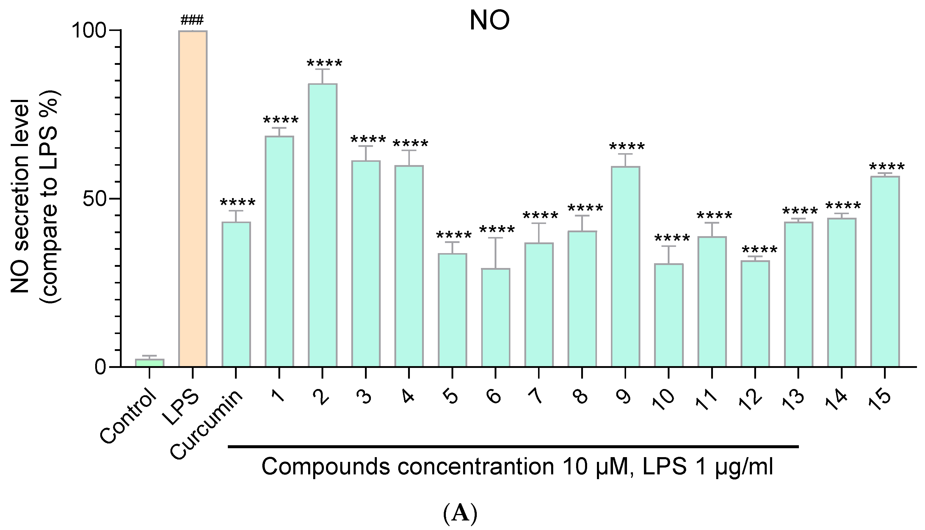
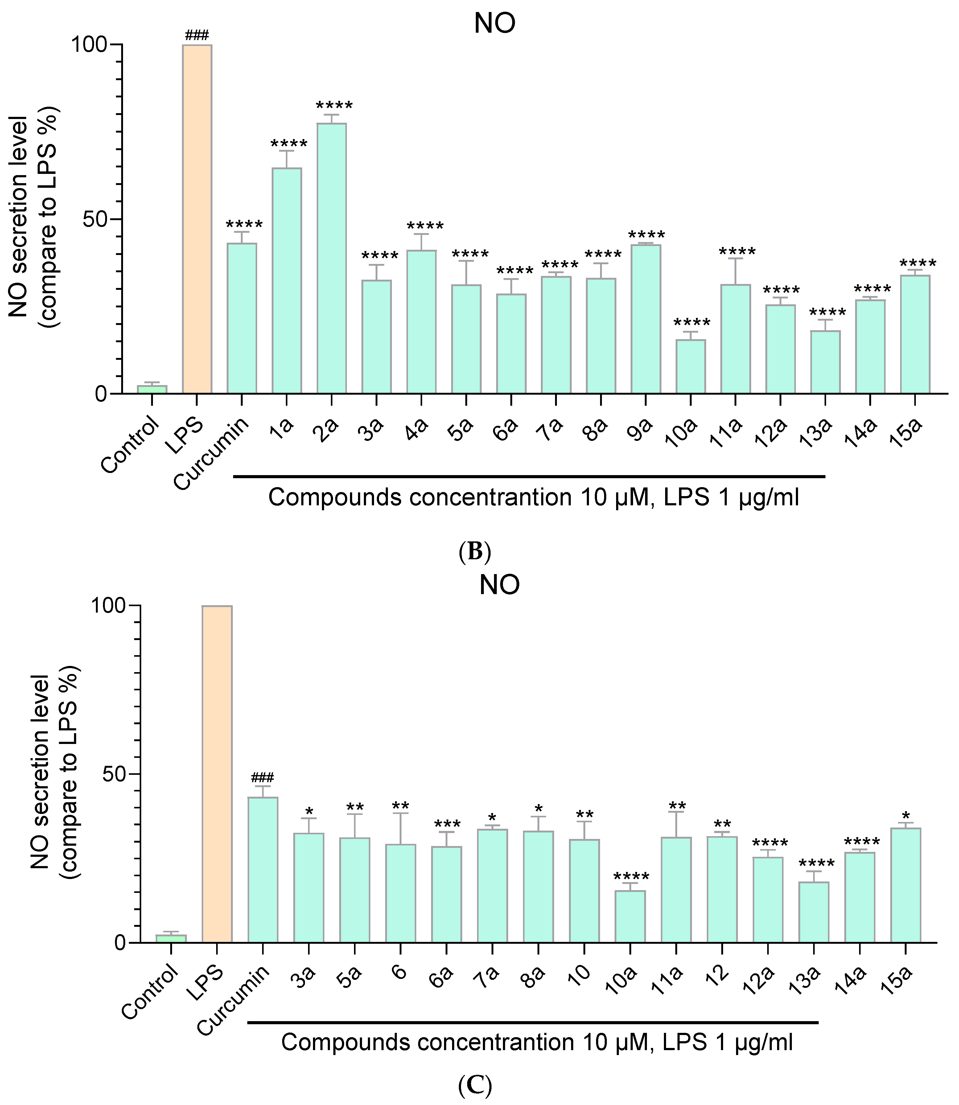
2.3. Anti–Inflammatory Activity against NO Release

The anti-inflammatory assays of curcumin derivatives 115 and their acetates 1a15a were evaluated in the LPS–stimulated RAW264.7 cell lines with curcumin as the positive control. Raw data of anti-inflammatory assays is shown in Table S1 in the Supplementary Material. Data were represented as mean ± SD of three independent experiments. The statistical analyses were performed using the one-way analysis of ANOVA in GraphPad Prism 8. As shown in Figure 4A,B, all the compounds exhibited a certain degree of inhibition of NO release at a concentration of 10 μM. Among the curcumin derivatives 115 and their acetates 1a15a, compound 10a exhibited the most significant inhibition effect (inhibition rate 92% at 15 μM) on NO release (Table S1). Compounds 6, 10, 12, 3a, 5a, 6a, 7a, 8a, 10a, 11a, 12a, 13a, 14a, and 15a exhibited better inhibitory activity against NO production compared with curcumin at a concentration of 10 μM (Figure 5C). Most of the target compounds inhibit the release of NO in a dose-dependent manner (Table S1).

2.4. Antioxidant Capacity Evaluation

The anti-oxidant assays of curcumin derivatives 115 and their acetates 1a15a were evaluated in H2O2–stimulated PC 12 cell lines with curcumin as the positive control. Raw data of anti-oxidant assays is shown in Table S2 in the Supplementary Material. Data were represented as mean ± SD of three independent experiments. The statistical analyses were performed using the one-way analysis of ANOVA in GraphPad Prism 8. As shown in Figure 5A,B, curcumin derivatives 115 and their acetates 1a15a exhibited a certain degree of antioxidant capacity at a concentration of 10 μM. Compounds 5, 1a, 5a, 8a, and 12a exhibited better anti-oxidant capacity compared with curcumin at a concentration of 10 μM (Figure 5C). Among the synthesized curcumin derivatives and their acetates, compound 5a showed the best anti-oxidant effect, with a cell survival rate of 106% at 20 μM (Table S2). Most of the target compounds shown an anti-oxidant capacity in a dose-dependent manner. Compounds 2a, 7a, and 10a had a relatively poor anti-oxidant capacity (with cell survival rate of 61%, 62%, and 64% at 10μM, respectively).

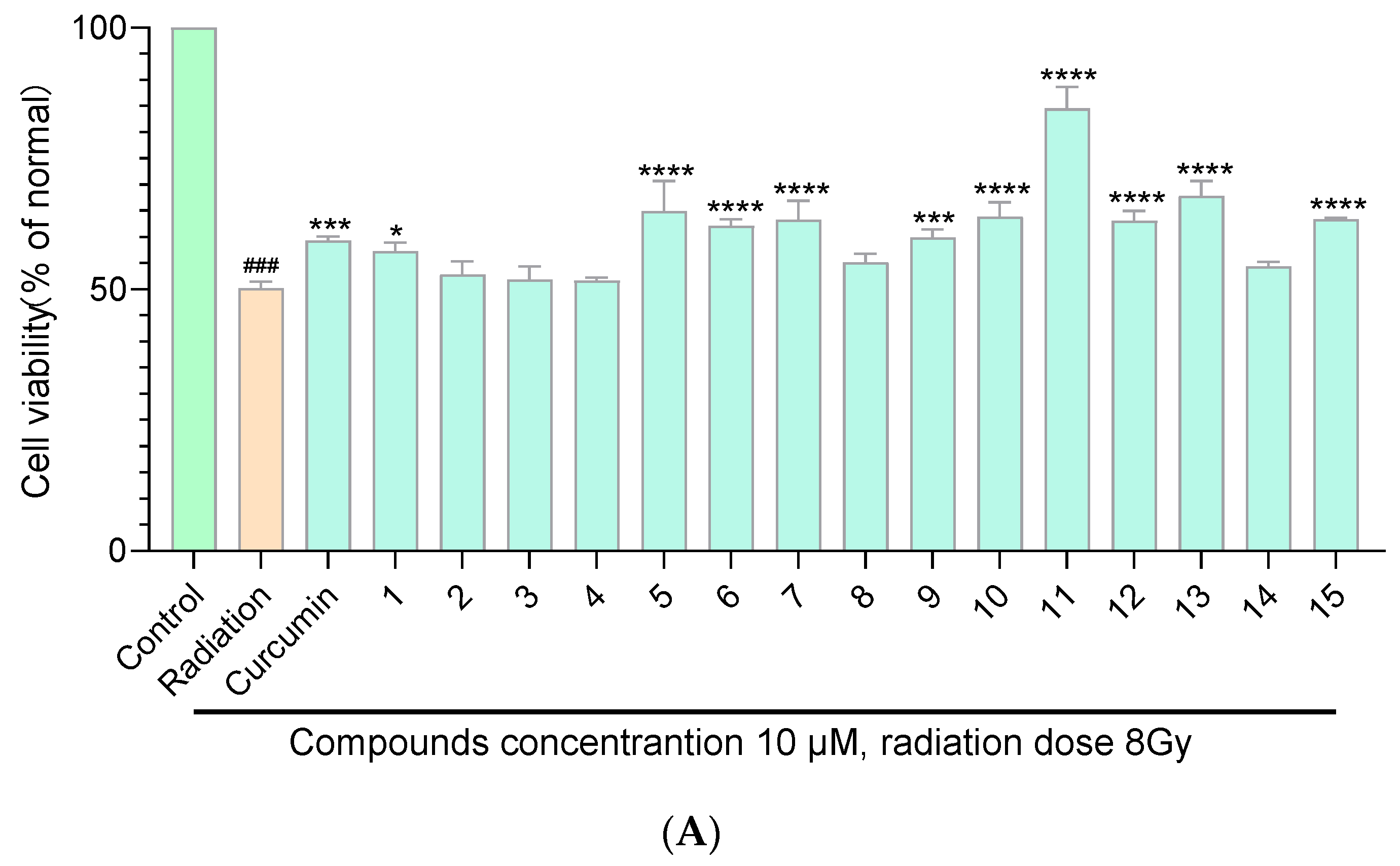
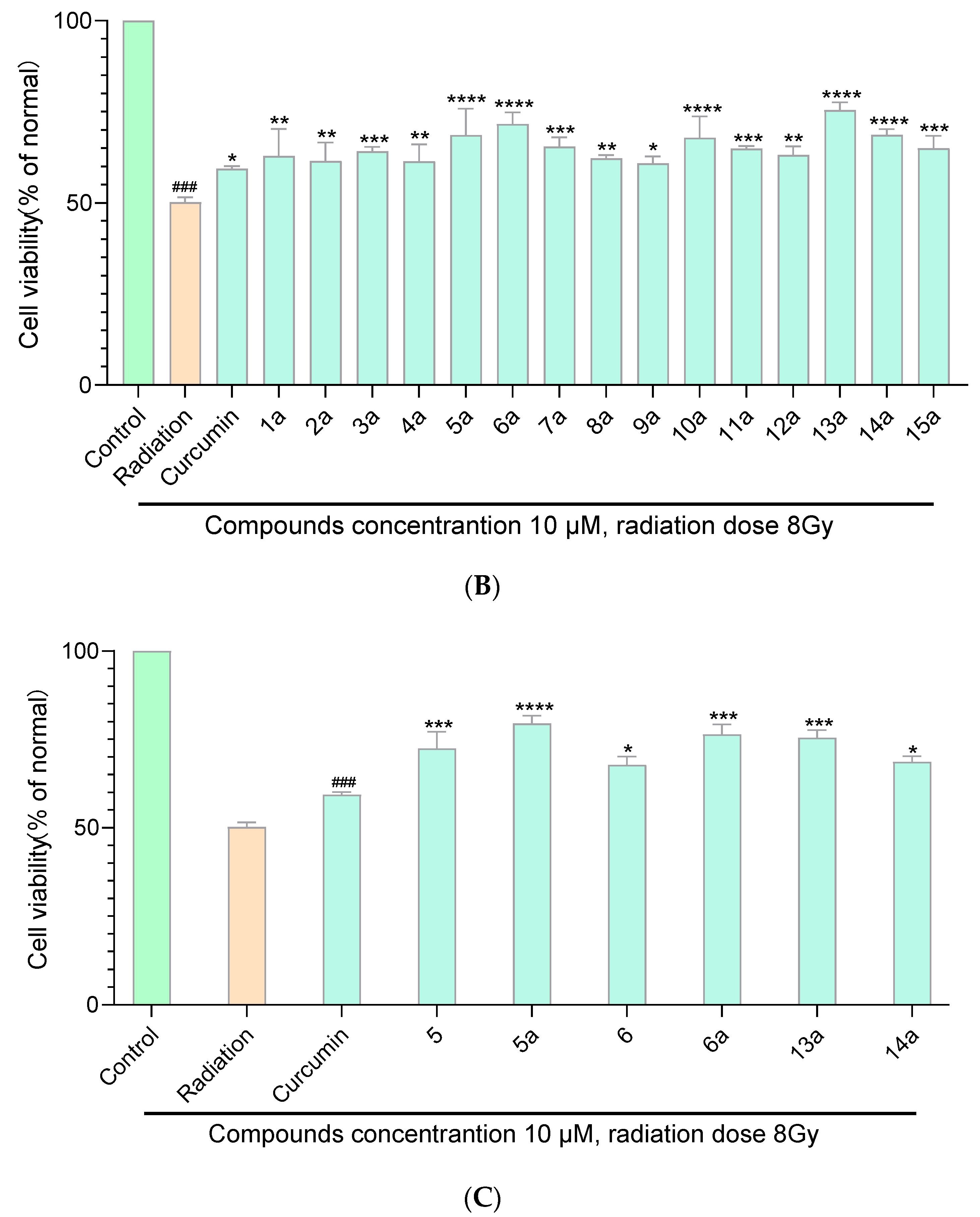
2.5. Radiation Protection Capability

The anti-radiation assays of curcumin derivatives 115 and their acetates 1a15a were evaluated in PC 12 cell lines with curcumin as the positive control. Raw data of anti-radiation assays is shown in Table S3 in the Supplementary Material. Data were represented as mean ± SD of three independent experiments. The statistical analyses were performed using the one-way analysis of ANOVA in GraphPad Prism 8. As shown in Figure 6A,B, compounds 1, 5, 6, 8, 11, 12, 1a, 5a, 6a, 8a, 11a, 12a, 13a, and 14a exhibited a certain degree of radiation protection capacity at a concentration of 10 μM. Compounds 6, 5a, 6a, 13a, and 14a exhibited better radiation protection capability compared with curcumin at a concentration of 10 μM (Figure 6C). Among the synthesized curcumin derivatives and their acetates, compound 11a has the best radiation protection ability, with a cell survival rate of 106% at 20 μM (Table S3 in the Supplementary Material). Most of the target compounds showed a radiation protection capacity in a dose-dependent manner.

2.6. Aqueous Solubility of Curcumin Derivative Acetates

Aqueous solubility of curcumin-derivative acetates 1a15a were measured using an UV spectrophotometer. The optical density–concentration curves of compounds 1a15a are shown in Figures S76–S90, and the saturated solution concentrations of curcumin derivative acetates 1a15a are shown in Table 2. Compared with the solubility of curcumin (<0.1 mg/mL), those of the curcumin-derivative acetates 1a15a were significantly improved. Among the test compounds, compound 10a has the best aqueous solubility, 335.16 mg/mL, which is similar to the solubility (367.88 mg/mL, 305.67 mg/mL) of dimethylaminomethyl substituted curcumin derivative salts in previous study [14].

3. Discussion

3.1. The Effect of Introducing Dimethylaminomethyl Group on the Bioactivity of Curcumin

Compound 15 is a derivative of curcumin where the hydroxyl ortho position is substituted by a dimethylaminomethyl group. Thus, the impact of introducing dimethylaminomethyl can be demonstrated by comparing the activity data of compound 15 and curcumin. It is found that the introduction of dimethylaminomethyl group did not increase the toxicity of curcumin derivatives on RAW264.7 and PC12 cell lines (Figure 3). The anti-oxidant (Table S2) and anti-radiation abilities (Table S3) of compound 15 and curcumin had no statistical difference. Fang X. et al. replaced the methoxy group in curcumin with dimethylaminomethyl group, and concluded that the compound’s in vitro free radical scavenging ability (IC50 18.6 μM, while that of curcumin was 26.5 μM) was enhanced [14], indicating that the different substitution positions of dimethylaminomethyl group may have an impact on anti-oxidant activity. It is worth noting that the anti-inflammatory activity was slightly decreased after the introduction of the dimethylaminomethyl group (Table S1).

3.2. Effect of Modification of Aromatic Ring Substituents on Biological Activity of Dimethylamino-Methyl Group Substituted Curcumin Derivatives

Comparing the anti-inflammatory activity of compound 10 (inhibition rate 71% at 10 μM) with that of compounds 9 (inhibition rate 41% at 10 μM) and 15 (inhibition rate 44% at 10 μM), it was found that methoxy substituent at the ortho and meta positions of the hydroxyl group may reduce the anti-inflammatory activity of the compound. In addition, when R1 and R5 (compound 14) or R2 and R4 (compound 2) were both substituted with methoxy, it could also lead to a decrease in anti-inflammatory activity.
There is no obvious rule that the antioxidant activity changes with the aromatic ring structure. Interestingly, compound 5a exhibited both excellent anti-oxidant (survival rate 106% at 20 μM) and anti-radiation activities (cell survival rate 104% at 15 μM), which may be due to its capacity to promote the proliferation of PC12 cell line (Figure 3). Similarly, compound 11 could promote the proliferation of the PC12 cell line, and showed good anti-radiation (cell survival rate 94% at 20 μM) and anti-oxidant activities (cell survival rate 96% at 20 μM).

3.3. Effect of Salt Formation on the Biological Activity and Aqueous Solubility of Curcumin Deriva-Tives

Comparing the bioactivity of curcumin-derivative acetates with that of curcumin derivatives themselves (Tables S1–S3), it was found that salt formation significantly increased the aqueous solubility of the compounds and had almost no negative effect on their physiological activities, such as their anti-inflammatory, anti-oxidant, and anti-radiation activities.
Although there is often no statistically significant difference between the bioactivity of curcumin-derivative acetates and curcumin derivatives themselves, salt formation increases the significance of difference in activity between some curcumin derivatives and curcumin. For example, the significance of difference in antioxidant activity between curcumin derivative 5 and curcumin was p = 0.0054, and the significance of difference between its acetate 5a and curcumin was p < 0.0001 (Figure 5C).

4. Materials and Methods

4.1. General Experimental Procedures

All chemicals were obtained from suppliers (Sigma-Adrich, St. Louis, MO, USA, TCI, Ark). NMR spectra were recorded on a Bruker ECA-400 MHz (Bruker Co. Billerica, MA, USA) spectrometer with TMS as an internal standard. Chemical shifts were in ppm (δ), and coupling constants (J) were reported in Hertz (Hz). High-resolution electrospray ionization mass spectra (HR-ESI-MS) was performed on the Agilent G6230A TOF LC/MS (Agilent Technologies Co. Ltd., Santa Clara, CA, USA) using ESI ion source. Absorbances in vitro experiments were measured using a spectrophotometer (Thermo, Singapore Republic of Singapore). Column chromatography (CC) was performed using silica gel (Qingdao Marine Chemical Inc., Qingdao, China). Thin-layer chromatography (TLC) was performed with silica gel GF254 (Yantai Huayang New Material Technology Co., Ltd., Yantai, China). Data of in vitro experiments were analyzed and plotted using Graghpad Prism (version 8, GraphPad Software, San Diego, California, 2020).

4.2. Procedure for the Synthesis of Target Compounds

4.2.1. Synthesis of 3-((Dimethylamino)methyl)-4-Hydroxy-5-Methoxybenzaldehyde (a)

Intermediate a was synthesized using the method from reference [19]. To a mixture of dimethylamine (40%, 1 g, 1 eq) and HCHO (37%, 1.1 g, 1.5 eq) at 70 °C, vanillin (0.68 g, 0.5 eq) in MeOH (20 mL) was added in batches. The reaction mixture was stirred at 70 °C in air for 12 h. After the completion of the reaction (monitored using TLC), the reaction mixture was evaporated under reduced pressure. The residue was quenched with H2O (20 mL) and extracted with EtOAc (3 × 20 mL). The organic layers were dried over anhydrous Na2SO4 and evaporated in vacuo. The residue was purified using flash column chromatography on silica gel (petroleum ether/EtOAc = 4:1) to afford the compound a.

4.2.2. Synthesis of Compounds b1b15

Intermediates b1b15 were synthesized by the method from reference [20].To a solution of acetylacetone (0.9 g, 1 eq) in EA (10 mL), boron trioxide (0.21 g, 0.33 eq) was added. The reaction mixture was stirred at 80 °C for 0.5 h. Then, the solution of benzaldehydes (0.33 eq) and trimethyl borate (0.31 g, 0.33 eq) in EA (10 mL) was added to the mixture above. The reaction mixture was stirred at 80 °C for 0.5 h before n-butylamine (0.22 g, 0.33 eq) was added. The reaction mixture was stirred at 100 °C for another 0.5 h before HCl (1 N, 5 mL) was added. After the completion of the reaction (monitored by TLC), the reaction was quenched with H2O (20 mL) and extracted with EtOAc (3 × 20 mL). The organic layers were dried over anhydrous Na2SO4 and evaporated in vacuo. The residue was purified by flash column chromatography on silica gel (petroleum ether/EtOAc = 4:1), respectively, to afford the compounds b1b15.

4.2.3. Synthesis of Compounds 115 and 1a15a

Compounds 115 were synthesized by the method from reference [20]. To a solution of compounds b1b15 (1 mmol, 1 eq) in EA (3 mL), boron trioxide (21 mg, 0.33 eq) was added, respectively. The reaction mixture was stirred at 80 °C for 0.5 h. Then, the solution of compound a (70 mg, 0.33 eq) and trimethyl borate (31 mg, 0.33 eq) in EA (3 mL) was added to the mixture above. The reaction mixture was stirred at 80 °C for 0.5 h before n-butylamine (22 mg, 0.33 eq) was added. The reaction mixture was stirred at 100 °C for another 0.5 h before HCl (1 N, 2 mL) was added. After completion of the reaction (monitored by TLC), the reaction was quenched with H2O (20 mL) and extracted with EtOAc (3 × 20 mL). The organic layers were dried over anhydrous Na2SO4 and evaporated in vacuo. The residue was purified by flash column chromatography on silica gel (petroleum ether/EtOAc = 4:1), respectively, to afford the compounds 115. Compounds 1a15a were obtained by treating compounds 115 with excessive CH3COOH in acetonitrile.

4.3. Cytotoxicity Assay

The cytotoxicity assay was performed using the Cell Counting Kit-8 (CCK-8, Applygen Technologies Co., Ltd., Beijing, China) assay on murine macrophage cell line (RAW264.7) and the rat adrenal pheochromocytoma cells (PC 12) using curcumin as positive controls, respectively. RAW264.7 cells were cultured in 96-well plates at a density of 3 × 10⁴ cells/mL in Dulbecco’s modified Eagle’s medium (DMEM, Gibco, Baltimore, MD, USA) with 10% fetal calf serum (Gibco, Baltimore, MD, USA), 100 U/mL penicillin, and 100 μg/mL streptomycin. Plates were incubated in an atmosphere containing 5% CO2 at 37 mL. PC 12 cells were cultured in 96-well plates at a density of 8000 cells/mL in Dulbecco’s modified Eagle’s medium (DMEM, Gibco, Baltimore, MD, USA) with 10% fetal calf serum (Gibco, Baltimore, MD, USA), 100 U/mL penicillin, and 100 μg/mL streptomycin. Plates were incubated in an atmosphere containing 5% CO2 at 37 °C. Cells were treated with different concentrations of test compounds for 48 h. Then, 10 μL CCK-8 solution were added to all the wells. After 4 h incubation, the absorbance at 450 nm of each well was measured in a multiscan photometer.

4.4. Determination of NO Content

RAW 264.7 cells were plated in 96-well plates at a density of 1 × 10⁴ cells/well and incubated overnight at 37 °C under 5% CO2. Then, the cells were treated with compounds 115 and 1a15a at a concentration of 10 µM, respectively for 1 h and then stimulated with LPS (1 µg/mL). Dexamethasone (Solarbio, D8040, Beijing, China) was used as the positive control. Then, 24 h later, NO production was determined by measuring the nitrite content using Griess reagents. Fifty microliters of culture supernatants were transferred to 96-well plates and mixed with 50 μL Griess reagent A. After incubation at room temperature for 5 min, the samples were mixed with an equal volume Griess reagent B for another 5 min. The absorbance of each mixture at 540 nm was measured using a microplate reader.

4.5. Antioxidant Activity Effect Evaluation

PC 12 cells were seeded in 96-well plates at a density of 8000 cells/well and incubated overnight. Then, the cells were treated with target compounds at a concentration of 10 μM, respectively for 1 h, and then stimulated with H2O2 (140 μM). After incubating under the same conditions for 24 h, 10 μL CCK-8 solution was added to each well. After 2 h incubation, the absorbance of each well was measured at 450 nm using a microplated reader (Multiskan MK-3, Thermo, Waltham, MA, USA).

4.6. Radiation Protection In Vitro

The rat adrenal pheochromocytoma cells (PC 12) were used for in vitro studies. A Cell Counting Kit-8 (CCK-8, Applygen Technologies Co., Ltd., Beijing, China) assay was used to determine cell viability. Cells were seeded in 96-well plates with a density of 8 × 10³ cells/ well and treated with curcumin and different concentrations of test compounds (10 µM) for 24 h before irradiation. Cells were irradiated with dose of 8.0 Gy at a dose rate of 0.8 Gy/min. Then, 10 μL CCK-8 solution was added to all the wells. After 4 h incubation, the absorbance at 450 nm of each well was measured in a multiscan photometer.

4.7. Aqueous Solubility Determination

A UV spectrophotometer was used to test the aqueous solubility of target compounds. Each compound to be tested was prepared into solutions with concentrations of 0.2 M, 0.1 M, 0.05 M, 0.025 M, 0.0125 M, 0.00625 M, and 0.003125 M. The optical density (OD) values of the prepared solutions were measured using an ultraviolet spectrophotometer, and concentration–OD-value standard curves were drawn for each compound to be tested. The saturated solutions of the target compounds were diluted by 5000 times and detected using an ultraviolet spectrophotometer to measure the OD values, respectively. The measured OD values were substituted into the corresponding concentration-OD value standard curve to calculate the diluted concentrations, which were multiplied by 5000 to obtain the saturated solution concentrations of the target compounds.

4.8. Statistical Analysis

In vitro experimental data were represented as mean ± SD of three independent experiments. The statistical analyses were performed using the one-way analysis of ANOVA in GraphPad Prism 8.

5. Conclusions

In summary, a series of dimethylaminomethyl-substituted curcumin derivatives were designed and synthesized (compounds 115). Acetate of these derivatives were prepared (compounds 1a15a). The various physiological activities of curcumin derivatives and their acetate were tested in vitro and compared with curcumin, including anti-inflammatory activity, anti-oxidant activity, anti-radiation activity, and cytotoxic activities. Finally, aqueous solubility of these curcumin derivative acetates was measured. Compounds 6, 10, 12, 3a, 5a, 6a, 7a, 8a, 10a, 11a, 12a, 13a, 14a, and 15a exhibited significantly better anti-inflammatory activity than curcumin, and could be further developed as anti-inflammatory lead compounds. Compounds 5, 1a, 5a, 8a, and 12a showed significantly better anti-oxidant activity than curcumin, and could be further developed as anti-oxidant lead compounds. Compounds 6, 5a, 6a, 13a, and 14a exhibited significantly better anti-radiation activity than curcumin, and could be further developed as anti-radiation lead compounds. Those compounds with more than two kinds of physiological activities deserve more attention of researchers, such as compounds 6, 5a, 6a, 8a, 12a, and 14a. The aqueous solubility of curcumin-derivative acetates (112.26–335.36 mg/mL) was greatly improved compared with that of curcumin (<0.1 mg/mL).

Supplementary Materials

The following supporting information can be downloaded at: https://0-www-mdpi-com.brum.beds.ac.uk/article/10.3390/molecules29091985/s1, Table S1. Inhibitory effects of the curcumin derivatives 115 and their acetates 1a15a on LPS-induced NO production. Table S2. Protection effects of the curcumin derivatives 115 and their acetates 1a15a on H2O2-induced oxidative damage. Table S3. Protection effects of the curcumin derivatives 115 and their acetates 1a15a on Radiation. 1 Figure S1: 1H-NMR spectrum of 1 in CD3OD. Figure S2: 13C-NMR spectrum of 1 in DMSO-d6. Figure S3: HR-ESI-MS spectrum of 1. Figure S4: 1H-NMR spectrum of 2 in DMSO-d6. Figure S5: 13C-NMR spectrum of 2 in DMSO-d6. Figure S6: HR-ESI-MS spectrum of 2. Figure S7: 1H-NMR spectrum of 3 in CD3OD. Figure S8: 13C-NMR spectrum of 3 in CD3OD. Figure S9: HR-ESI-MS spectrum of 3. Figure S10: 1H-NMR spectrum of 4 in CD3OD. Figure S11: 13C-NMR spectrum of 4 in CD3OD. Figure S12: HR-ESI-MS spectrum of 4. Figure S13: 1H-NMR spectrum of 5 in CD3OD. Figure S14: 13C-NMR spectrum of 5 in CD3OD. Figure S15: HR-ESI-MS spectrum of 5. Figure S16: 1H-NMR spectrum of 6 in CD3OD. Figure S17: 13C-NMR spectrum of 6 in DMSO-d6. Figure S18: HR-ESI-MS spectrum of 6. Figure S19: 1H-NMR spectrum of 7 in DMSO-d6. Figure S20: 13C-NMR spectrum of 7 in DMSO-d6. Figure S21: HR-ESI-MS spectrum of 7. Figure S22: 1H-NMR spectrum of 8 in CD3OD. Figure S23: 13C-NMR spectrum of 8 in CD3OD. Figure S24: HR-ESI-MS spectrum of 8. Figure S25: 1H-NMR spectrum of 9 in CD3OD. Figure S26: 13C-NMR spectrum of 9 in DMSO-d6. Figure S27: HR-ESI-MS spectrum of 9. Figure S28: 1H-NMR spectrum of 10 in CD3OD. Figure S29: 13C-NMR spectrum of 10 in CD3OD. Figure S30: HR-ESI-MS spectrum of 10. Figure S31: 1H-NMR spectrum of 11 in CD3OD. Figure S32: 13C-NMR spectrum of 11 in CD3OD. Figure S33: HR-ESI-MS spectrum of 11. Figure S34: 1H-NMR spectrum of 12 in DMSO-d6. Figure S35: 13C-NMR spectrum of 12 in CD3OD. Figure S36: HR-ESI-MS spectrum of 12. Figure S37: 1H-NMR spectrum of 13 in CD3OD. Figure S38: 13C-NMR spectrum of 13 in CD3OD. Figure S39: HR-ESI-MS spectrum of 13. Figure S40: 1H-NMR spectrum of 14 in CD3OD. Figure S41: 13C-NMR spectrum of 14 in CD3OD. Figure S42: HR-ESI-MS spectrum of 14. Figure S43: 1H-NMR spectrum of 15 in DMSO-d6. Figure S44: 13C-NMR spectrum of 15 in DMSO-d6. Figure S45: HR-ESI-MS spectrum of 15. Figure S46: 1H-NMR spectrum of 1a in CD3OD. Figure S47: HR-ESI-MS spectrum of 1a. Figure S48: 1H-NMR spectrum of 2a in CD3OD. Figure S49: HR-ESI-MS spectrum of 2a. Figure S50: 1H-NMR spectrum of 3a in CD3OD. Figure S51: HR-ESI-MS spectrum of 3a. Figure S52: 1H-NMR spectrum of 4a in CD3OD. Figure S53: HR-ESI-MS spectrum of 4a. Figure S54: 1H-NMR spectrum of 5a in CD3OD. Figure S55: HR-ESI-MS spectrum of 5a. Figure S56: 1H-NMR spectrum of 6a in CD3OD. Figure S57: HR-ESI-MS spectrum of 6a. Figure S58: 1H-NMR spectrum of 7a in CD3OD. Figure S59: HR-ESI-MS spectrum of 7a. Figure S60: 1H-NMR spectrum of 8a in CD3OD. Figure S61: HR-ESI-MS spectrum of 8a. Figure S62: 1H-NMR spectrum of 9a in CD3OD. Figure S63: HR-ESI-MS spectrum of 9a. Figure S64: 1H-NMR spectrum of 10a in CD3OD. Figure S65: HR-ESI-MS spectrum of 10a. Figure S66: 1H-NMR spectrum of 11a in CD3OD. Figure S67: HR-ESI-MS spectrum of 11a. Figure S68: 1H-NMR spectrum of 12a in DMSO-d6. Figure S69: HR-ESI-MS spectrum of 12a. Figure S70: 1H-NMR spectrum of 13a in CD3OD. Figure S71: HR-ESI-MS spectrum of 13a. Figure S72: 1H-NMR spectrum of 14a in CD3OD. Figure S73: HR-ESI-MS spectrum of 14a. Figure S74: 1H-NMR spectrum of 15a in CD3OD. Figure S75: HR-ESI-MS spectrum of 15a. Figure S75. HR-ESI-MS spectrum of 15a. Figure S76. Concentration-absorbance curve of 1a. Figure S77. Concentration-absorbance curve of 2a. Figure S78. Concentration-absorbance curve of 3a. Figure S79. Concentration-absorbance curve of 4a. Figure S80. Concentration-absorbance curve of 5a. Figure S81. Concentration-absorbance curve of 6a. Figure S82. Concentration-absorbance curve of 7a. Figure S83. Concentration-absorbance curve of 8a. Figure S84. Concentration-absorbance curve of 9a. Figure S85. Concentration-absorbance curve of 10a. Figure S86. Concentration-absorbance curve of 11a. Figure S87. Concentration-absorbance curve of 12a. Figure S88. Concentration-absorbance curve of 13a. Figure S89. Concentration-absorbance curve of 14a. Figure S90. Concentration-absorbance curve of 15a.

Author Contributions

Conceptualization, B.L. and S.L. (Shuchen Liu); methodology, S.L. (Sifan Liu) and H.G.; investigation, H.G. and K.L.; resources, Z.X. and K.L.; validation, B.L.; data curation, G.Z.; writing—original draft preparation, S.L. (Sifan Liu); writing—review and editing, B.L. and S.L. (Shuchen Liu). All authors have read and agreed to the published version of the manuscript.

Funding

The authors (Bin Li and Shuchen Liu) acknowledge the following agency for funding this project: National Natural Science Foundation of China (81903497).

Institutional Review Board Statement

Not applicable.

Informed Consent Statement

Not applicable.

Data Availability Statement

The data presented in this study are available in the Supplementary Materials.

Conflicts of Interest

The authors declare no conflicts of interest.

References

  1. Basham, S.A.; Waldman, H.S.; Krings, B.M.; Lamberth, J.; Smith, J.W.; McAllister, M.J. Effect of Curcumin Supplementation on Exercise-Induced Oxidative Stress, Inflammation, Muscle Damage, and Muscle Soreness. J. Diet. Suppl. 2020, 17, 401–414. [Google Scholar]
  2. Alvarenga, L.; Cardozo, L.; Da Cruz, B.O.; Paiva, B.R.; Fouque, D.; Mafra, D. Curcumin supplementation improves oxidative stress and inflammation biomarkers in patients undergoing hemodialysis: A secondary analysis of a randomized controlled trial. Int. Urol. Nephrol. 2022, 54, 2645–2652. [Google Scholar] [CrossRef] [PubMed]
  3. Johnson, S.; Shaikh, S.B.; Muneesa, F.; Rashmi, B.; Bhandary, Y.P. Radiation induced apoptosis and pulmonary fibrosis: Curcumin an effective intervention? Int. J. Radiat. Biol. 2020, 96, 709–717. [Google Scholar] [CrossRef] [PubMed]
  4. Li, W.; Jiang, L.; Lu, X.; Liu, X.; Ling, M. Curcumin protects radiation-induced liver damage in rats through the NF-κB signaling pathway. BMC Complement. Med. Ther. 2021, 21, 10. [Google Scholar] [CrossRef] [PubMed]
  5. Jagetia, G.C. Antioxidant activity of curcumin protects against the radiation-induced micronuclei formation in cultured human peripheral blood lymphocytes exposed to various doses of γ-Radiation. Int. J. Radiat. Biol. 2021, 97, 485–493. [Google Scholar] [CrossRef] [PubMed]
  6. Tønnesen, H.H.; Másson, M.; Loftsson, T. Studies of curcumin and curcuminoids. XXVII. Cyclodextrin complexation: Solubility, chemical and photochemical stability. Int. J. Pharm. 2002, 244, 127–135. [Google Scholar] [CrossRef] [PubMed]
  7. Fan, J.; de Lannoy, I.A.M. Pharmacokinetics. Biochem. Pharmacol. 2014, 87, 93–120. [Google Scholar] [CrossRef]
  8. Fleisher, D.; Li, C.; Zhou, Y.; Pao, L.H.; Karim, A. Drug, meal and formulation interactions influencing drug absorption after oral administration. Clinical implications. Clin. Pharmacokinet. 1999, 36, 233–254. [Google Scholar] [CrossRef] [PubMed]
  9. Nair, R.; Lamare, I.; Tiwari, N.K.; Ravi, P.R.; Pillai, R. In Situ Salification in Polar Solvents: A Paradigm for Enabling Drug Delivery of Weakly Ionic Drugs as Amorphous Solid Dispersion. AAPS PharmSciTech 2018, 19, 326–337. [Google Scholar] [CrossRef] [PubMed]
  10. Meng, Z.; Lv, Q.; Lu, J.; Yao, H.; Lv, X.; Jiang, F.; Lu, A.; Zhang, G. Prodrug Strategies for Paclitaxel. Int. J. Mol. Sci. 2016, 17, 796. [Google Scholar] [CrossRef] [PubMed]
  11. Ling, M.; Yan, C.; Huang, X.; Xu, Y.; He, C.; Zhou, Z. Phosphorylated walnut protein isolate as a nanocarrier for enhanced water solubility and stability of curcumin. J. Sci. Food Agric. 2022, 102, 5700–5710. [Google Scholar] [CrossRef] [PubMed]
  12. Sharma, A.; Arora, K.; Mohapatra, H.; Sindhu, R.K.; Bulzan, M.; Cavalu, S.; Paneshar, G.; Elansary, H.O.; El-Sabrout, A.M.; Mahmoud, E.A.; et al. Supersaturation-Based Drug Delivery Systems: Strategy for Bioavailability Enhancement of Poorly Water-Soluble Drugs. Molecules 2022, 27, 2969. [Google Scholar] [CrossRef] [PubMed]
  13. Tnnesen, H.H. Chemistry of Curcumin and Curcuminoids. In Phenolic Compounds in Food and Their Effects on Health I; American Chemical Society: Washington, DC, USA, 1992. [Google Scholar]
  14. Fang, X.; Fang, L.; Gou, S.; Cheng, L. Design and synthesis of dimethylaminomethyl-substituted curcumin derivatives/analogues: Potent antitumor and antioxidant activity, improved stability and aqueous solubility compared with curcumin. Bioorganic Med. Chem. Lett. 2013, 23, 1297–1301. [Google Scholar] [CrossRef] [PubMed]
  15. Kurnia, A.; Saputri, F.C.; Hayun. Synthesis and anticancer potential of aminomethyl derivatives of methyl-substituted asymmetrical curcumin mono-carbonyl. J. Appl. Pharm. Sci. 2019, 9, 18–24. [Google Scholar]
  16. Liu, B.-M.; Zhang, M.-H.; Tao, S.-J.; Xia, M.-Y.; Dong, J.-H. Synthesis and antiproliferative effect of Mannich bases of demethoxycurcumin derivatives. Zhongguo Yaowu Huaxue Zazhi 2014, 24, 271–278. [Google Scholar]
  17. Dong, J.; Liu, B.; Xu, L.; Xia, M.; Yin, X.; Chen, N.; Zhang, T. Curcumin Analogs Useful in Treatment of Cancer and Their Preparation. Patent CN103848747, 9 September 2014. [Google Scholar]
  18. Fang, L.; Gou, S.; Liu, X.; Cao, F.; Cheng, L. Design, synthesis and anti-Alzheimer properties of dimethylaminomethyl-substituted curcumin derivatives. Bioorganic Med. Chem. Lett. 2014, 24, 40–43. [Google Scholar] [CrossRef] [PubMed]
  19. Sinhababu, A.K.; Borchardt, R. Selective ring C-Methylation of hydroxybenzaldehydes via their mannich bases. ChemInform 1983, 14, 677–683. [Google Scholar]
  20. Gottlieb, O.R.; Mors, W.B. The Chemistry of Rosewood. III. Isolation of 5,6-Dehydrokavain and 4-Methoxyparacotoin from Aniba firmula Mez. J. Org. Chem. 1959, 24, 36–47. [Google Scholar] [CrossRef]
  21. Shi, L.; Wang, Z. Preparation of 2-alkoxyl-4-((1E,6E)-7-(4-oxophenyl)-3,5-dioxo-1,6-heptadien-1-yl)phenol Metal Salt Derivative and Application as Anticancer Agents. Patent CN105884597, 2016. [Google Scholar]
  22. Peng, S.; Zhao, M.; Chen, H. 1,7-Diaryl-1,6-trans-diene-3,5-diones Useful in the Treatment of Cancer and Their Preparation. Patent CN102807566, 5 December 2012. [Google Scholar]
  23. Venugopalan, P.; Krishnankutty, K. Metal chelates of 6-aryl-5-hexene-2,4-diones. J. Indian Chem. Soc. 2001, 78, 472–473. [Google Scholar]
  24. Balaji, N.V.; Hari, B.B.; Subbaraju, G.V.; Purna, N.K.; Murali, K.K.M. Synthesis, screening and docking analysis of hispolon analogs as potential antitubercular agents. Bioorganic Med. Chem. Lett. 2017, 27, 11–15. [Google Scholar] [CrossRef]
  25. Sugimoto, H.; Takahashi, T.; Hijikuro, I.; Okuda, M. Preparation of Diketone Compounds as β-Secretase Inhibitors. Patent WO2009145219, 3 December 2009. [Google Scholar]
  26. Sundaryono, A.; Nourmamode, A.; Gardrat, C.; Fritsch, A.; Castellan, A. Synthesis and complexation properties of two new curcuminoid molecules bearing a diphenylmethane linkage. J. Mol. Struct. 2003, 649, 177–190. [Google Scholar] [CrossRef]
Figure 1. Design strategy of curcumin derivatives.
Molecules 29 01985 g001
Figure 2. Synthesis of curcumin derivatives 115 and their acetates 1a15a. Reagents and conditions: (i) CH3OH, 50 °C, overnight; (ii) Acetylacetone, Butylamine, B(OCH3)3, B2O3, EtOAc, 80 °C, 4 h; (iii) compound a, piperidine, B(OCH3)3, B2O3, EtOAc, 80 °C, 4 h; and (iv) CH3COOH, rt, overnight.
Molecules 29 01985 g002
Figure 3. Cytotoxic activities of curcumin derivatives 115 and their acetates 1a15a to RAW264.7 cell line and PC 12 cell line at a concentration of 20 μM. Cytotoxic activities of compounds 115 to RAW264.7 cell line (A). Cytotoxic activities of compounds 1a15a to RAW264.7 cell line (B). Cytotoxic activities of compounds 115 to PC 12 cell line (C). Cytotoxic activities of compounds 1a15a to PC 12 cell line (D).
Molecules 29 01985 g003
Figure 4. Inhibitory effects of the curcumin derivatives 115 (A) and their acetates 1a15a (B) on LPS-induced NO production. ### The comparison object in the one-way analysis of ANOVA. **** p < 0.0001 compared with the LPS-treated group. Comparation of inhibitory effects on LPS-induced NO production between curcumin derivatives (acetates) and curcumin (C). ### The comparison object in the one-way analysis of ANOVA. * p < 0.1, ** p < 0.01, *** p < 0.001, **** p < 0.0001 compared with curcumin.
Molecules 29 01985 g004aMolecules 29 01985 g004b
Figure 5. Protection effects of the curcumin derivatives 115 (A) and their acetates 1a15a (B) on H2O2-induced oxidative damage. ### The comparison object in the one-way analysis of ANOVA. ** p < 0.01, *** p < 0.001, **** p < 0.0001 compared with the LPS-treated group. Comparation of protection effects on H2O2-induced oxidative damage between curcumin derivatives (acetates) and curcumin (C). ### The comparison object in the one-way analysis of ANOVA. ** p < 0.01, *** p < 0.001, **** p < 0.0001 compared with curcumin.
Molecules 29 01985 g005
Figure 6. Protection effects of the curcumin derivatives 115 (A) and their acetates 1a15a (B) on radiation. ### The comparison object in the one-way analysis of ANOVA. * p < 0.1, ** p < 0.01, *** p < 0.001, **** p < 0.0001 compared with the LPS-treated group. Comparation of protection effects on H2O2-induced oxidative damage between curcumin derivatives (acetates) and curcumin (C). ### The comparison object in the one-way analysis of ANOVA. * p < 0.1, *** p < 0.001, **** p < 0.0001 compared with curcumin.
Molecules 29 01985 g006aMolecules 29 01985 g006b
Table 1. Synthesis of curcumin derivatives 115 and their acetates 1a15a.
CompoundThe Left Arene RingCompoundThe Left Arene RingCompoundThe Left Arene Ring
1, 1aMolecules 29 01985 i0016, 6aMolecules 29 01985 i00211, 11aMolecules 29 01985 i003
2, 2aMolecules 29 01985 i0047, 7aMolecules 29 01985 i00512, 12aMolecules 29 01985 i006
3, 3aMolecules 29 01985 i0078, 8aMolecules 29 01985 i00813, 13aMolecules 29 01985 i009
4, 4aMolecules 29 01985 i0109, 9aMolecules 29 01985 i01114, 14aMolecules 29 01985 i012
5, 5aMolecules 29 01985 i01310, 10aMolecules 29 01985 i01415, 15aMolecules 29 01985 i015
Table 2. Aqueous solubility of curcumin derivative acetates 1a15a.
CompoundOD 1Solubility (mg/mL)
1a0.34201.06
2a0.71148.19
3a0.66254.95
4a0.51314.20
5a0.30117.88
6a0.23112.26
7a0.58257.90
8a0.61118.16
9a0.94167.04
10a0.84335.16
11a0.51132.20
12a0.43206.04
13a0.74285.48
14a0.47126.01
15a0.72283.81
1 The optical density values were recorded at 384 nm, and the values of compounds 1a15a were measured at the 5 × 10³ fold diluted saturated concentration, since the values of the saturated solution were too high to be measured.
Disclaimer/Publisher’s Note: The statements, opinions and data contained in all publications are solely those of the individual author(s) and contributor(s) and not of MDPI and/or the editor(s). MDPI and/or the editor(s) disclaim responsibility for any injury to people or property resulting from any ideas, methods, instructions or products referred to in the content.

