
Meningococcal Disease Presenting with Acute Myopericarditis and Concurrent Acute Meningitis
Department of Emergency Medicine, Alameda Health System–Wilma Chan Highland Hospital Campus, Oakland, CA 94602, USA
Emerg. Care Med. 2024, 1(2), 95-102; https://0-doi-org.brum.beds.ac.uk/10.3390/ecm1020013
Submission received: 4 March 2024
/
Revised: 12 April 2024
/
Accepted: 17 April 2024
/
Published: 26 April 2024
Abstract
:Emergency department physicians uncommonly associate myopericarditis with bacterial infection and, even more rarely, encounter myopericarditis caused by meningococcal infection. This case report describes a 38-year-old man who presented with chest pain, electrocardiographic changes, and cardiac biomarkers consistent with acute myopericarditis and rapidly developed central nervous system symptoms that were concerning for acute bacterial meningitis. The diagnosis of Neisseria meningitidis infection was confirmed by blood cultures. Once identified, the patient made a full recovery following a course of intravenous antibiotic therapy. This case underscores the difficulty of diagnosing this rare, but potentially life-threatening, condition in the emergency department.
1. Introduction
Disseminated meningococcal infection is a rare, life-threatening disease caused by the gram-negative aerobic diplococcus bacteria Neisseria meningitidis [1,2]. Although meningococcal meningitis and meningococcemia are the two most common clinical syndromes associated with primary infection, uncommonly, extra-meningeal manifestations, such as acute bacterial pericarditis or myopericarditis, have been reported [2]. Acute bacterial myopericarditis from N. meningitidis can be classified into three groups based on the pathophysiologic process, including isolated (primary) meningococcal pericarditis, disseminated (secondary) meningococcal pericarditis, and reactive (immune-reactive) meningococcal pericarditis [3]. Herein, we describe a case of a 38-year-old man who initially presented to the emergency department with concern for acute ST-segment elevation myocardial infarction. Following a negative cardiac catheterization, he was subsequently diagnosed with acute myopericarditis and concomitant acute bacterial meningitis due to hematogenous disseminated N. meningitidis infection.
2. Case Report
A 38-year-old Hispanic male, who was employed as a restaurant server, presented with acute chest pain via ambulance to our emergency department. He had no past medical problems and denied tobacco use, significant alcohol consumption, or recreational drug use. His chest pain was reported to be in the substernal and upper epigastric region without radiation to other areas, which had begun less than 24 h prior to arrival. The pain was intermittent, was rated six out of ten in pain intensity, and lasted one to two hours at a time prior to spontaneous resolution. It was accompanied by subjective fever, neck pain, and nausea and overall, felt “like I was getting the flu”. The patient’s chest pain had acutely worsened at 3:00 am the morning of presentation, and when dyspnea started hours later, he called an ambulance to bring him to the closest emergency department. He was unsure if the pain improved with prehospital-administered aspirin and sublingual nitroglycerin.
A physical examination revealed a blood pressure of 140/70 mm Hg, a heart rate of 90/min, a respiratory rate of 20/min, and a temperature of 100.5 °F (38.1 °C). He appeared to be in mild distress from pain, but otherwise, appeared well. The examination was notable for moderate tenderness in the epigastrium, and his skin was noted to be mildly flushed and diaphoretic. The remainder of his examination (including neck, cardiopulmonary, vascular, and neurologic) was unremarkable.
The patient’s initial electrocardiogram (ECG) (Figure 1) showed concave-upward ST segment elevation in all leads, except leads III, aVR, and V1, and was interpreted as suspicious for acute ST-segment elevation myocardial infarction (STEMI). An emergency department point-of-care cardiac ultrasound revealed no significant pericardial fluid, normal left ventricular contractility, and normal chamber sizes. A portable chest radiograph showed normal heart size and mediastinum and was without pneumothorax, pulmonary infiltrates, or effusions. Initial labs were notable for a white blood cell count of 10.6 × 10³/mcL (normal range 4.5 to 11.5 × 10³/mcL) with 61% segmented neutrophils and 23% bands, potassium 2.8 mmol/L (normal range 3.6 to 5.0 mmol/L), total bilirubin 4.5 mg/dL (normal range 0.2 to 1.2 mg/dL), and cardiac troponin I 0.11 ng/mL (normal < 0.04 ng/mL). His cardiac troponin levels eventually peaked at 4.54 ng/mL and then declined to 2.98 mg/mL prior to discharge. Cardiac catheterization was emergently performed on admission but revealed no flow-limiting coronary artery disease. Subsequent labs were notable for an erythrocyte sedimentation rate of 82 mm/h (normal < 20 mm/h) and a C-reactive protein level of 31.4 mg/L (normal < 5 mg/L). The patient was admitted with the presumptive diagnosis of viral myopericarditis and started on Colchicine and a non-steroidal anti-inflammatory.
On hospital day 2, our patient complained of back and neck pain. Blood cultures, which were obtained 1 day earlier, returned positive for gram-negative diplococci, prompting the addition of ceftriaxone 2 g IV to his treatment.
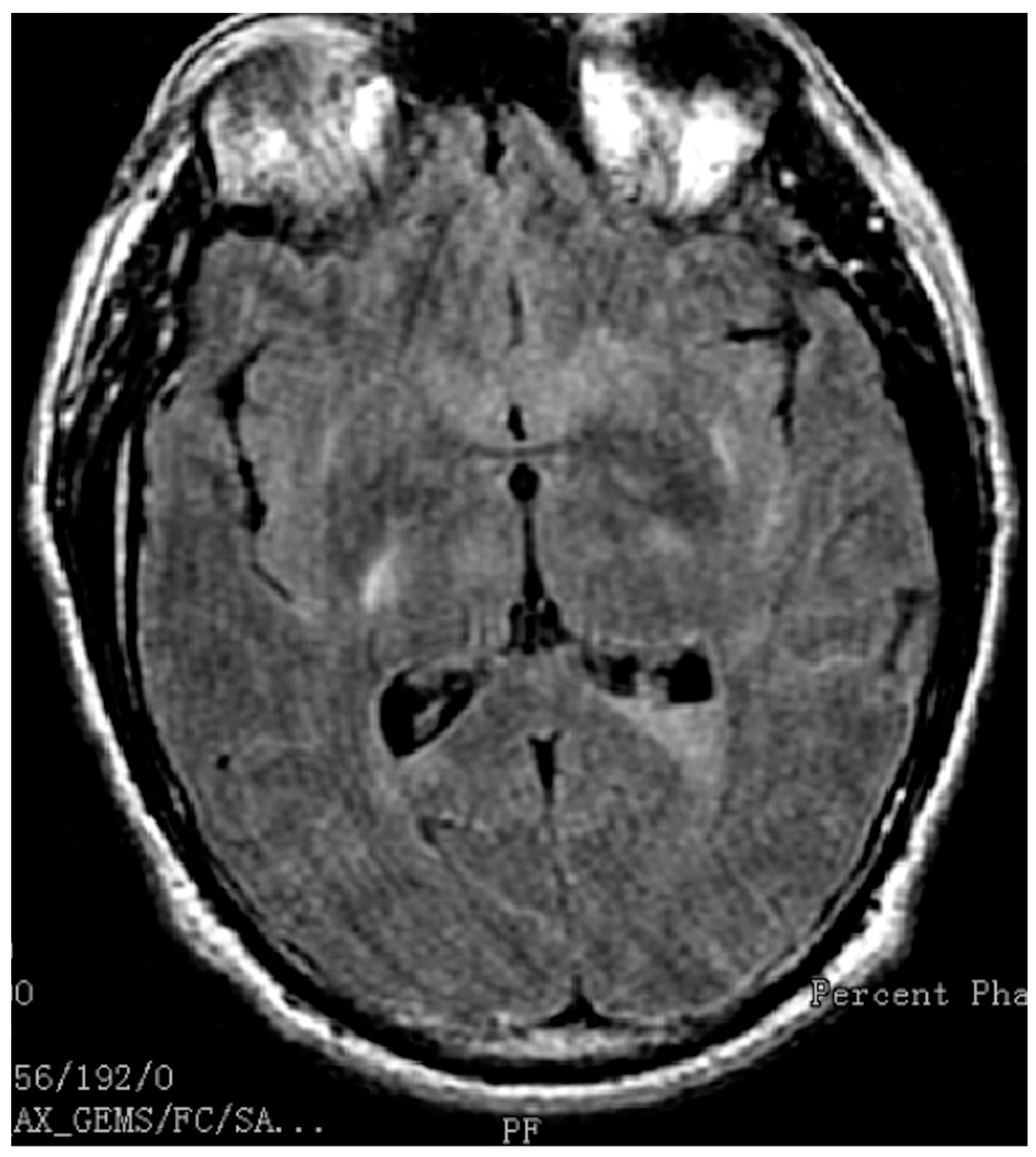
On hospital day 3, he complained of diplopia. A magnetic resonance image (MRI) of his brain was obtained, revealing several areas of mild diffuse leptomeningeal enhancement, as well as restricted diffusion within the posterior horns of the lateral ventricles (Figure 2) and frontal subcortical region, consistent with meningitis with ventriculitis. A lumbar puncture was performed, revealing a white blood cell count of 3640 cells/mcL (normal 0–5 cells/mcL), a red blood cell count of 135 cells/mcL (normal 0–5 cells/mvL), a protein level of 300.5 mg/dL (normal 15–40 mg/dL), and a glucose level of <10 mg/dL (normal 50–80 mg/dL). His final blood cultures, confirmed by the California Emerging Infections Program, which performs active bacterial core surveillance for our county, grew Neisseria meningitidis (serotype unavailable). Transthoracic echocardiography did not reveal a significant pericardial effusion; thus, no pericardial fluid sample was obtained. He was continued on ceftriaxone for two weeks and colchicine for three months. The Department of Public Health was notified so that the patient’s family, hospital staff, and the patient’s coworkers were able to receive post-exposure chemoprophylaxis for meningococcal infection. Additional information included that the patient revealed that he had not received a meningococcal vaccine in the past. Clinic visits from two months after discharge showed full and complete recovery, including the absence of diplopia. His final diagnosis was disseminated meningococcal infection involving the pericardium, myocardium, and meninges (i.e., meningococcal myopericarditis).
3. Discussion
N. meningitidis is a gram-negative diplococci with 13 serotypes and is capable of colonizing the nasopharyngeal surface membrane in humans. Whereas half a million cases of infection with N. meningitidis occur worldwide every year [4], the rates of meningococcal infection in the United States have significantly declined since the 1990s and remain low [2]. In 2019, there were about 375 total cases of meningococcal disease reported (incidence rate of 0.11 cases per 100,000 persons) [5]. Six serotypes account for the majority of cases of life-threatening disease worldwide (serogroups A, B, C, W-135, X, and Y), with serogroups B, C, and Y causing most of the illness seen in the United States [5]. Meningeal infection, resulting from hematogenous spread, is similar to other forms of acute bacterial meningitis, with a sudden onset of headache, fever, and neck stiffness, sometimes accompanied by nausea, vomiting, photophobia, and altered mental status. Less commonly, N. meningitidis can produce a devastating septicemia (i.e., meningococcemia), resulting in skin and soft tissue necrosis, hearing loss, and multi-organ system failure, along with the ultra-rapid development of shock and death, sometimes within 12–24 h of the initial symptoms. Additionally, extra-meningeal infection due to N. meningitidis can result in pneumonia, septic arthritis, conjunctivitis, otitis media, epiglottitis, urethritis, and pericarditis [2]. The overall mortality associated with N. meningitidis infection is approximately 10–15% [2,6]. In addition, significant morbidity is seen in an additional 11–19% of patients [2,6] and includes digit and limb loss, hearing loss, visual impairment, motor nerve deficits, seizures, and cognitive dysfunction, including educational difficulties, developmental delays, and behavioral problems [7].
N. meningitidis infection most often affects pediatric or adult patients in close living quarters, such as school dormitories, military barracks, homeless shelters, long-term care facilities, and prisons [2,8]. Although people of any age are at risk for infection, the rates of infection are highest in children younger than 1 year old, with a second peak in adolescence. Among adolescents and young adults, those 16 through 23 years old have higher rates of meningococcal disease [8]. Human exposure to meningococcal infection depends on having close contact with someone who is infected or can arise from close contact with a healthy person colonized with N. meningitidis within the environment. Person-to-person transmission occurs through respiratory, saliva, or throat secretions.
The prevalence of nasopharyngeal carriage is 5–10% under non-epidemic conditions in the United States [9]. In closed populations, such as amongst military recruits, carriage rates can reach up to 50% [10]. Long-term asymptomatic carriage is an important factor that may lead to the development of invasive disease by increasing the risk of transmission of the bacteria to susceptible individuals. The results of research that evaluated the effectiveness of immunoprophylaxis in suppressing the carriage of N. meningitidis in a military environment showed that asymptomatic carriers are a major source of meningococcal infection [11].
Because of the risks of severe morbidity and death, effective antibiotics should be administered promptly to patients suspected of having meningococcal disease. Empirical therapy for suspected meningococcal disease should include an extended-spectrum cephalosporin, such as cefotaxime or ceftriaxone [5]. Depending on the severity of illness, patients with meningococcemia may require breathing support and vasopressors, as well as wound care and, in some cases, surgery to remove necrotic tissue.
The most powerful weapon we have to prevent meningococcal disease is meningococcal vaccines. In the United States, there are three available, including the meningitis A C W Y vaccine (MenACWY), the meningitis B vaccine (MenB), and the meningitis A B C W Y vaccine (MenABCWY). The Centers for Disease Control and Prevention (CDC) recommends that all 11- to 12-year-olds should obtain a MenACWY vaccine, with a booster dose at 16 years old [12]. Teens and young adults (16 through 23 years old) may also obtain a MenB vaccine. Those who are receiving MenACWY and MenB vaccines at the same visit may instead obtain a MenABCWY vaccine. The CDC also recommends meningococcal vaccination for other children and adults who are at an increased risk of meningococcal disease. Routine immunization against meningococcal disease has resulted in over a 90% drop in the incidence of this infection [12]. Close contacts, including people in the same household, roommates, and anyone with direct contact with a patient’s oral secretions, of someone with meningococcal disease should receive prophylactic antibiotic treatment with a single dose of ceftriaxone or ciprofloxacin or be prescribed rifampin (four doses over 2 days) to help prevent infection transmission [5].
The combination of myopericarditis and meningitis is rare; however, it has been previously reported in the literature [13,14,15,16,17,18,19,20,21,22,23,24,25,26,27,28,29,30,31,32,33,34,35]. Such a combination has historically been the result of untreated meningococcal bacteremia; thus, it has become remarkably unusual with appropriate antibiotic treatment and routine vaccination. Meningococcal pericardial disease was first reported in 1918. In this initial report, 12 out of 280 cases of meningococcal meningitis were noted to have concurrent pericarditis [36]. In a larger case series involving military recruits at a United States Army Hospital in Fort Dix, New Jersey, which was published in 1971, 17 cases of pericarditis were found in 334 cases of meningococcal meningitis [37]. A review of laboratory samples at the National Reference Center for the Meningococci in Paris, France, which was conducted from 1999 to 2002, identified that out of 2089 cases of meningococcal infections, only 6 cases showing isolated myopericarditis and an additional 7 cases having both myopericarditis and meningitis were identified [38]. Our case report is only one of a dozen or so in which acute myopericarditis was the presenting symptom in a patient who was subsequently diagnosed with acute bacterial meningitis due to N. meningitidis.
There are three proposed mechanisms in which N. meningitidis can result in pericarditis, including isolated (primary) meningococcal pericarditis, disseminated (secondary) meningococcal pericarditis, and reactive (immune-reactive) meningococcal pericarditis [3]. Isolated meningococcal pericarditis is a purulent pericarditis with positive pericardial or blood cultures for N. meningitidis without evidence of meningeal involvement or clinical manifestations of meningococcemia. Pericardial effusion and/or tamponade can be seen in cases of isolated meningococcal pericarditis. Disseminated meningococcal pericarditis is a consequence of direct pericardial invasion secondary to bacteremia. It appears as a complication of widespread, clinically manifested meningococcal disease; however, rarely, as in the case presented, it can be the presenting symptom. Significant pericardial effusion with tamponade is uncommon. The prognosis of both isolated and disseminated meningococcal pericarditis is good when antibiotics are initiated promptly and in cases without significant pericardial effusion [3,20]. Reactive meningococcal pericarditis typically occurs 1–2 weeks after the treatment of an N. meningitidis infection. A sterile pericardial effusion may result from the progressive accumulation of fluid around the heart. Simultaneous foci of aseptic inflammation, such as pleura, joints, and skin, may be present. Reactive meningococcal pericarditis is usually more severe than the other two forms of meningococcal pericarditis and is often complicated by tamponade [3].
The clinical presentation of meningococcal infection is largely dependent on the host, environment, and pathogen factors. The most common presenting symptoms of acute meningococcal pericarditis are chest pain (80%), fever (70%), and dyspnea (50%) [3]. Chest pain is typically abrupt in onset, pleuritic and/or sharp in character, substernal or left precordial in location, may radiate to the neck, arm, or jaw, and is improved by leaning forward and worsened by lying supine [39,40]. Pain that radiates to the trapezial ridge is likely pericarditis because the phrenic nerve that innervates these muscles traverses the pericardium [40]. On physical examination, patients may appear anxious or uncomfortable and may have sinus tachycardia and low-grade fever. Heart sounds may be muffled if a pericardial effusion is present, and a pericardial friction rub caused by friction between the two inflamed pericardial layers, may be appreciated [39,40].
Classic electrocardiographic findings in acute pericarditis include diffuse, concave-upward ST segment elevation with an upright T wave and PR segment depression (typically in all leads except aVR and V1). A downsloping TP segment (Spodick’s sign) has been associated with acute pericarditis [41,42]. The lead aVR may display PR segment elevation and ST segment depression (“knuckle sign”) [43]. Supporting laboratory features include an elevated white blood cell count, ESR, and CRP. An abnormal cardiac troponin indicates myocardial involvement (i.e., myopericarditis). Cardiac function is not impaired on echocardiography; however, a small, medium, or large pericardial effusion may be seen. Although pericardial inflammation can be identified on a cardiac CT or MRI, acute pericarditis remains a clinical diagnosis based on the presence of two out of four cardinal features, including (1) characteristic chest pain, (2) pericardial friction rub, (3) electrocardiographic findings, and (4) pericardial effusion on echocardiography [39].
As seen with our case, it can be challenging to distinguish acute myocardial infarction from acute myopericarditis in the emergency department. There is significant overlap in age (although myopericarditis is generally seen in younger adults), clinical features, initial electrocardiogram (ECG), and cardiac biomarkers [40]. The presence of a low-grade temperature and flu-like symptoms and the lack of anatomically reciprocal ST segment depression in our patient were subtle clues that were not appreciated initially. In our patient, the emergency department providers reasonably, but hastily, activated the cardiac catheterization lab to exclude the life-threatening diagnosis of acute ST-segment elevation myocardial infarction. Once flow-limiting coronary artery disease as a cause for the presentation was eliminated, the diagnosis of acute myopericarditis became more apparent. Our patient continued to have a fever with worsening neck and back pain and developed diplopia, which resulted in an MRI, followed by a lumbar puncture and the correct diagnosis. In our patient, there was no definitive evidence of Neisseria in the CSF (likely sterilized from the administration of antibiotics); however, the cerebrospinal fluid profile was consistent with acute bacterial meningitis, and the patient’s blood cultures grew this organism. The patient ultimately had a benign course with full and complete recovery. A clinic visit two months after his initial illness found this patient to be asymptomatic without any recurrent cardiac or neurologic symptoms.
4. Conclusions
Although infection with N. meningitidis typically presents with acute bacterial meningitis or, less commonly, meningococcemia, hematogenous spread to other organs is possible and can pose a diagnostic challenge. We present a case of a 38-year-old male whose initial symptoms were believed to be consistent with acute ST-segment elevation myocardial infarction and who underwent cardiac catheterization.
Following a negative result, he was subsequently diagnosed with acute myopericarditis, which was presumed idiopathic or viral in etiology. During hospitalization, blood culture and lumbar puncture results were consistent with concomitant meningococcemia and acute bacterial meningitis. Emergency medicine, internal medicine, pediatric, cardiology, and infectious disease physicians should consider this rare association when confronted by patients with simultaneous cardiac, neurologic, and infectious symptoms. In our patient, the early recognition and treatment of meningococcal infection resulted in a favorable clinical outcome.
Funding
This research received no external funding.
Informed Consent Statement
Written informed consent has been obtained from the patient to publish this paper.
Data Availability Statement
Author has electronic versions of data images. Medical records are unavailable due to patient privacy.
Conflicts of Interest
The author declares no conflict of interest.
References
- Available online: https://www.cdc.gov/meningococcal/index.html (accessed on 20 March 2024).
- Rosenstein, N.E.; Perkins, B.A.; Stephens, D.S.; Popovic, T.; Hughes, J.M. Meningococcal disease. N. Engl. J. Med. 2001, 344, 1378–1388. [Google Scholar] [CrossRef] [PubMed]
- Finkelstein, Y.; Adler, Y.; Nussinovitch, M.; Varsano, I.; Amir, J. A new classification for pericarditis associated with meningococcal infection. Eur. J. Pediatr. 1997, 156, 585–588. [Google Scholar] [CrossRef]
- Almeida-González, L.; Franco-Paredes, C.; Pérez, L.F.; Santos-Preciado, J.I. Enfermedad por meningococo, Neisseria meningitidis: Perspectiva epidemiológica, clínica y preventiva [Meningococcal disease caused by Neisseria meningitidis: Epidemiological, clinical, and preventive perspectives]. Salud Publica Mex. 2004, 46, 438–450. (In Spanish) [Google Scholar] [CrossRef]
- Available online: https://www.cdc.gov/meningococcal/clinical-info.html (accessed on 20 March 2024).
- Available online: https://www.cdc.gov/meningococcal/about/diagnosis-treatment.html (accessed on 20 March 2024).
- Edwards, M.S.; Baker, C.J. Complications and sequelae of meningococcal infections in children. J. Pediatr. 1981, 99, 540–545. [Google Scholar] [CrossRef] [PubMed]
- Available online: https://www.cdc.gov/meningococcal/about/risk-factors.html (accessed on 20 March 2024).
- Christensen, H.; May, M.; Bowen, L.; Hickman, M.; Trotter, C.L. Meningococcal carriage by age: A systematic review and meta-analysis. Lancet Infect. Dis. 2010, 10, 853–861, Erratum in Lancet Infect. Dis. 2011, 11, 584. [Google Scholar] [CrossRef] [PubMed]
- Andersen, J.; Berthelsen, L.; Bech Jensen, B.; Lind, I. Dynamics of the meningococcal carrier state and characteristics of the carrier strains: A longitudinal study within three cohorts of military recruits. Epidemiol. Infect. 1998, 121, 85–94. [Google Scholar] [CrossRef] [PubMed]
- Korzeniewski, K.; Skoczyńska, A.; Guzek, A.; Konior, M.; Chciałowski, A.; Waśko, I.; Markowska, M.; Zwolińska, E. Effectiveness of immunoprophylaxis in suppressing carriage of Neisseria meningitidis in the military environment. Adv. Exp. Med. Biol. 2015, 836, 19–28. [Google Scholar] [CrossRef] [PubMed]
- Available online: https://www.cdc.gov/vaccines/vpd/mening/hcp/about-vaccine.html (accessed on 20 March 2024).
- Saslaw, S.; Diserens, R.V. Purulent pericardial effusion complicating meningococcal meningitis. N. Engl. J. Med. 1960, 263, 1074–1075. [Google Scholar] [CrossRef] [PubMed]
- Penny, J.L.; Grace, W.J.; Kennedy, R.J. Meningococcic pericarditis. A case report and review of the literature. Am. J. Cardiol. 1966, 18, 281–285. [Google Scholar] [CrossRef]
- Morse, J.R.; Oretsky, M.I.; Hudson, J.A. Pericarditis as a complication of meningococcal meningitis. Ann. Intern. Med. 1971, 74, 212–217. [Google Scholar] [CrossRef]
- Novak, S.F.; Samuelson, C.W. An unusual case of meningococcal meningitis. J. Pediatr. 1971, 78, 310–312. [Google Scholar] [CrossRef] [PubMed]
- Herman, R.A.; Rubin, H.A. Meningococcal pericarditis without meningitis presenting as tamponade. N. Engl. J. Med. 1974, 290, 143–144. [Google Scholar] [CrossRef] [PubMed]
- Chow, A.W.; Culver, B.; Yoshikawa, T.T.; Guze, L.B. Primary meningococcal pericarditis presenting with tamponade. Chest 1975, 67, 611–612. [Google Scholar] [CrossRef] [PubMed]
- Kwa, R.; Kumar, R.; Varriale, P. Primary meningococcal pericarditis with tamponade. N. Y. State J. Med. 1981, 81, 79–81. [Google Scholar]
- Blaser, M.J.; Reingold, A.L.; Alsever, R.N.; Hightower, A. Primary meningococcal pericarditis: A disease of adults associated with serogroup C Neisseria meningitidis. Rev. Infect. Dis. 1984, 6, 625–632. [Google Scholar] [CrossRef] [PubMed]
- Ramsdale, D.R.; Kaul, T.K.; Coulshed, N. Primary meningococcal pericarditis with tamponade. Postgrad. Med. J. 1985, 61, 1067–1068. [Google Scholar] [CrossRef]
- Hardy, D.J.; Bartholomew, W.R.; Amsterdam, D. Pathophysiology of primary meningococcal pericarditis associated with Neisseria meningitidis group C. A case report and review of the literature. Diagn. Microbiol. Infect. Dis. 1986, 4, 259–265. [Google Scholar] [CrossRef] [PubMed]
- Brasier, A.R.; Macklis, J.D.; Vaughan, D.; Warner, L.; Kirshenbaum, J.M. Myopericarditis as an initial presentation of meningococcemia. Unusual manifestation of infection with serotype W135. Am. J. Med. 1987, 82, 641–644. [Google Scholar] [CrossRef]
- Ejlertsen, T.; Vesterlund, T.; Schmidt, E.B. Myopericarditis with cardiac tamponade caused by Neisseria meningitidis serogroup W135. Eur. J. Clin. Microbiol. Infect. Dis. 1988, 7, 403–404. [Google Scholar] [CrossRef]
- Wilks, D.; Sutters, M.; Sissons, J.G.; Rubenstein, D. Group C meningococcal meningitis presenting as acute pericarditis. Eur. J. Clin. Microbiol. Infect. Dis. 1993, 12, 478–479. [Google Scholar] [CrossRef]
- Moss, W.; Prince, A. Pericarditis complicating meningococcal meningitis in a 7-month-old boy. Clin. Pediatr. 1994, 33, 169–171. [Google Scholar] [CrossRef] [PubMed]
- Baevsky, R.H. Primary meningococcal pericarditis. Clin. Infect. Dis. 1999, 29, 213–215. [Google Scholar] [CrossRef] [PubMed]
- Blom, J.R.; Andriesse, G.I.; Hoorntje, J.C. Passager hartfalen als gevolg van een infectieuze myocarditis [Transient cardiac failure due to infectious myocarditis]. Ned. Tijdschr. Geneeskd. 2003, 147, 1621–1624. [Google Scholar] [PubMed]
- Zeidan, A.; Tariq, S.; Faltas, B.; Urban, M.; McGrody, K. A case of primary meningococcal pericarditis caused by Neisseria meningitidis serotype Y with rapid evolution into cardiac tamponade. J. Gen. Intern. Med. 2008, 23, 1532–1535. [Google Scholar] [CrossRef] [PubMed]
- Nkosi, J.; Thakrar, A.; Kumar, K.; Ahmadie, R.; Fang, T.; Lytwyn, M.; Francis, A.; Kasper, K.; Kirkpatrick, I.; Jassal, D.S. Meningococcal serotype Y myopericarditis. Diagn. Microbiol. Infect. Dis. 2009, 63, 223–227. [Google Scholar] [CrossRef] [PubMed]
- Taldir, G.; Parize, P.; Arvis, P.; Faisy, C. Acute right-sided heart failure caused by Neisseria meningitidis. J. Clin. Microbiol. 2013, 51, 363–365. [Google Scholar] [CrossRef]
- Hoppe, B.P.; Wattel-Louis, G.H.; IJzerman, E.P.; Voogel, A.J. Een 24-jarige man met pijn op de borst [A 24-year-old man with chest pain]. Ned. Tijdschr. Geneeskd. 2015, 159, A9077. [Google Scholar] [PubMed]
- Woudstra, O.I.; Boink, G.J.; Winkelman, J.A.; van Stralen, R. A Rare Case of Primary Meningococcal Myopericarditis in a 71-Year-Old Male. Case Rep. Cardiol. 2016, 2016, 1297869. [Google Scholar] [CrossRef] [PubMed]
- Dawson, L.P.; Hare, J.; Duffy, S.J. Myopericarditis with preserved left ventricular function secondary to Neisseria meningitidis. Diagn. Microbiol. Infect. Dis. 2018, 92, 241–244. [Google Scholar] [CrossRef]
- Durkin, S.M.; Britton, C.; Cooke, G.S.; Mehta, R. A case of invasive meningococcal disease presenting as myopericarditis. Clin. Infect. Pract. 2021, 12, 100082. [Google Scholar] [CrossRef]
- Herrick, W.W. Meningococcic pericarditis, with report of 12 cases. Med. Clin. N. Am. 1918, 2, 411–426. [Google Scholar]
- Dixon, L.M.; Sanford, H.S. Meningococcal pericarditis in the antibiotic era. Mil. Med. 1971, 136, 433–438. [Google Scholar] [CrossRef] [PubMed]
- Vienne, P.; Ducos-Galand, M.; Guiyoule, A.; Pires, R.; Giorgini, D.; Taha, M.K.; Alonso, J.M. The role of particular strains of Neisseria meningitidis in meningococcal arthritis, pericarditis, and pneumonia. Clin. Infect. Dis. 2003, 37, 1639–1642. [Google Scholar] [CrossRef] [PubMed]
- Chiabrando, J.G.; Bonaventura, A.; Vecchié, A.; Wohlford, G.F.; Mauro, A.G.; Jordan, J.H.; Grizzard, J.D.; Montecucco, F.; Berrocal, D.H.; Brucato, A.; et al. Management of Acute and Recurrent Pericarditis: JACC State-of-the-Art Review. J. Am. Coll. Cardiol. 2020, 75, 76–92. [Google Scholar] [CrossRef]
- Lange, R.A.; Hillis, L.D. Clinical practice. Acute pericarditis. N. Engl. J. Med. 2004, 351, 2195–2202, Erratum in N. Engl. J. Med. 2005, 352, 1163. [Google Scholar] [CrossRef]
- Chaubey, V.K.; Chhabra, L. Spodick’s sign: A helpful electrocardiographic clue to the diagnosis of acute pericarditis. Perm. J. 2014, 18, e122. [Google Scholar] [CrossRef]
- Witting, M.D.; Hu, K.M.; Westreich, A.A.; Tewelde, S.; Farzad, A.; Mattu, A. Evaluation of Spodick’s Sign and Other Electrocardiographic Findings as Indicators of STEMI and Pericarditis. J. Emerg. Med. 2020, 58, 562–569. [Google Scholar] [CrossRef]
- George, A.; Arumugham, P.S.; Figueredo, V.M. aVR-the forgotten lead. Exp. Clin. Cardiol. 2010, 15, e36–e44, Erratum in Exp. Clin. Cardiol. 2010, 15, 32. [Google Scholar]
Figure 1.
Presenting EKG with diffuse ST segment elevation and PR segment depression consistent with acute pericarditis. Note: downsloping TP segment in V3 (Spodick’s sign) and PR segment elevation followed by deep QS wave in aVR (“knuckle sign”).
Figure 1.
Presenting EKG with diffuse ST segment elevation and PR segment depression consistent with acute pericarditis. Note: downsloping TP segment in V3 (Spodick’s sign) and PR segment elevation followed by deep QS wave in aVR (“knuckle sign”).

Figure 2.
MRI showing leptomeningeal inflammation and restricted diffusion in the posterior horns of the lateral ventricles consistent with meningitis with ventriculitis.
Figure 2.
MRI showing leptomeningeal inflammation and restricted diffusion in the posterior horns of the lateral ventricles consistent with meningitis with ventriculitis.

Disclaimer/Publisher’s Note: The statements, opinions and data contained in all publications are solely those of the individual author(s) and contributor(s) and not of MDPI and/or the editor(s). MDPI and/or the editor(s) disclaim responsibility for any injury to people or property resulting from any ideas, methods, instructions or products referred to in the content. |
© 2024 by the author. Licensee MDPI, Basel, Switzerland. This article is an open access article distributed under the terms and conditions of the Creative Commons Attribution (CC BY) license (https://creativecommons.org/licenses/by/4.0/).
Share and Cite
MDPI and ACS Style
Singh, A. Meningococcal Disease Presenting with Acute Myopericarditis and Concurrent Acute Meningitis. Emerg. Care Med. 2024, 1, 95-102. https://0-doi-org.brum.beds.ac.uk/10.3390/ecm1020013
AMA Style
Singh A. Meningococcal Disease Presenting with Acute Myopericarditis and Concurrent Acute Meningitis. Emergency Care and Medicine. 2024; 1(2):95-102. https://0-doi-org.brum.beds.ac.uk/10.3390/ecm1020013
Chicago/Turabian StyleSingh, Amandeep. 2024. "Meningococcal Disease Presenting with Acute Myopericarditis and Concurrent Acute Meningitis" Emergency Care and Medicine 1, no. 2: 95-102. https://0-doi-org.brum.beds.ac.uk/10.3390/ecm1020013