Case Report

Pleural Effusion following Yoga: A Report of Delayed Spontaneous Chylothorax and a Brief Review of Unusual Cases in the Literature

by
Gabriel Hunduma
1,
Paolo Albino Ferrari
2,*,
Farouk Alreshaid
1,3,
Tayyeba Kiran
1,
Aiman Alzetani
1 and
Alessandro Tamburrini
1
1
Department of Thoracic Surgery of Wessex Cardiothoracic Centre, University Hospital Southampton NHS Foundation Trust, Southampton SO16 6YD, UK
2
Division of Thoracic Surgery, Oncology Hospital “A. Businco”, Azienda di Rilievo Nazionale ed Alta Specializzazione “G. Brotzu”, 09121 Cagliari, Italy
3
Department of Surgery, King Fahd Hospital of the University, Imam Abdulrahman Bin Faisal University, Dammam 34212, Saudi Arabia
*
Author to whom correspondence should be addressed.
Submission received: 6 March 2024 / Revised: 4 April 2024 / Accepted: 18 April 2024 / Published: 25 April 2024
(This article belongs to the Special Issue Cardiothoracic Surgery)

Abstract

:
Chylothorax is a rare condition where the extravasated chyle accumulates into the pleural space. It is most commonly associated with malignancies, infective or inflammatory disorders and iatrogenic causes. Extremely rarely, it could occur spontaneously. We present the case of a healthy 40-year-old woman who presented with acute right shoulder and neck pain associated with shortness of breath and loss of consciousness. This was preceded by a yoga class two weeks prior. Chest imaging showed right pleural effusion, and tapping revealed a milky fluid which was confirmed to be chylothorax. Conservative management failed and the patient was successfully treated with video-assisted thoracoscopic drainage, thoracic duct ligation and mechanical pleurodesis. Chylothorax association with yoga is not reported in the literature.

1. Introduction

Chylothorax is the accumulation of the extravasated chyle into the pleural space which may result from malignant disorders, inflammatory, and infective diseases and thoracic duct injury [1]. Injury of the duct can be caused by indirect trauma as a result of transmission of external forces but, more often, it results from iatrogenic during surgeries, and this contributes to almost 50% of the cases. Esophageal, aortic mediastinal, neck, and thoracic operations such as lobectomies or pneumonectomy are most associated with chyle leak [2]. Moreover, it may occur spontaneously, but this is a far rarer scenario and the most common causes need to be ruled out first (inflammatory, infective, neoplastic processes) [3].
A mimicking case of chylothorax is the pseudo-chylothorax, also described as a cholesterol effusion and chyliform effusion, which is a cholesterol-rich fluid often accompanied by chronic inflammatory disorders [4]. The clinical definition of a pseudo-chylothorax is the association of a milky pleural effusion, pleural cholesterol level greater than 200 mg/dL, pleural triglyceride level generally below 110 mg/dL, a pleural cholesterol/triglyceride ratio of greater than one, and often, the microscopic findings of cholesterol crystals [5].
On the contrary, chyle is an alkaline fluid that mainly consists of triglycerides, proteins, and lymphocytes, with a plasma-like electrolyte content [6]. The pleural fluid appears milky and is characterized by elevated triglycerides > 110 mg/dL or the presence of chylomicrons. Chylothorax is associated with significant morbidity, is potentially lethal, and can result in respiratory, immunologic, respiratory, and nutritional impairment predominantly due to poor fat absorption [7].
Clinically, patients can be asymptomatic or present with moderate respiratory symptoms only since the chyle does not irritate the pleural cavity. Once chyle is infected, they may present with sepsis [8]. In order to prevent severe complications, effective treatments are therefore urgently required. Herein, we describe and discuss the clinical presentation, diagnosis, and treatment of chylothorax, presumably caused by a yoga class in a juvenile lady.

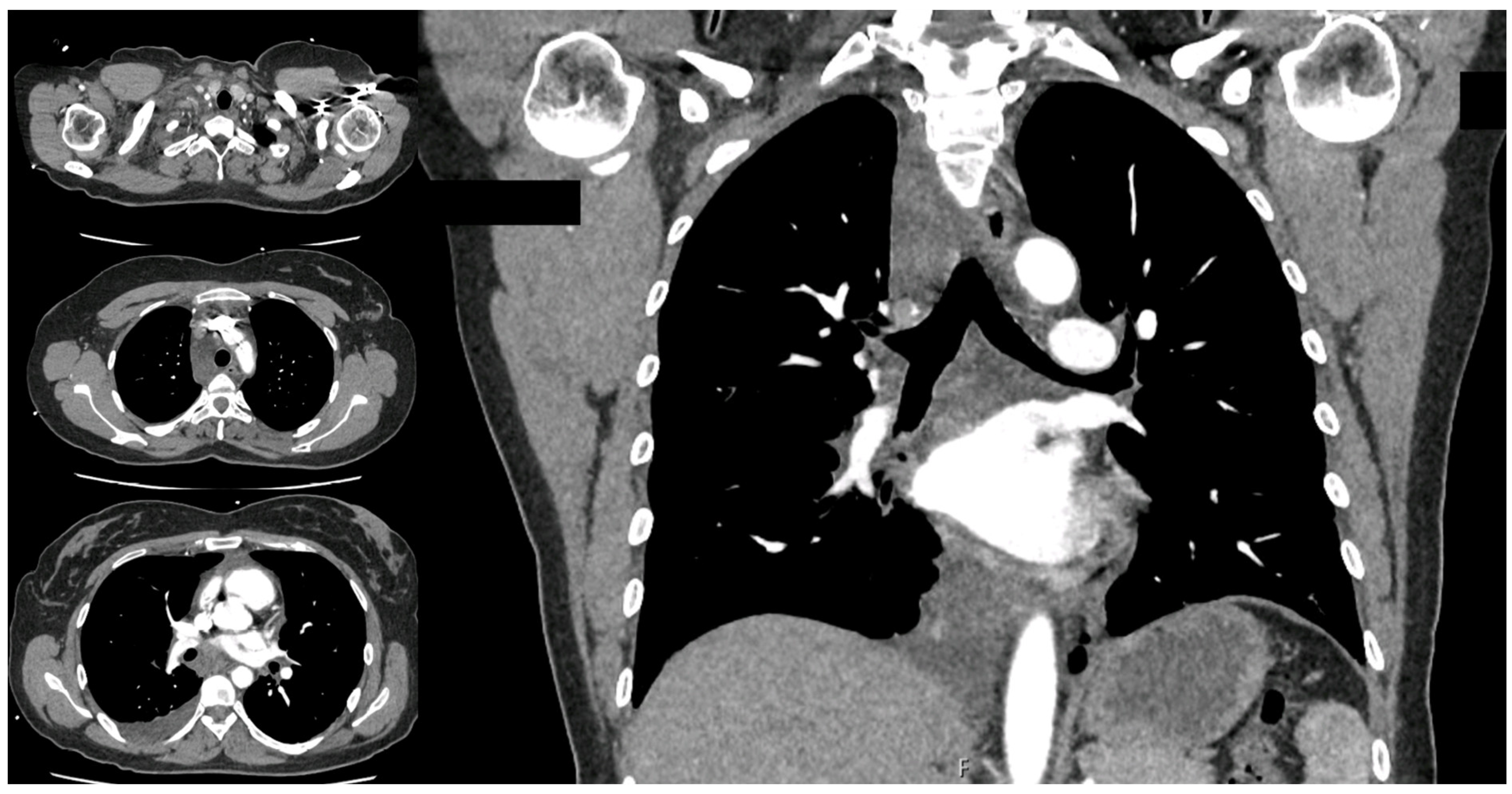
2. Case Presentation

A 40-year-old woman attended the emergency department complaining of acute onset shortness of breath, right sided shoulder pain, and neck swelling. She had no previous medical history nor comorbidities and only reported attendance to her first yoga class 2 weeks prior. Clinical examination revealed lateral neck swelling. Her blood pressure and heart rate were within normal range. However, she did not require additional oxygen support to maintain saturations above 92% and her respiration rate was at 14 breaths per minute. Routine laboratory tests were all within normal limits. A chest X-ray showed minimal right pleural effusion which was conservatively managed, and she was discharged home with analgesia as she was clinically well. Only a few hours later, she was readmitted with an increasing shortness of breath, worsening pain, dysphagia, and an episode of collapse with a loss of consciousness. Within the following 48 h, she had three chest computed tomography (CT) scans including an oral contrast that showed a very interesting sequence of events. Initially, diffuse infiltration and non-specific soft tissue thickening of the right supraclavicular area and the whole mediastinum was found, particularly around the esophagus with radiological suspicion of esophageal perforation (Figure 1).
The barium swallow scan, however, did not show any extravasation of contrast ruling out esophageal perforation. The third scan showed a significant increase in size of the right pleural effusion (Figure 2).
A diagnostic ultrasound-guided thoracocentesis revealed milky pleural effusion (Figure 3), which tested positive for chylomicrons, and the unexpected diagnosis of spontaneous chylothorax was made.
The patient was referred to the thoracic surgery department for treatment. Initially, conservative management was established with intravenous octreotide (300 μg/ daily, up to 600 μg/daily after three days of ineffective treatment) and a medium chain triglyceride (MCT) diet. Since the chylous drain output remained high (over 1000 mL for five consecutive days), surgical exploration was then scheduled and a 2-port right video-assisted thoracoscopic surgery (VATS) was performed. The pleural cavity was debrided and the chylothorax fully evacuated. No damage to thoracic duct nor obvious leak were found. We decided to perform the mass control of the thoracic duct together with the azygos vein by ligating both vessels and performing total pleurectomy to achieve pleurodesis. Satisfactory lung expansion was observed prior to skin closure. The patient remained on the MCT diet and octreotide for 5 days as the chyle output drastically reduced from post-operative day (POD) 2 and turned serous on POD 4. On POD 5, a normal diet was restarted and a further 48 h observation did not reveal an increase in drain output nor chyle leak. The drain was therefore removed, and the patient discharged home.
The patient did not exhibit malnutrition or weight loss and remained in a good general health condition throughout the treatment. At 3 months follow up, she was asymptomatic and chest X-ray showed complete resolution of the effusion and full lung expansion.

3. Discussion

Chylothorax is a rare cause of pleural effusion with several etiologies grouped broadly into traumatic and nontraumatic (spontaneous) causes. Most cases of spontaneous chylothorax are associated with minor trauma such as coughing, vomiting, stretching, and hiccups after a high-fat meal [2]. Infiltrating neoplasms located in the mediastinum or neck, or expansive solid lesions of another nature, can cause thoracic duct lesions and result in chyle loss, some of which are anecdotal, as reported by rare cases in the literature [9]. Trauma-related chylothorax contributes to almost 50% of all cases. Surgeries that involve posterior or superior mediastinum such as a foregut or aortic surgery or mediastinal lymph nodes sampling and lymph node dissection are also common causes. Idiopathic and medical causes account for 6% and 44%, respectively [2]. About 50–60% of chylothorax occur on the right side, 33.3% on the left side and about 16.6% will present with bilateral pleural effusions [8,10].
Chylothorax can be diagnosed either clinically or biochemically. Clinically, milky pleural aspirate suggests the presence of chyle; however, it has to be confirmed biochemically to exclude pseudochylothorax [10]. Triglyceride levels with a value of greater than 110 mg/dL is highly suggestive (99%) of chylothorax, whereas a value of <50 mg/dL is unlikely to be chyle [10,11]. Fluid cholesterol-to-triglyceride ratio of <1 is considered diagnostic as well [12]. Usually, careful history taking is the key to reaching the offending cause in patients with chylothorax. However, a CT chest scan is a very useful tool to assess for underlying malignancy as a cause of chylothorax.
Further diagnostic approaches, such as MR-lymphangiography, should be critically considered regarding feasibility, clinical benefit, and patient safety. Lymphatic system imaging studies remain challenging, though recently, non-enhanced MR-lymphography has been attempted in nontraumatic chylothorax [13]. It has also been reported that combining intermittent digital X-rays and live near-infrared imaging with microsurgical techniques, the “dual imaging lymphangiography” might be an option for refractory chylothorax [10]. However, such imaging techniques should be reserved for unresponsive or complicated cases [14,15].
Patients with chylothorax may be asymptomatic or present with non-specific symptoms such as chest discomfort, shortness of breath, and fatigue [12]. Moreover, if there was a chronic chyle loss, this could lead to nutrients, electrolytes, and volume loss in addition to the depletion of lymphocytes [10]. Ultimately, this will lead to malnutrition and immunosuppression.
The cisterna chyli is a lymphatic sac located at the lumbar level to the right of the abdominal aorta and behind the right diaphragmatic crus, at the level of L1–L2. The lymphatic flow from the lower half of the body converges to this saccular structure [16,17]. The cisterna chyli receives multiple lymphatic afferents and gives rise to the thoracic duct to the right of the aorta at the level of T12–L2. This primary lymphatic duct crosses the aortic diaphragmatic hiatus to enter the thorax, passing by the aorta, the esophagus, and the azygos vein. In 50% of people, the thoracic duct crosses from right to left at the level of T5–T6, passing the left brachiocephalic vein on the transverse plane and rotates inferiorly to the end in the subclavian vein at the confluence of the left jugular vein [16,17]. Therefore, a thoracic duct injury below the T-5 level results in a right chylous effusion, while a thoracic duct injury above T-5 results in a left chylous effusion. Because of the usual transition from the right to the left side of the thorax at the level of T3–6, a vessel injury at this point is expected to result in a chyle effusion in both pleural cavities.
Although there are a few reported cases of chylothorax after light exercises or a workout (Table 1), chylous effusion resulting from only the hyperextension of the neck secondary to the yoga exercise has not been reported in the literature [18,19,20,21,22,23]. In our case, the patient presented with respiratory symptoms 2 weeks following a yoga class, hence this was also a delayed presentation. Upon detailed history taking, there was no other obvious cause for the chyle leakage. We are raising the potential correlation between the yoga hyperextension of the neck and a spontaneous rupture of the thoracic duct. The duct more often drains into the confluence of the left internal jugular and subclavian veins which is located at the cervical base in close relationship to the stretched cervical muscles over the left side. Interestingly, however, our case of chylothorax was on the right side. In this setting, one must be aware than anatomical variations of the thoracic duct pathway have indeed been described and that a right lymphatic duct that drains the right upper trunk, arm, and neck does exist as well [24].
According to a brief literature review, the leading causes of thoracic duct rupture related to light physical activity are associated with forced postures of the backbone or overstretched junctions. The first report was presented by Meade [25], describing five cases of spontaneous unilateral chylothorax, believed to be correlated to the hyperextension of the spine with the rupture of an inherently weakened thoracic duct. In 1975, Reilly and Tsou [26] reported a case of bilateral chylothorax apparently associated with stretching exercises and yawning, assuming that a thoracic duct disruption was created by the hyperextension of the spine. Similarly, Gullane and Marsh [27] reported a case of bilateral spontaneous chylothorax presenting as an anterior neck mass after swimming and assumed the mechanism to be the same as that reported by Reilly and Tsou. In another reported case report, due to the absence of any demonstrable pathology, trauma, or structural abnormality, Tankanow et al. considered the sudden hyperextension of the spine while climbing into the bathtub as the cause that resulted in injury to a weakened portion of the thoracic duct, with the leakage of chyle in both pleural spaces [28]. An unusual presentation of chylothorax was also reported by Fehr et al., who described a left-side neck mass and bilateral chylous pleural effusion occurred in a patient during routine vacuum cleaning, potentially related to an unnatural movement of the scapular girdle and consequent extension of the cervical spine while using the cleaning tool [29]. In all the reported cases, a conservative treatment (cessation of fatty food consumption and medium chain triglyceride diet supplement) was the choice that resulted in complete patient healing.
However, the management of chylothorax depends on the etiology. Other than conservative treatment, there are different approaches, such as non-surgical operative intervention or surgery [8].
Dietary fat restriction and the use of octreotide to reduce the production of chyle constitute conservative management. In clinically stable patients, it seems reasonable to begin therapy with a noninvasive, fat-free oral diet, and MCT supplement. Saturated fatty acids of eight-to-twelve carbon chain lengths are absorbed through the portal venous system, overriding the lymphatic drainage [28]. In our algorithm, we used octreotide early, as diet alterations alone did not lower the fluid patient’s output.
Compared to somatostatin, octreotide has a longer half-life in circulation and can be administered subcutaneously [30]. The concurrent administration of octreotide or somatostatin with chest tube drainage and enteric rest in postoperative adult patients with chyle leaks has been shown to decrease the need for surgical intervention [31]. Although the octreotide mechanism of action is not entirely clear [32], it is known that its vasoconstrictor effect reduces intestinal blood flow due to the vasoconstriction of the splanchnic circulation, thus reducing gastric, pancreatic, and biliary secretions. Furthermore, fat absorption from the intestine is also reduced [33,34]. It has also been reported that the expression of somatostatin receptors SSTR2 and SSTR5 in the human thoracic duct and their stimulation may decrease lymphatic flow and lymph production [35,36]. In most reports, the benefit of octreotide treatment was seen within 2–3 days [37]. As recently published in a case series, orally administered propranolol, commonly used to treat childhood hemangiomas, appears to be an alternative treatment for chylothorax, especially in children [38]. The advantages of propranolol therapy include ease of administration, broad experience, and easy accessibility.
It is a matter of debate among authors as to when to shift the treatment from a nonoperative approach to operative means [39]. Once introduced by Cope et al., thoracic duct embolization (TDE) was accepted as the treatment of choice or at least as a viable alternative to surgical ligation in treating intractable postoperative chylothorax [40,41,42,43]. Thoracic duct cannulation (TDC) is the primary determining factor for the technical success of TDE due to its technical difficulty [40]. Different studies reported the conventional antegrade TDC’s technical success rate to be about 70% [41,44,45]. Hence, significant numbers of patients for whom TDC had failed were forced to settle for the unproven therapeutic effect of Lipiodol during lymphangiography or thoracic duct disruption. However, neither procedure is as clinically efficient as TDE is [46,47]. Recently, novel retrograde transvenous or percutaneous cervical TDC techniques, with the aid of a bail-out retrograde approach, significantly improved overall technical success [48]. The main limitation of this nonsurgical operative approach lies in the availability of the necessary equipment and devices. For this reason, although recommended in selected cases, thoracic duct embolization techniques are to be reserved for high-volume and experienced centers [49].
Surgical interventions for managing chylothorax include thoracic duct ligation or clipping by thoracotomy or VATS, mass ligation, pleurodesis, pleurectomy, or pleuroperitoneal shunt. Thoracic duct ligation can be performed at the site of leakage or in the supradiaphragmatic region; in the latter case, success rates of up to 90% were reported [50]. In our case, the conservative treatment failed, and because leakage from the thoracic duct could not be identified intraoperatively, the mass ligation of the duct and azygos vein was performed by minimally invasive technique, followed by total pleurectomy. The aim of pleurectomy is to reduce the risk of recurrence and need for repetitive exploration. Alternatively, some authors report high success rates with talc pleurodesis for cases in which the thoracic duct could not be identified [51]. Others suggest pleurodesis with iodopovidone in cases where leakage was identified and ligated [9]. With the adoption of surgical measures, the mortality rate of chylothorax decreased from 50% with conservative management to 10% [52].

4. Conclusions

Chylothorax, although rare, should be treated with a high index of suspicion. It should be considered as a differential diagnosis in any patient presenting with spontaneous pleural effusion in the context of very light physical exercise. Our case was quite unique in that it occurred after a prolonged time (2 weeks) following modest exercise after a neck extension only, during introductory beginner yoga techniques. Other cases of spontaneous chylothorax after exercise have been described, but more likely to be attributable to sudden, stressful movements than those predicted by yoga relaxation techniques. As defined by experiences reported in the literature, surgical management aided by conservative therapy was also successful in our case.

Author Contributions

Conceptualization, all authors; methodology, G.H., F.A., T.K., A.A. and A.T.; investigation, G.H., F.A., T.K., A.A. and A.T.; resources, G.H., F.A., T.K., A.A. and A.T.; data curation, G.H., F.A., T.K., A.A. and A.T.; writing—original draft preparation, G.H., P.A.F., F.A., T.K., A.A. and A.T.; writing—review and editing, G.H., P.A.F., F.A., T.K., A.A. and A.T.; visualization, G.H., P.A.F., F.A., T.K., A.A. and A.T.; supervision, P.A.F. and A.T. All authors have read and agreed to the published version of the manuscript.

Funding

This research received no external funding.

Institutional Review Board Statement

The study was conducted according to the guidelines of the Declaration of Helsinki. Ethical review and approval were waived for this study due to its retrospective illustrative nature. It does not meet the national policy for the protection of human subjects’ definition of research, which requires an investigation that contributes to generalizable knowledge about disease or condition.

Informed Consent Statement

Written informed consent has been obtained from the patient to publish this paper.

Data Availability Statement

Further inquiries can be directed to the corresponding authors.

Conflicts of Interest

The authors declare no conflicts of interest.

References

  1. Doerr, C.H.; Miller, D.L.; Ryu, J.H. Chylothorax. Semin. Respir. Crit. Care Med. 2001, 22, 617–626. [Google Scholar] [CrossRef] [PubMed]
  2. Doerr, C.H.; Allen, M.S.; Nicholas, F.C. Etiology of chylothorax in 203 patients. Mayo Clin. Proc. 2005, 80, 867. [Google Scholar] [CrossRef] [PubMed]
  3. Bender, B.; Murthy, V.; Chamberlin, R.S. The changing management of chylothorax in in the modern era. Eur. J. Cardiothorac. Surg. 2016, 49, 18–24. [Google Scholar] [CrossRef]
  4. Lama, A.; Ferreiro, L.; Toubes, M.E.; Golpe, A.; Gude, F.; Álvarez-Dobaño, J.M.; González-Barcala, F.J.; San José, E.; Rodríguez-Núñez, N.; Rábade, C.; et al. Characteristics of patients with pseudochylothorax—A systematic review. J. Thorac. Dis. 2016, 8, 2093–2101. [Google Scholar] [CrossRef] [PubMed]
  5. Sassoon, C.S.; Light, R.W. Chylothorax and pseudochylothorax. Clin. Chest Med. 1985, 6, 163–171. [Google Scholar] [CrossRef]
  6. McCray, S.P.C. Nutritional management of chyle leaks: An update. Pract. Gastroentrol. Ser. 2011, 94, 12–32. [Google Scholar]
  7. Huggins, J.T. Chylothorax and cholesterol pleural effusion. Semin. Respir. Crit. Care Med. 2010, 31, 743–750. [Google Scholar] [CrossRef] [PubMed]
  8. Riley, L.E.; Ataya, A. Clinical approach and review of causes of chylothorax. Respir. Med. 2019, 157, 7–13. [Google Scholar] [CrossRef]
  9. Ferrari, P.A.; Fusaro, F.; Ferrari, A.; Tamburrini, A.; Grimaldi, G.; Santoru, M.; Zappadu, S.; Tanda, E.; Nemolato, S.; Comelli, S.; et al. Refractory Chylothorax Secondary to Sizeable Azygos Vein Hemangioma: Tailored Multimodal Treatment of a Challenging Case Report. Medicina 2022, 59, 91. [Google Scholar] [CrossRef]
  10. McGrath, E.E.; Blades, Z.; Anderson, P.B. Chylothorax: Aetiology, diagnosis and therapeutic options. Respir. Med. 2010, 104, 1–8. [Google Scholar] [CrossRef]
  11. Staats, B.A.; Ellefson, R.D.; Budahn, L.L.; Dines, D.E.; Prakash, U.B.; Offord, K. The lipoprotein profile of chylous and nonchylous pleural effusion. Mayo Clin. Proc. 1980, 55, 700–704. [Google Scholar]
  12. Madaniah, A.A. Spontaneous idiopathic chylothorax in adults. Saudi Med. J. 2005, 26, 145–146. [Google Scholar] [PubMed]
  13. Cholet, C.; Delalandre, C.; Monnier-Cholley, L.; Le Pimpec-Barthes, F.; El Mouhadi, S.; Arrivé, L. Nontraumatic Chylothorax: Nonenhanced MR Lymphography. Radiographics 2020, 40, 1554–1573. [Google Scholar] [CrossRef] [PubMed]
  14. Savla, J.J.; Itkin, M.; Rossano, J.W.; Dori, Y. Post-Operative Chylothorax in Patients with Congenital Heart Disease. J. Am. Coll. Cardiol. 2017, 69, 2410–2422. [Google Scholar] [CrossRef] [PubMed]
  15. Weissler, J.M.; Cho, E.H.; Koltz, P.F.; Carney, M.J.; Itkin, M.; Laje, P.; Levin, L.S.; Dori, Y.; Kanchwala, S.K.; Kovach, S.J. Lymphovenous Anastomosis for the Treatment of Chylothorax in Infants: A Novel Microsurgical Approach to a Devastating Problem. Plast. Reconstr. Surg. 2018, 141, 1502–1507. [Google Scholar] [CrossRef] [PubMed]
  16. Aalami, O.O.; Allen, D.B.; Organ, C.H., Jr. Chylous ascites: A collective review. Surgery 2000, 128, 761–778. [Google Scholar] [CrossRef] [PubMed]
  17. Breslin, J.W.; Yang, Y.; Scallan, J.P.; Sweat, R.S.; Adderley, S.P.; Murfee, W.L. Lymphatic vessel network structure and physiology. Compr. Physiol. 2018, 9, 207–299. [Google Scholar] [PubMed]
  18. Torrejais, J.C.; Rau, C.B.; de Barros, J.A.; Torrejais, M.M. Spontaneous chylothorax associated with light physical activity. J. Bras. Pneumol. 2006, 32, 599–602. [Google Scholar] [CrossRef] [PubMed]
  19. Kolbaş, I.; Tezel, Y.; Coşgun, T.; Baysungur, V.; Tezel, C. Chylothorax Due to Weight Lifting: A Rare Etiology. South. Clin. Ist. Euras. 2020, 31, 75–77. [Google Scholar] [CrossRef]
  20. Akbar, S.; Advani, R.; Aggarwal, R. Bilateral spontaneous chylothorax presenting as a left-sided neck mass. BMJ Case Rep. 2021, 14, e240320. [Google Scholar] [CrossRef]
  21. Garcia-Aparicio, J.; Herrero-Herrero, J.-I.; Corral-Gudine, L.; Jorge-Sanchez, R.-J. Bilateral idiopathic chylothorax associated with light exercise. Resp. Med. CME 2009, 2, 68–69. [Google Scholar] [CrossRef]
  22. Bottet, B.; Melki, J.; Levesque, H.; Baste, J.M.; Roussel, E.; Peillon, C. Stretching et chylothorax. Rev. Mal. Respir. 2019, 36, 742–746. [Google Scholar] [CrossRef] [PubMed]
  23. Bocquel, V.; Girard, P.; Fournel, P.; Vergnon, J.M. Spontaneous chylothorax. Apropos of a further case. Rev. Mal. Respir. 1997, 14, 395–396. [Google Scholar] [PubMed]
  24. Hematti, H.; Mehran, R.J. Anatomy of the thoracic duct. Thorac. Surg. Clin. 2011, 21, 229–238. [Google Scholar] [CrossRef] [PubMed]
  25. Meade, R.H. Spontaneous chylothorax. Arch. Intern. Med. 1972, 90, 30–36. [Google Scholar] [CrossRef] [PubMed]
  26. Reilly, K.M.; Tsou, F. Bilateral chylothorax: A case report following episodes of stretching. JAMA 1975, 233, 536–537. [Google Scholar] [CrossRef] [PubMed]
  27. Gullane, P.J.; Marsh, A.S. Bilateral spontaneous chylothorax presenting as a neck mass. J. Otolaryngol. 1984, 13, 4. [Google Scholar]
  28. Tankanow, L.B.; Petrozzi, C.; Ward, J.C.; Bower, G.C. Idiopathic Bilateral Chylothorax Presenting as a Left-Sided Neck Swelling. Henry Ford. Hosp. Med. J. 1986, 34, 130–131. [Google Scholar] [PubMed]
  29. Fehr, M.; Lehmann, T.; Kuhn, M. Idiopathic chylothorax while vacuum cleaning. Schweiz Med. Forum. 2007, 7, 630–632. [Google Scholar]
  30. Bellini, C.; Cabano, R.; De Angelis, L.C.; Bellini, T.; Calevo, M.G.; Gandullia, P.; Ramenghi, L.A. Octreotide for congenital and acquired chylothorax in newborns: A systematic review. J. Paediatr. Child. Health 2018, 54, 840–847. [Google Scholar] [CrossRef]
  31. Anger, M.; Hofmann, J.; Ruf, B.; Steinborn, M.; Reber, D.; Warncke, K.; Rieber, N. Cough-induced chylothorax in a two-year-old boy—case report and review of the literature. BMC Pediatr. 2023, 23, 416. [Google Scholar] [CrossRef] [PubMed]
  32. Madhavan, S.; Nakao, M. How efficacious are Octreotide and Somatostatin in the management of chylothorax in congenital cardiac surgical patients? Interact. Cardiovasc. Thorac. Surg. 2021, 33, 773–778. [Google Scholar] [CrossRef] [PubMed]
  33. Al-Zubairy, S.A.; Al-Jazairi, A.S. Octreotide as a therapeutic option for management of chylothorax. Ann. Pharmacother. 2003, 37, 679–682. [Google Scholar] [CrossRef] [PubMed]
  34. Kalomenidis, I. Octreotide and chylothorax. Curr. Opin. Pulm. Med. 2006, 12, 264–267. [Google Scholar] [CrossRef] [PubMed]
  35. Jiang, H.; Deng, X.F.; Duan, C.M.; Chen, C.; Xiang, J.L.; Lu, Y.L.; Ma, O.F. Somatostatin receptors SSTR2 and SSTR5 are expressed in the human thoracic duct. Lymphology 2011, 44, 21–28. [Google Scholar] [PubMed]
  36. Buettiker, V.; Hug, M.I.; Burger, R.; Baenziger, O. Somatostatin: A new therapeutic option for the treatment of chylothorax. Intensive Care Med. 2001, 27, 1083–1086. [Google Scholar] [CrossRef] [PubMed]
  37. Ismail, N.A.; Gordon, J.; Dunning, J. The use of octreotide in the treatment of chylothorax following cardiothoracic surgery. Interact. Cardiovasc. Thorac. Surg. 2015, 20, 848–854. [Google Scholar] [CrossRef]
  38. Mitchell, K.; Weiner, A.; Ramsay, P.; Sahni, M. Use of Propranolol in the Treatment of Chylous Effusions in Infants. Pediatrics 2021, 148, e2020049699. [Google Scholar] [CrossRef] [PubMed]
  39. Jindal, R.; Singh, J.; Garg, L.; Gupta, M. Diagnosis and management of traumatic bilateral chylothorax: A clinical conundrum. BMJ Case Rep. 2019, 12, e229400. [Google Scholar] [CrossRef] [PubMed]
  40. Cope, C.; Kaiser, L.R. Management of unremitting chylothorax by percutaneous embolization and blockage of retroperitoneal lymphatic vessels in 42 patients. J. Vasc. Interv. Radiol. 2002, 13, 1139–1148. [Google Scholar] [CrossRef]
  41. Itkin, M.; Kucharczuk, J.C.; Kwak, A.; Trerotola, S.O.; Kaiser, L.R. Nonoperative thoracic duct embolization for traumatic thoracic duct leak: Experience in 109 patients. J. Thorac. Cardiovasc. Surg. 2010, 139, 584–589. [Google Scholar] [CrossRef] [PubMed]
  42. Nadolski, G.J.; Itkin, M. Thoracic duct embolization for nontraumatic chylous effusion: Experience in 34 patients. Chest 2013, 143, 158–163. [Google Scholar] [CrossRef] [PubMed]
  43. Boffa, D.J.; Sands, M.J.; Rice, T.W.; Murthy, S.C.; Mason, D.P.; Geisinger, M.A.; Blackstone, E.H. A critical evaluation of a percutaneous diagnostic and treatment strategy for chylothorax after thoracic surgery. Eur. J. Cardiothorac. Surg. 2013, 33, 435–439. [Google Scholar] [CrossRef] [PubMed]
  44. Laslett, D.; Trerotola, S.O.; Itkin, M. Delayed complications following technically successful thoracic duct embolization. J. Vasc. Interv. Radiol. 2012, 23, 76–79. [Google Scholar] [CrossRef] [PubMed]
  45. Kim, P.H.; Tsauo, J.; Shin, J.H. Lymphatic interventions for chylothorax: A systematic review and meta-analysis. J. Vasc. Interv. Radiol. 2018, 29, 194–202. [Google Scholar] [CrossRef]
  46. Pamarthi, V.; Stecker, M.S.; Schenker, M.P.; Baum, R.A.; Killoran, T.P.; Han, A.S.; O’Horo, S.K.; Rabkin, D.J.; Fan, C.M. Thoracic duct embolization and disruption for treatment of chylous effusions: Experience with 105 patients. J. Vasc. Interv. Radiol. 2014, 25, 1398–1404. [Google Scholar] [CrossRef] [PubMed]
  47. Binkert, C.A.; Yucel, E.K.; Davison, B.D.; Sugarbaker, D.J.; Baum, R.A. Percutaneous treatment of high-output chylothorax with embolization or needle disruption technique. J. Vasc. Interv. Radiol. 2005, 16, 1257–1262. [Google Scholar] [CrossRef] [PubMed]
  48. Jun, H.; Hur, S.; Jeong, Y.S.; Kang, C.H.; Lee, H. Thoracic duct embolization in treating postoperative chylothorax: Does bail-out retrograde access improve outcomes? Eur. Radiol. 2022, 32, 377–383. [Google Scholar] [CrossRef] [PubMed]
  49. Higgins, M.C.; Park, A.W.; Angle, J.F. Chylothorax: Percutaneous Embolization of the Thoracic Duct. Oper. Tech. Thorac. Cardiovasc. Surg. 2015, 20, 402–412. [Google Scholar] [CrossRef]
  50. Pillay, T.G.; Singh, B. A review of traumatic chylothorax. Injury 2016, 47, 545–550. [Google Scholar] [CrossRef]
  51. Fernández Alvarez, J.R.; Kalache, K.D.; Graŭel, E.L. Management of spontaneous congenital chylothorax: Oral medium-chain triglycerides versus total parenteral nutrition. Am. J. Perinatol. 1999, 16, 0415–0420. [Google Scholar] [CrossRef] [PubMed]
  52. Seitelman, E.; Arellano, J.J.; Takabe, K.; Barrett, L.; Faust, G.; Angus, L.G. Chylothorax after blunt trauma. J. Thorac. Dis. 2012, 4, 327–330. [Google Scholar] [PubMed]
Figure 1. First CT scan axial and coronal views showing predominantly neck and diffuse mediastinal fat and soft tissue swelling with minimal pleural effusion.
Surgeries 05 00026 g001
Figure 2. Further CT scan axial and coronal views showing the significant reduction in neck and mediastinal soft tissue swelling and the increase in the pleural effusion.
Surgeries 05 00026 g002
Figure 3. Aspirated sample of Chyle from thoracocentesis.
Surgeries 05 00026 g003
Table 1. Unusual cases of chylothorax following nontraumatic physical activity.
Authors, YearSubjects (n)Supposed CauseLocation of Chyle LeakageIntervention
Meade RH, 1972 [25]5Hyperextension of the spineUnilateral chylothoraxN/A
Reilly et al., 1975 [26]1Hyperextension of the spine during stretching exercise and yawningBilateral chylothoraxConservative
Gullane et al., 1984 [27]1Hyperextension of the spine while swimmingBilateral chylothoraxConservative
Tankanow et al., 1986 [28]1Hyperextension of the spine while climbing into a bathtubBilateral chylothoraxConservative
Bocquel et al., 1997 [23]1Overstretch of subclavius and anterior scalenus muscle during stretching exerciseUnilateral chylothoraxConservative
Torrejais et al., 2006 [18]1Hyperextension of the neck during light physical activity at a fitness centerBilateral neck swelling and bilateral chylothoraxConservative
Fehr et al., 2007 [29]1Hyperextension of the neck and overstretch of thoracic outlet during routine vacuum cleaningLeft-sided neck swelling and bilateral chylothoraxConservative
Garcìa-Aparicio et al., 2009 [21]1Hyperextension of the neck during light physical activityBilateral chylothoraxConservative
Bottet et al., 2019 [22]1Overstretch of subclavius and anterior scalenus muscle during stretching exerciseBilateral chylothoraxConservative
Kolbas et al., 2020 [19]1Overstretch of subclavius and anterior scalenus muscle while lifting 40 kg at a fitness centerLeft-sided chylothoraxConservative
Akbar et al., 2021 [20]1Hyperextension of the spine during stretching exerciseLeft-sided neck swelling and bilateral chylothoraxConservative
Disclaimer/Publisher’s Note: The statements, opinions and data contained in all publications are solely those of the individual author(s) and contributor(s) and not of MDPI and/or the editor(s). MDPI and/or the editor(s) disclaim responsibility for any injury to people or property resulting from any ideas, methods, instructions or products referred to in the content.

