Article

A Lateral Flow-Recombinase Polymerase Amplification Method for Colletotrichum gloeosporioides Detection

1
Institute of Botany, Jiangsu Province and Chinese Academy of Sciences, Nanjing 210014, China
2
Jiangsu Key Laboratory for the Research and Utilization of Plant Resources, Nanjing 210014, China
3
Jiangsu Provincial Science and Technology Resources Coordination Platform (Agricultural Germplasm Resources) Germplasm Resources Nursery of Medicinal Plants, Nanjing 210014, China
*
Author to whom correspondence should be addressed.
These authors contributed equally to this work.
Submission received: 26 February 2024 / Revised: 17 April 2024 / Accepted: 22 April 2024 / Published: 26 April 2024

Abstract

:
The greater yam (Dioscorea alata), a widely cultivated and nutritious food crop, suffers from widespread yield reduction due to anthracnose caused by Colletotrichum gloeosporioides. Latent infection often occurs before anthracnose phenotypes can be detected, making early prevention difficult and causing significant harm to agricultural production. Through comparative genomic analysis of 60 genomes of 38 species from the Colletotrichum genus, this study identified 17 orthologous gene groups (orthogroups) that were shared by all investigated C. gloeosporioides strains but absent from all other Colletotrichum species. Four of the 17 C. gloeosporioides-specific orthogroups were used as molecular markers for PCR primer designation and C. gloeosporioides detection. All of them can specifically detect C. gloeosporioides out of microbes within and beyond the Colletotrichum genus with different sensitivities. To establish a rapid, portable, and operable anthracnose diagnostic method suitable for field use, specific recombinase polymerase amplification (RPA) primer probe combinations were designed, and a lateral flow (LF)-RPA detection kit for C. gloeosporioides was developed, with the sensitivity reaching the picogram (pg) level. In conclusion, this study identified C. gloeosporioides-specific molecular markers and developed an efficient method for C. gloeosporioides detection, which can be applied to the prevention and control of yam anthracnose as well as anthracnose caused by C. gloeosporioides in other crops. The strategy adopted by this study also serves as a reference for the identification of molecular markers and diagnosis of other plant pathogens.

1. Introduction

Dioscorea alata, commonly known as greater yam, is a widely cultivated and highly important crop in tropical and subtropical areas. It is the most widely distributed and most-produced yam species globally, except for D. polystachya [1]. Greater yam is a widely cultivated tuber crop due to its high yield potential even in low soil fertility conditions, as well as its ease of propagation, early vigor in competing with weeds, being rich in starch and protein [2], and tuber storability [3]. However, the yields of greater yam have been hindered by various biotic and abiotic constraints. Among the diseases caused by microbial infection, anthracnose caused by Colletotrichum gloeosporioides is the main problem affecting greater yam yields and post-harvest quality [4]. Greater yam anthracnose can induce symptoms such as leaf necrosis, premature shedding, and wilting of young shoots in yam plants, resulting in yield reductions of up to 90% [5] and leading to significant economic losses, and this has necessitated frequent fungicide applications for chemical control. However, this approach poses environmental risks and potential fungicide resistance, and it is often cost-prohibitive for many farmers in tropical regions [6].
Moreover, recent studies have revealed high genotypic diversity within C. gloeosporioides [7]. It is a species complex of more than 20 subspecies with a rapid rate of intraspecific variation and a wide geographic distribution [8], suggesting potential for the evolution of new virulent strains capable of overcoming plant resistance. In addition, C. gloeosporioides is not only a widespread causal agent of greater yam anthracnose but also a pathogen infecting over 470 different host genera from monocotyledons to dicotyledons [9]. Controlling C. gloeosporioides disease has been hampered by its rapid spread and long-term survival in natural environments. The pathogen exhibits a relatively extended incubation period following its infection of the host plant, thereby impeding the activation of the plant’s immune responses [10]. Therefore, the rapid and accurate detection of C. gloeosporioides during the early infection stages is critical for disease management.
Traditionally, the identification and characterization of Colletotrichum spp. relied on morphological features such as the colony color, conidia and appressorium shape, and growth rate. Molecular techniques now provide alternative methods for taxonomic studies and are important tools for species delimitation [11,12]. The selection of appropriate target genes is essential for the successful detection of specific pathogens. The target genes should exhibit a high degree of conservation within the pathogen species to ensure broad applicability across different strains and isolates [13,14]. One established PCR-based method for detecting C. gloeosporioides is to detect the internally transcribed spacer (ITS) 1 region of ribosomal DNA (rDNA) [15]. However, this region has a low specificity for species discrimination within the Colletotrichum genera because of the high sequence identity of the ITS sequence among closely related species [16]. Therefore, mining new C. gloeosporioides-specific targets is needed for rapid and precise detection of this pathogen.
Comparative genomics is a powerful approach used to identify specific target genes for various applications, including diagnostics [17], classification [18], and evolutionary [19] studies. By comparing the genomes of different species or individuals within a species, researchers can identify genes that are unique to specific taxa [13,14], which may serve as ideal targets for designing species-specific diagnostic methods. Molecular techniques, including the polymerase chain reaction (PCR) and reverse transcription quantitative PCR (RT-qPCR), are highly sensitive DNA-based methods that have been applied in C. gloeosporioides detection [20,21]. However, conventional PCR-based methods are not suitable for field detection due to the need for bulky and expensive lab equipment and complicated protocols for amplification. Recombinase polymerase amplification (RPA) has been proven to be a promising alternative to traditional PCR-based methods for the detection of plant pathogens in recent studies [22]. Unlike the PCR, RPA does not require complicated temperature cycling conditions, making it more conducive for field applications. This isothermal amplification technique offers several advantages, including field mobility, rapid results, and the ability to perform amplification directly on a sample material without the need for DNA extraction [23]. Furthermore, the combination of RPA with lateral flow (LF) assays allows for rapid detection and easy visualization of results, enhancing its practicality for on-site testing [24]. The application of this technology also reduces the cost requirements for specialist and complicated instruments. Moreover, because the detection kit facilitates industrial production, its cost will be further reduced.
This study conducted comparative genomics to identify C. gloeosporioides-specific genes. The designed primer pairs targeting 4 out of the 17 C. gloeosporioides-specific genes display high specify and sensitivity in amplifying the genomic DNA of C. gloeosporioides. Using one gene as a target, we developed a rapid and simple LF-RPA method for the detection of C. gloeosporioides. The LF-RPA method enables detection of C. gloeosporioides in resource-limited laboratories and field environments at a low cost. Furthermore, the strategy adopted by this study may serve as a reference for the molecular mark development and identification of different kinds of plant pathogens.

2. Materials and Methods

2.1. Materials

In this study, 60 genomes from 38 species of the genus Colletotrichum were used in the comparative genomic analysis. The C. gloeosporioides strain CgDa01 (GCA_021650765.1), which was recently isolated from greater yam, was sequenced by our lab [25]. The other 59 Colletotrichum genomes were downloaded from the NCBI database (Table S1).
The fungal species included in this research for DNA extraction were Fusarium graminearum, F. oxysporum, Sclerotinia sclerotiorum, Botrytis cinerea, Blumeria graminis, C. fructicola, and C. gloeosporioides strains CgDa01 and CgDaM3. The CgDa01 and CgDaM3 strains were isolated in our laboratory. The C. gloeosporioides strains CgDa01 and CgDaM3 were kept in our lab. The C. fructicola strain was a gift from Dr. Yuqiang Zhao of the Fruit Tree Research Centre of the Institute of Botany at the Jiangsu Province and Chinese Academy of Sciences. The other strains were kindly provided by Professor Dao-Long Dou’s laboratory at Nanjing Agricultural University.

2.2. Comparative Genomic Analysis

The OrthoFinder software (version 2.5.2) was employed to analyze orthologous relationships across all coding genes of the genomes used in this study with the default settings [26]. Genes shared by all C. gloeosporioides strains but absent in the genomes of all other Colletotrichum species were categorized as C. gloeosporioides-specific orthogroups. The coding sequences (CDSs) of each orthogroup were aligned by using the ClustalW program (v1.4) integrated into BioEdit (v7.2.5) [27]. The resulting alignments were used for the next step: molecular marker design.

2.3. Fungal Cultivation and Genomic DNA Extraction

All fungal strains were cultivated in a potato dextrose agar (PDA) medium and incubated at 28 °C for 5 days. Mycelium from the actively growing margin of the culture was carefully transferred to fresh plates to establish pure, isolated colonies through incubation under the same conditions. From these colonies, mycelium was introduced into a potato dextrose broth (PDB) medium and incubated for 2 days. Then, the culture was filtered, and the mycelium was collected, promptly frozen in liquid nitrogen, and ground to a fine powder. The genomic DNA was then extracted from the powdery mycelium using the modified hexadecyltrimethylammonium bromide (CTAB) method [28]. All reagents in this section were purchased from Solarbio Technology Co., Ltd. (Beijing, China). The DNA concentration was quantified using a spectrophotometer, and the DNA was stored at −20 °C for later use.

2.4. Primer Design and Specificity Identification

The primers (Table S2) were designed against the conserved region of the CDSs of the C. gloeosporioides-specific orthogroups based on the alignments [29]. To evaluate the specificity of the primers, PCR assays were performed. The total reaction volume for each PCR was 20 μL, including 10 μL of Taq Green Mix (sourced from Vazyme Biotechnology Co., Ltd., Nanjing, China), 0.5 μL of each forward and reverse primer (concentration of 10 μmol/L), 1 μL of the template DNA (concentration at 100 ng/μL), and 8 μL of deionized distilled water (ddH2O). The PCR conditions included an initial denaturation step at 95 °C for 5 min followed by 35 cycles, with each cycle consisting of denaturation at 95 °C for 15 s, annealing at 58 °C for 15 s, and extension at 72 °C for 30 s. A final extension was carried out at 72 °C for 10 min. The PCR products were then subjected to analysis via 1% agarose gel electrophoresis. The primers that demonstrated a specific affinity for targeting C. gloeosporioides were selected for further study.

2.5. Sensitivity Testing of Primers

For the sensitivity assessment, DNA extracted from the C. gloeosporioides strain CgDa01 was used as the template. A series of serial dilutions was performed to obtain DNA concentrations of 10 ng/μL, 1 ng/μL, 0.1 ng/μL, and down to 10−5 ng/μL. The conditions for the amplification and gel analysis were kept consistent as described above.

2.6. Preparing DNA Extraction of Yam Anthracnose Samples

The plant inoculation protocol [30] involved harvesting conidia from 7 day-old CgDa01 cultures. For this, 3 mL of sterile water was added to a 60 mm Petri dish, and the mycelium surface was gently scraped until it was detached with the aid of a spreader. The resulting slurry was filtered through four layers of gauze into a centrifuge tube, repeating the process as many times as necessary to ensure comprehensive capture of the conidia. The conidial concentration was quantified using a hemocytometer and adjusted to 105 conidia/mL. Tween-20 (Solarbio Technology Co., Ltd., Beijing, China), at a final concentration of 0.1%, was added to the suspension as an emulsifying agent. Subsequently, the leaves of greater yam were disinfected with alcohol, punctured with a syringe, and inoculated with 10 µL of the conidial emulsion at the puncture site. These leaves were then maintained in a humidified environment inside plastic containers padded with moist cotton and incubated at 25 °C for 5 days. Tissue samples from the inoculated area were collected on days 0 (immediately after inoculation), 1, 3, and 5 post inoculation. After washing with ddH2O to remove the conidia at the surface, the leaf samples were subjected to DNA extraction and subsequent PCR analysis. For comparison, the control group was inoculated with sterile water alone.

2.7. Development of a Rapid RPA Detection Kit against C. gloeosporioides

For RPA detection, the TwistAmp® DNA amplification kit (TwistDx, Maidenhead, UK) was employed. The primers and probes were designed while following the recommendations of the kit’s manual. The template DNA was serially diluted to a final concentration of 1 fg/μL. The RPA reaction was configured in a 50 μL mixture containing 2.1 μL of both forward and reverse primers at a concentration of 10 μmol/L, 0.6 μL of the Twist Amp LF probe at a 10 μmol/L concentration, 29.5 μL of the provided buffer, 5 μL of the target DNA, 8.2 μL of ddH2O, and finally 2.5 μL of magnesium acetate at a concentration of 280 mmol/L. After thoroughly mixing the components, the mixture was incubated at 37 °C for 4 min. Following another round of mixing, the sample was incubated again at 37 °C for an additional 20 min. Detection was then executed using a lateral flow strip. Specifically, 0.5 mL of the reaction mixture was applied to the sample well of the lateral flow strip and allowed to react at ambient temperature for 10 min before assessing the results. Similarly, we applied this protocol to the DNA samples obtained in Section 2.6, which were extracted from leaves of greater yam inoculated by C. gloeosporioides at different time points.

3. Results

3.1. Comparative Genomic Analsysis of 60 Genomes Revealed C. gloeosporioides-Specific Orthogroups

To identify genes that were shared by all C. gloeosporioides strains but absent in all other Colletotrichum species, comparative genomic analysis was conducted on 4 genomes of C. gloeosporioides and 56 genomes from 37 other species of the Colletotrichum genus. The results showed that the genes from the four C. gloeosporioides strains could be assigned to 19,050 orthogroups (Figure 1A and Table S3). Among them, 12,504 orthogroups were shared by all of the four investigated C. gloeosporioides genomes, whereas 2393 and 1356 orthogroups were shared by three and two genomes, respectively, and 2797 orthogroups were strain-specific (Figure 1A). Further comparative analysis of the 12,504 C. gloeosporioides-conserved orthogroups with the 59,370 orthogroups identified from other Colletotrichum genomes revealed that 12,487 orthogroups were present in both C. gloeosporioides and other Colletotrichum species, while 17 orthogroups were C. gloeosporioides-specific (Figure 1B and Table S3). A detailed analysis of the genes belonging to the 17 orthogroups revealed that 14 of them were presented as single-copy genes in all C. gloeosporioides strains (Figure 1C), suggesting their potential application in molecular detection of C. gloeosporioides.

3.2. Screening of Specific Primers for C. gloeosporioides Detection

By aligning the CDSs of the genes belong to the 14 C. gloeosporioides-specific orthogroups, highly conserved regions that are suitable for PCR primer design were identified from the four orthogroups, namely OG0034811, OG0034817, OG0034823, and OG0034840. The protein encoded by genes belonging to the orthogroup OG0034817 was annotated as Ribonuclease H, whereas proteins encoded by genes from the remaining orthogroups were all annotated as uncharacterized proteins. The specific primers were then designed against genes belonging to these orthogroups by targeting the conserved regions. Within these orthogroups, the lengths of the target sequences were 542 bp (OG0034840), 156 bp (OG0034823), 691 bp (OG0034817), and 327 bp (OG0034811).
Initially, we performed a PCR assay using genomic DNA from eight strains as templates, including two C. gloeosporioides strains, one C. fructicola strain from the Colletotrichum genus, and five strains from fungal species outside of the Colletotrichum genus. The target products of the primer pairs targeting all of the four orthogroups successfully amplified the products from the genomic DNA of two C. gloeosporioides strains, with one specific band corresponding to each gene being detected in the agarose gel electrophoresis. In contrast, no bands were visualized in the PCR products of other species within and beyond the Colletotrichum genus. The results were consistent among the three replications, suggesting that the primer pairs targeting the four C. gloeosporioides-specific orthogroups OG0034840 (Figure 2A), OG0034823 (Figure 2B), OG0034817 (Figure 2C), and OG0034811 (Figure 2D) can specifically amplify DNA fragments from C. gloeosporioides.

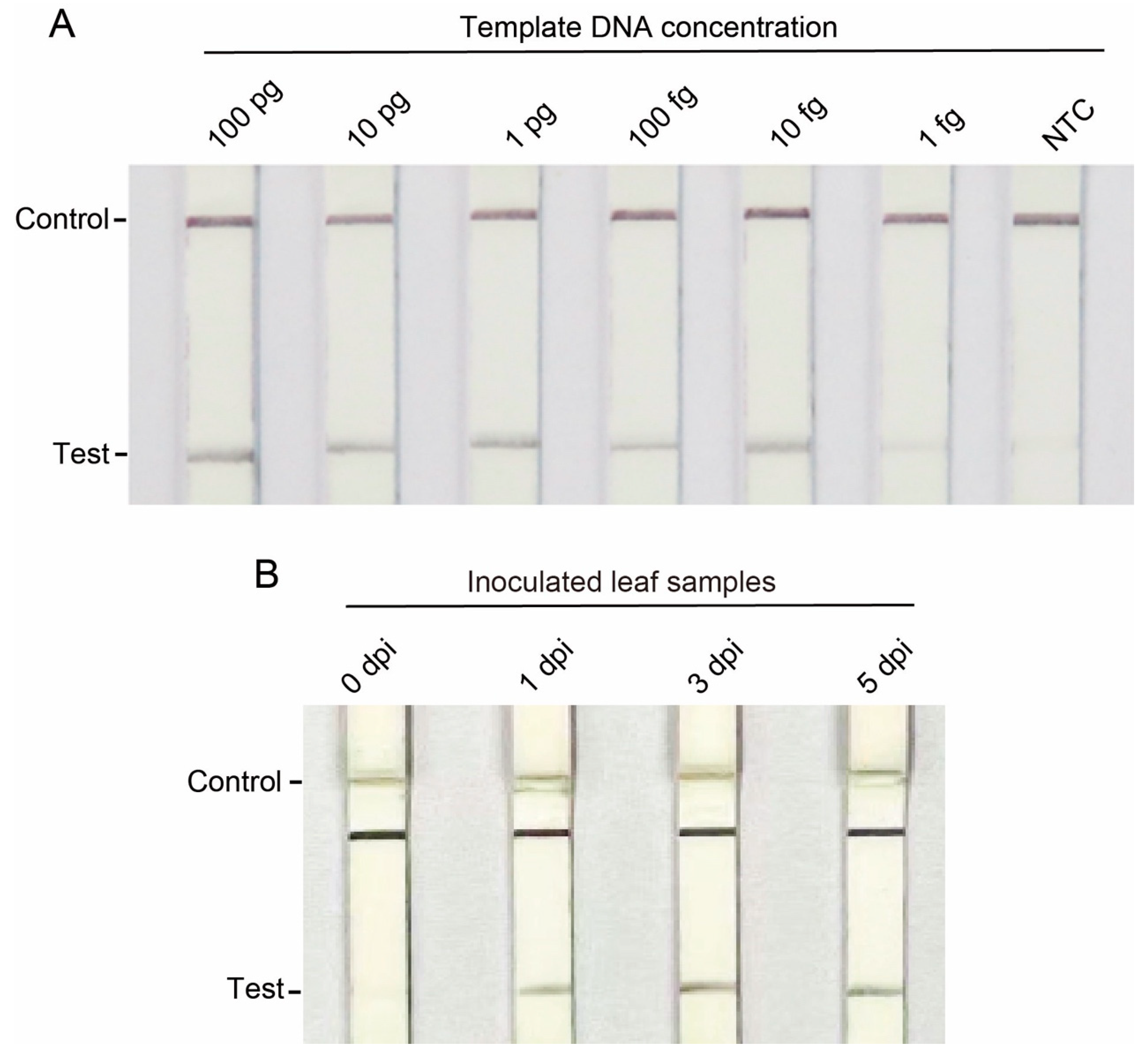
3.3. Sensitivity Testing of Primers for C. gloeosporioides Detection

To evaluate the sensitivity of the primers targeting the OG0034840, OG0034823, OG0034817, and OG0034811 orthogroup genes, PCR analyses were performed using C. gloeosporioides strain CgDa01 genomic DNA as templates by diluting them into different concentrations from 1 ng/μL to 1 fg/μL. The results showed that the primer pairs of OG0034840 and OG0034823 could successfully amplify the target fragments at a genomic DNA concentration of 10 pg/μL (Figure 3A,B), whereas the primer pairs of OG0034817 and OG0034811 could only amplify the target fragments at a genomic DNA concentration of 100 pg/μL (Figure 3C,D). This experiment was replicated three times with similar results, which indicated that the primer pairs of OG0034840 and OG0034823 were more sensitive than those of OG0034817 and OG0034811 and might be more suitable for the development of rapid detection methods.

3.4. Evaluation of Specific Primers for C. gloeosporioides Detection in Infected Plant Tissues

To evaluate the efficiency of the four pairs of specific primers in the anthracnose yam samples, greater yam leaves exhibiting different anthracnose phenotypes were collected at one, three, and five days post inoculation with the C. gloeosporioides strain CgDa01 (Figure 4A). The greater yam leaves that were not inoculated with C. gloeosporioides were used as controls. DNA from the CgDa01-inoculated or non-inoculated leaves were extracted and used as templates for the PCR analysis. The results show that all of the four primer pairs failed to amplify any products when using the DNA of the yam leaves not inoculated with CgDa01 or the DNA of yam leaves inoculated with CgDa01 at 0 dpi as templates (Figure 4B). In contrast, all of the four primer pairs successfully amplified the target fragments from the DNA samples of yam leaves infected with C. gloeosporioides at 1, 3, and 5 dpi (Figure 4C–E). The results indicate that these specific primer pairs can detect the infection of yams by C. gloeosporioides as early as the first day post infection.

3.5. Development of the LF-RPA Method for Rapid Detection of C. gloeosporioides

Having observed that all of the four primer pairs could specifically detect C. gloeosporioides, and the primer pairs of OG0034840 and OG0034823 were more sensitive than those of OG0034817 and OG0034811, we selected OG0034840 as a target for developing an LF-RPA based C. gloeosporioides detection method. The detection threshold of the LF-RPA assay was measured using serial dilutions of genomic DNA of C. gloeosporioides as templates. The results reveal that a clear band could be detected at a template concentration of 10 fg/μL and a faint band was present for the 1 fg/μL template, with all quality control lines being clearly discernible (Figure 5A). Therefore, the detection threshold of the LF-RPA assay was determined to be down toward 10 fg and even 1 fg of genomic DNA, which is more sensitive than conventional PCR in the detection of C. gloeosporioides genomic DNA. Next, we used a lateral flow strip to detect the DNA obtained from the greater yam leaves infected or not infected by C. gloeosporioides as described in Section 3.4. Test bands were clearly observed in the DNA extracted from the greater yam leaves inoculated with C. gloeosporioides at 1, 3, and 5 dpi but not in the DNA of the greater yam leaves that were inoculated with C. gloeosporioides at 0 dpi. The experiments were repeated three times with consistent results, suggesting that the LF-RPA assay can detect C. gloeosporioides in infected leaves after the first day of inoculation (Figure 5B).

4. Discussion and Conclusions

C. gloeosporioides is a causal agent of anthracnose, a plant disease that is economically important and widespread [31]. Developing a rapid and user-friendly anthracnose diagnosis method is highly important and valuable for the cultivation of greater yam and other cash crops. In this study, we performed a genome-wide screening of C. gloeosporioides-specific sequences and designed amplification primers, which were then validated for specificity, sensitivity, and C. gloeosporioides-infected plant sample detection. Finally, we developed a practical LF-RPA detection kit.
Species-specific genes are ideal targets for developing molecular detection kits against microbes [13,14]. However, rapid evolution of a microbe genome often causes the presence or absence of variation in genes among different strains of the same species [32]. To overcome this problem, comparative genomic analysis has been adopted by independent studies to identify species-specific genes. For example, a recent study identified species-specific genes, namely ComFA, group_14348, group_26190, and group_26478 from Staphylococcus aureus, S. epidermidis, S. haemolyticus, and S. hominis, respectively, as molecular markers and used these markers for developing molecular detection methods against these pathogens [33]. Similarly, a series of species-specific genes was obtained for seven species belonging to the Cronobacter genus through pan-genome analysis, including 4, 10, 31, 30, 70, 93, and 331 genes specific to Cronobacter sakazakii, Cr. malonaticus, Cr. dublinensis, Cr. turicensis, Cr. muytjensii, Cr. universalis, and Cr. condimenti, respectively [34]. Inspired by these previous reports, this study included genomes of four C. gloeosporioides strains that were isolated from different hosts by independent studies (Table S1). The identification of orthogroups shared by all investigated strains suggests their conservation during the evolution of C. gloeosporioides. Considering C. gloeosporioides has dozens of closely related species within the same genus [35], we included the genomes of 37 other species belonging to the Colletotrichum genus. This step enabled us to identify 17 orthogroups that are specific to and conservatively shared by all C. gloeosporioides strains. These orthogroups provide a raw resource for the screening of gene targets for developing molecular detection methods. Although these 17 orthogroups display the same feature of being shared by all C. gloeosporioides strains, they may exhibit different evolutionary patterns and show differential sequence variation. Therefore, a further sequence alignment assay enabled us to identify conserved sequence regions from the four orthogroup genes.
Our results show that while the primer pairs of all of the above four orthogroups could specifically detect C. gloeosporioides out of the pathogens within the Colletotrichum genus, the primer pairs of OG0034840 and OG0034823 exhibited greater sensitivity with a detection threshold of 10 pg/μL. These primers produced clear, discrete bands at this concentration. In contrast, the other two primer pairs targeting OG0034817 and OG0034811 could only detect the template DNA at a concentration of 100 pg/μL, which may have been due to their higher annealing temperature and easier-to-form secondary structures. These two primer pairs can be used as an alternative to those targeting OG0034840 and OG0034823 for the specific detection of C. gloeosporioides. By using the above primer pairs to detect C. gloeosporioides in yam leaves, we found that all of them could detect infections of the C. gloeosporioides strain CgDa01 as early as the first day post incubation when the leaves did not yet have any obvious external symptoms. Therefore, our approach can serve as an effective method for the early diagnosis of yam anthracnose, which is conducive to reducing the spread of the disease and improving the yield and quality of greater yam.
Currently, methods for identifying C. gloeosporioides and its close relatives can be technically divided into two main categories: microscopic detection techniques based on the morphology of the organism and detection techniques based on PCR amplification of nucleic acid sequences [15]. Because morphological identification is easily influenced by environmental factors, the latter is used more nowadays. In the host greater yam, Mithun Raj et al. previously used nested PCR to detect a C. gloeosporioides limit of 200 fg/μL [36], and a recent study showed that RT-qPCR in other hosts such as Arabidopsis thaliana could increase the detection limit to 5 fg/μL [37]. Compared with the above PCR techniques, the LF-RPA detection method developed in this study can recognize C. gloeosporioides DNA at even a mere 1 fg/μL. Moreover, a recent study proposed a DNA extraction protocol that does not require complicated instruments and does not rely on the participation of specialists. The protocol can be completed in only 30 s and costs only USD 0.06 per session [38]. This method can be jointly applied to our study to improve the efficiency of extracting DNA in a resource-limited environment. In addition, the LF-RPA detection kit also exhibits the advantage of a low cost, being as low as USD 0.61 per reaction [39]. Because the industrial production of lateral flow strips, the key component, is well established, they can easily be applied in the field and poor rural areas.
In summary, through the utilization of comprehensive comparative genomic analysis, we successfully obtained distinct molecular markers specific to C. gloeosporioides and conservatively shared by different strains. In taking advantage of these molecular markers, we developed an efficient, rapid, transportable, and simple method by introducing RPA technology to detect C. gloeosporioides in greater yam for the first time. This enables early diagnosis of yam anthracnose and establishes the groundwork for the prevention and control of this disease. The strategy adopted by this study also provides new research ideas for the development of detection and diagnosis tools for different fungal diseases.

Supplementary Materials

The following supporting information can be downloaded at https://0-www-mdpi-com.brum.beds.ac.uk/article/10.3390/jof10050315/s1. Table S1: Genomes used in this study. Table S2: Candidate gene primer sequences. Table S3: Orthogroups identified from 60 Colletotrichum genomes.

Author Contributions

W.-T.X. and X.-Y.L. performed the experiments; Y.W., M.-H.L. and X.-Q.S. analyzed the data; Y.-M.Z. designed the experiments; W.-T.X., X.-Y.L., K.H. and Z.-J.S. wrote the manuscript; Y.-M.Z. and X.-Q.S. revised the manuscript. All authors have read and agreed to the published version of the manuscript.

Funding

This research was funded by the National Natural Science Foundation of China (32172089 and 32302330), the Jiangsu Key Laboratory for the Research and Utilization of Plant Resources (JSPKLB202207), and The Jiangsu Institute of Botany Talent Fund (JIBTF202303).

Institutional Review Board Statement

Not applicable.

Informed Consent Statement

Not applicable.

Data Availability Statement

Data are contained within the article and Supplementary Materials.

Acknowledgments

We thank Daolong Dou (Nanjing Agricultural University) for providing the Fusarium graminearum, Fusarium oxysporum, Sclerotinia sclerotiorum, Botrytis cinerea, and Blumeria graminis strains and Yuqiang Zhao (Fruit Tree Research Centre, Institute of Botany, Jiangsu Province and Chinese Academy of Sciences) for providing the Colletotrichum fructicola strain.

Conflicts of Interest

The authors declare no conflicts of interest.

References

  1. Wang, P.; Shan, N.; Ali, A.; Sun, J.; Luo, S.; Xiao, Y.; Wang, S.; Hu, R.; Huang, Y.; Zhou, Q. Comprehensive Evaluation of Functional Components, Biological Activities, and Minerals of Yam Species (Dioscorea polystachya and D. alata) from China. LWT 2022, 168, 113964. [Google Scholar] [CrossRef]
  2. Lebot, V.; Lawac, F.; Legendre, L. The Greater Yam (Dioscorea alata L.): A Review of Its Phytochemical Content and Potential for Processed Products and Biofortification. J. Food Compos. Anal. 2023, 115, 104987. [Google Scholar] [CrossRef]
  3. More, S.J.; Ravi, V.; Raju, S.; de Freitas, S.T.; Pareek, S. Tropical Tuber Crops; CRC Press: Boca Raton, FL, USA, 2019; Volume 1, pp. 719–758. [Google Scholar]
  4. Ntui, V.O.; Uyoh, E.A.; Ita, E.E.; Markson, A.A.A.; Tripathi, J.N.; Okon, N.I.; Akpan, M.O.; Phillip, J.O.; Brisibe, E.A.; Ene-Obong, E.O.E. Strategies to Combat the Problem of Yam Anthracnose Disease: Status and Prospects. Mol. Plant Pathol. 2021, 22, 1302–1314. [Google Scholar] [CrossRef] [PubMed]
  5. Agre, P.A.; Darkwa, K.; Olasanmi, B.; Kolade, O.; Mournet, P.; Bhattacharjee, R.; Lopez-Montes, A.; De Koeyer, D.; Adebola, P.; Kumar, L.; et al. Identification of QTLs Controlling Resistance to Anthracnose Disease in Water Yam (Dioscorea alata). Genes 2022, 13, 347. [Google Scholar] [CrossRef] [PubMed]
  6. Pornprapa, K.; Suriyasit, S.; Mongkutkarn, U.; Panisa, P.; Tanimnun, J. Cross-Resistance to Benzimidazole Group and Mancozeb Fungicides in Colletotrichum spp. Causing Anthracnose Disease. Sci. Technol. Asia 2022, 27, 400–408. [Google Scholar]
  7. Liu, F.; Ma, Z.; Hou, L.; Diao, Y.; Wu, W.; Damm, U.; Song, S.; Cai, L. Updating Species Diversity of Colletotrichum, with a Phylogenomic Overview. Stud. Mycol. 2022, 101, 1–56. [Google Scholar] [CrossRef] [PubMed]
  8. Weir, B.S.; Johnston, P.R.; Damm, U. The Colletotrichum gloeosporioides species complex. Stud. Mycol. 2012, 73, 115–180. [Google Scholar] [CrossRef]
  9. Talhinhas, P. Hosts of Colletotrichum. Mycosphere 2023, 14, 158–261. [Google Scholar] [CrossRef]
  10. De Silva, D.D.; Crous, P.W.; Ades, P.K.; Hyde, K.D.; Taylor, P.W.J. Life Styles of Colletotrichum Species and Implications for Plant Biosecurity. Fungal Biol. Rev. 2017, 31, 155–168. [Google Scholar] [CrossRef]
  11. Luo, A.; Ling, C.; Ho, S.Y.W.; Zhu, C.D. Comparison of Methods for Molecular Species Delimitation Across a Range of Speciation Scenarios. Syst. Biol. 2018, 67, 830–846. [Google Scholar] [CrossRef]
  12. Attaluri, S.; Dharavath, R. Novel plant disease detection techniques-a brief review. Mol. Biol. Rep. 2023, 50, 9677–9690. [Google Scholar] [CrossRef]
  13. Kim, E.; Yang, S.M.; Kim, I.S.; Kim, H.Y. Identification of novel molecular targets for Weissella species-specific real-time PCR based on pangenome analysis. Appl. Microbiol. Biotechnol. 2022, 106, 4157–4168. [Google Scholar] [CrossRef] [PubMed]
  14. Chelkha, N.; Jardot, P.; Moussaoui, I.; Levasseur, A.; La Scola, B.; Colson, P. Core gene-based molecular detection and identification of Acanthamoeba species. Sci. Rep. 2020, 10, 1583. [Google Scholar] [CrossRef] [PubMed]
  15. Mills, P.R.; Sreenivasaprasad, S.; Brown, A.E. Detection and differentiation of Colletotrichum gloeosporioides isolates using PCR. FEMS Microbiol. Lett. 1992, 77, 137–143. [Google Scholar] [CrossRef] [PubMed]
  16. Vieira, W.A.d.S.; Bezerra, P.A.; Silva, A.C.d.; Veloso, J.S.; Câmara, M.P.S.; Doyle, V.P. Optimal Markers for the Identification of Colletotrichum Species. Mol. Phylogenetics Evol. 2020, 143, 106694. [Google Scholar] [CrossRef] [PubMed]
  17. Toth, I.K.; Pritchard, L.; Birch, P.R. Comparative Genomics Reveals what Makes an Enterobacterial Plant Pathogen. Annu. Rev. Phytopathol. 2006, 44, 305–336. [Google Scholar] [CrossRef] [PubMed]
  18. Gupta, R.S.; Patel, S.; Saini, N.; Chen, S. Robust Demarcation of 17 Distinct Bacillus Species Clades, Proposed as Novel Bacillaceae Genera, by Phylogenomics and Comparative Genomic Analyses: Description of Robertmurraya kyonggiensis sp. nov. and Proposal for an Emended Genus Bacillus Limiting It Only to the Members of the Subtilis and Cereus Clades of Species. Int. J. Syst. Evol. Microbiol. 2020, 70, 5753–5798. [Google Scholar] [PubMed]
  19. Miyauchi, S.; Kiss, E.; Kuo, A.; Drula, E.; Kohler, A.; Sánchez-García, M.; Morin, E.; Andreopoulos, B.; Barry, K.W.; Bonito, G. Large-Scale Genome Sequencing of Mycorrhizal Fungi Provides Insights into the Early Evolution of Symbiotic Traits. Nat. Commun. 2020, 11, 5125. [Google Scholar] [CrossRef] [PubMed]
  20. Yang, J.; Duan, K.; Liu, Y.; Song, L.; Gao, Q.H. Method to Detect and Quantify Colonization of Anthracnose Causal Agent Colletotrichum gloeosporioides Species Complex in Strawberry by Real-time PCR. J. Phytopathol. 2022, 170, 326–336. [Google Scholar] [CrossRef]
  21. Syafitri, L.M.; Wibowo, A.; Widiastuti, A.; Subandiyah, S.; Harper, S. Molecular Identification of Colletotrichum gloeosporioides Causing Anthracnose on Shallot in Bantul, Yogyakarta, Indonesia. J. Biol. Divers. 2023, 24. [Google Scholar] [CrossRef]
  22. Babu, B.; Ochoa-Corona, F.M.; Paret, M.L. Recombinase polymerase amplification applied to plant virus detection and potential implications. Anal. Biochem. 2018, 546, 72–77. [Google Scholar] [CrossRef] [PubMed]
  23. Li, J.; Macdonald, J.; Von Stetten, F. A Comprehensive Summary of a Decade Development of the Recombinase Polymerase Amplification. Analyst 2019, 144, 31–67. [Google Scholar] [CrossRef] [PubMed]
  24. Hu, S.; Yan, C.; Yu, H.; Zhang, Y.; Zhang, C.Q. Establishment of the Recombinase Polymerase Amplification-Lateral Flow Dipstick Detection Technique for Fusarium oxysporum. Plant Dis. 2023, 107, 2665–2672. [Google Scholar] [CrossRef] [PubMed]
  25. Wang, Y.; Xu, W.T.; Lu, R.S.; Chen, M.; Liu, J.; Sun, X.Q.; Zhang, Y.M. Genome Sequence Resource for Colletotrichum gloeosporioides, an Important Pathogenic Fungus Causing Anthracnose of Dioscorea alata. Plant Dis. 2023, 107, 893–895. [Google Scholar] [CrossRef] [PubMed]
  26. Nguyen, H.D.; Sultana, T.; Kesanakurti, P.; Hambleton, S. Genome Sequencing and Comparison of Five Tilletia Species to Identify Candidate Genes for the Detection of Regulated Species Infecting Wheat. IMA Fungus 2019, 10, 1–17. [Google Scholar] [CrossRef]
  27. Alzohairy, A.M. BioEdit: An Important Software for Molecular Biology. Gerf Bull. Biosci. 2011, 2, 60–61. [Google Scholar]
  28. Aamir, S. A Rapid and Efficient Method of fungal Genomic DNA Extraction, Suitable for PCR Based Molecular Methods. Plant Pathol. Quar. 2015, 5, 74–81. [Google Scholar] [CrossRef]
  29. Lalitha, S.J.B.S. Primer Premier 5. Biotech Softw. Internet Rep. Comput. Softw. J. Sci. 2000, 1, 270–272. [Google Scholar] [CrossRef]
  30. Huang, L.; Li, Q.C.; Zhang, Y.; Li, D.W.; Ye, J.R. Colletotrichum gloeosporioides Sensu Stricto is a Pathogen of Leaf Anthracnose on Evergreen Spindle Tree (Euonymus japonicus). Plant Dis. 2016, 100, 672–678. [Google Scholar] [CrossRef]
  31. Dean, R.; Van Kan, J.A.; Pretorius, Z.A.; Hammond-Kosack, K.E.; Di Pietro, A.; Spanu, P.D.; Rudd, J.J.; Dickman, M.; Kahmann, R.; Ellis, J.; et al. The Top 10 Fungal Pathogens in Molecular Plant Pathology. Mol. Plant Pathol. 2012, 13, 414–430. [Google Scholar] [CrossRef]
  32. Hu, Z.J.; Huang, Y.Y.; Lin, X.Y.; Feng, H.; Zhou, S.X.; Xie, Y.; Liu, X.X.; Liu, C.; Zhao, R.M.; Zhao, W.S.; et al. Loss and Natural Variations of Blast Fungal Avirulence Genes Breakdown Rice Resistance Genes in the Sichuan Basin of China. Front. Plant Sci. 2022, 13, 788876. [Google Scholar] [CrossRef] [PubMed]
  33. Zhou, B.; Ye, Q.; Chen, M.; Li, F.; Xiang, X.; Shang, Y.; Wang, C.; Zhang, J.; Xue, L.; Wang, J.; et al. Novel species-specific targets for real-time PCR detection of four common pathogenic Staphylococcus spp. Food Control 2022, 131, 108478. [Google Scholar] [CrossRef]
  34. Shang, Y.; Ye, Q.; Wu, Q.; Pang, R.; Zhou, B.; Wang, C.; Xiang, X.; Li, F.; Wang, J.; Zhang, Y.; et al. PCR and multiplex PCR assays for the detection of Cronobacter species using specific targets obtained by a bioinformatics approach. Food Control 2021, 125, 107896. [Google Scholar] [CrossRef]
  35. da Silva, L.L.; Moreno, H.L.A.; Correia, H.L.N.; Santana, M.F.; de Queiroz, M.V. Colletotrichum: Species complexes, lifestyle, and peculiarities of some sources of genetic variability. Appl. Microbiol. Biotechnol. 2020, 104, 1891–1904. [Google Scholar] [CrossRef] [PubMed]
  36. Raj, M.; Jeeva, M.; Hegde, V.; Vidyadharan, P.; Archana, P.; Senthil alias Sankar, M.; Nath, S.V. Polymerase Chain Reaction Assay for Rapid, Sensitive Detection, and Identification of Colletotrichum gloeosporioides Causing Greater Yam Anthracnose. Mol. Biotechnol. 2012, 52, 277–284. [Google Scholar] [CrossRef] [PubMed]
  37. Wan, M.; Yang, L.; Zhang, S.; Gao, J.; Jiang, L.; Luo, L. Real-time PCR for Detection and Quantification of C. gloeosporioides s.l. Growth in Stylosanthes and Arabidopsis. Crop Prot. 2022, 159, 106021. [Google Scholar] [CrossRef]
  38. Zou, Y.; Mason, M.G.; Wang, Y.; Wee, E.; Turni, C.; Blackall, P.J.; Trau, M.; Botella, J.R. Nucleic acid purification from plants, animals and microbes in under 30 seconds. PLoS Biol. 2017, 15, e2003916. [Google Scholar] [CrossRef]
  39. Gootenberg, J.S.; Abudayyeh, O.O.; Lee, J.W.; Essletzbichler, P.; Dy, A.J.; Joung, J.; Verdine, V.; Donghia, N.; Daringer, N.M.; Freije, C.A.; et al. Nucleic acid detection with CRISPR-Cas13a/C2c2. Science 2017, 356, 438–442. [Google Scholar] [CrossRef]
Figure 1. Statistics of orthologous gene clustering analyses. (A) Number of orthogroups occupied by genes of different C. gloeosporioides strains, Venn diagram shows the number of orthogroups which were strain-specific (1) or shared by 2, 3, or 4 strains. (B) Comparison of orthogroups shared by all C. gloeosporioides strains with all non-redundant orthogroups of the other 56 Colletotrichum genomes. (C) Details of C. gloeosporioides-specific orthogroups, including gene IDs and length of each genome. The four colored orthogroups were eventually the molecular markers used in this study.
Jof 10 00315 g001
Figure 2. Determination of the primers targeting four C. gloeosporioides-specific orthogroups by PCR. M = 2000 bp DNA Ladder; 1 = C. gloeosporioides strain CgDa01; 2 = C. gloeosporioides strain CgDaM3; 3 = Fusarium graminearum; 4 = F. oxysporum; 5 = Sclerotinia sclerotiorum; 6 = Botrytis cinerea; 7 = Blumeria graminis; and 8 = C. fructicola. (AD) PCR results of different primer pairs.
Jof 10 00315 g002
Figure 3. Sensitivity testing of four pairs of specific primers. M = 2000 bp DNA Ladder, and 1~8 = different DNA concentrations: 1 ng/μL, 100 pg/μL, 10 pg/μL, 1 pg/μL, 100 fg/μL, 10 fg/μL, 1 fg/μL, and NTC (no-template control), respectively. (AD) PCR results of different primer pairs.
Jof 10 00315 g003
Figure 4. Evaluation of specific primers for C. gloeosporioides detection of infected plant tissues. (A) Phenotypes of leaves post inoculation with C. gloeosporioide on different days. Dpi = days post infection. (BE) PCR results of DNA extracted from plant leaves at 0, 1, 3, and 5 days post inoculation (dpi) with C. gloeosporioides, respectively. M = 2000 bp DNA Ladder; CK = control check, namely greater yam leaves that were not inoculated with C. gloeosporioides; Lanes 1~3 = independent samples infected by C. gloeosporioide at the same time point.
Jof 10 00315 g004
Figure 5. Sensitivity test of LF-RPA C. gloeosporioides detection methods. NTC = no template control; Dpi = days post infection. (A) Sensitivity of LF-RPA at different concentrations of C. gloeosporioides genomic DNA. (B) Results of LF-RPA conducted on DNA samples extracted from greater yam leaves infected by C. gloeosporioides from 0 to 5 dpi.
Jof 10 00315 g005
Disclaimer/Publisher’s Note: The statements, opinions and data contained in all publications are solely those of the individual author(s) and contributor(s) and not of MDPI and/or the editor(s). MDPI and/or the editor(s) disclaim responsibility for any injury to people or property resulting from any ideas, methods, instructions or products referred to in the content.

