
Kidney Function Tests and Continuous eGFR Decrease at Six Months after SARS-CoV-2 Infection in Patients Clinically Diagnosed with Post-COVID Syndrome
 ,
,  , ,
, ,  ,
,
Abstract
:1. Introduction
2. Materials and Methods
2.1. Study Design and Ethics
2.2. Patients’ Inclusion and Exclusion Criteria
2.3. Study Variables
2.4. Statistical Analysis
3. Results
3.1. Patients’ Background
3.2. Laboratory Data
3.3. Risk Assessment
4. Discussion
4.1. Literature Findings
4.2. Study Limitations
5. Conclusions
Author Contributions
Funding
Institutional Review Board Statement
Informed Consent Statement
Data Availability Statement
Conflicts of Interest
References
- ReferencesHope, A.A.; Evering, T.H. Postacute Sequelae of Severe Acute Respiratory Syndrome Coronavirus 2 Infection. Infect. Dis. Clin. N. Am. 2022, 36, 379–395. [Google Scholar] [CrossRef] [PubMed] [PubMed Central]
- O’Donnell, J.S.; Chappell, K.J. Chronic SARS-CoV-2, a Cause of Post-acute COVID-19 Sequelae (Long-COVID)? Front. Microbiol. 2021, 12, 724654. [Google Scholar] [CrossRef] [PubMed] [PubMed Central]
- Davis, H.E.; McCorkell, L.; Vogel, J.M.; Topol, E.J. Long COVID: Major findings, mechanisms and recommendations. Nat. Rev. Microbiol. 2023, 21, 133–146, Erratum in: Nat. Rev. Microbiol. 2023, 21, 408. [Google Scholar] [CrossRef] [PubMed] [PubMed Central]
- Long, J.D.; Strohbehn, I.; Sawtell, R.; Bhattacharyya, R.; Sise, M.E. COVID-19 Survival and its impact on chronic kidney disease. Transl. Res. 2022, 241, 70–82. [Google Scholar] [CrossRef] [PubMed] [PubMed Central]
- Yeung, M.L.; Yao, Y.; Jia, L.; Chan, J.F.; Chan, K.H.; Cheung, K.F.; Chen, H.; Poon, V.K.; Tsang, A.K.; To, K.K.; et al. MERS coronavirus induces apoptosis in kidney and lung by upregulating Smad7 and FGF2. Nat. Microbiol. 2016, 1, 16004. [Google Scholar] [CrossRef] [PubMed] [PubMed Central]
- Menon, T.; Sharma, R.; Kataria, S.; Sardar, S.; Adhikari, R.; Tousif, S.; Khan, H.; Rathore, S.S.; Singh, R.; Ahmed, Z. The Association of Acute Kidney Injury With Disease Severity and Mortality in COVID-19: A Systematic Review and Meta-Analysis. Cureus 2021, 13, e13894. [Google Scholar] [CrossRef] [PubMed] [PubMed Central]
- Chong, W.H.; Saha, B.K. Relationship Between Severe Acute Respiratory Syndrome Coronavirus 2 (SARS-CoV-2) and the Etiology of Acute Kidney Injury (AKI). Am. J. Med Sci. 2021, 361, 287–296. [Google Scholar] [CrossRef] [PubMed] [PubMed Central]
- Chávez-Valencia, V.; Orizaga-de-la-Cruz, C.; Lagunas-Rangel, F.A. Acute Kidney Injury in COVID-19 Patients: Pathogenesis, Clinical Characteristics, Therapy, and Mortality. Diseases 2022, 10, 53. [Google Scholar] [CrossRef] [PubMed] [PubMed Central]
- Sabaghian, T.; Kharazmi, A.B.; Ansari, A.; Omidi, F.; Kazemi, S.N.; Hajikhani, B.; Vaziri-Harami, R.; Tajbakhsh, A.; Omidi, S.; Haddadi, S.; et al. COVID-19 and Acute Kidney Injury: A Systematic Review. Front. Med. 2022, 9, 705908. [Google Scholar] [CrossRef] [PubMed] [PubMed Central]
- Assiri, A.M.; Alamaa, T.; Elenezi, F.; Alsagheir, A.; Alzubaidi, L.; TIeyjeh, I.; Alhomod, A.S.; Gaffas, E.M.; Amer, S.A. Unveiling the Clinical Spectrum of Post-COVID-19 Conditions: Assessment and Recommended Strategies. Cureus 2024, 16, e52827. [Google Scholar] [CrossRef] [PubMed] [PubMed Central]
- Maltezou, H.C.; Pavli, A.; Tsakris, A. Post-COVID Syndrome: An Insight on Its Pathogenesis. Vaccines 2021, 9, 497. [Google Scholar] [CrossRef] [PubMed] [PubMed Central]
- Moeinzadeh, F.; Mortazavi, M.; Shahidi, S.; Mansourian, M.; Yazdani, A.; Zamani, Z.; Seirafian, S. Chronic Kidney Disease and COVID-19 Infection: A Case-Control Study. Adv. Biomed Res. 2022, 11, 112. [Google Scholar] [CrossRef] [PubMed] [PubMed Central]
- Huang, W.; Li, B.; Jiang, N.; Zhang, F.; Shi, W.; Zuo, L.; Liu, S.; Tang, B. Impact of the COVID-19 pandemic on patients with chronic kidney disease: A narrative review. Medicine 2022, 101, e29362. [Google Scholar] [CrossRef] [PubMed] [PubMed Central]
- Pecly, I.M.D.; Azevedo, R.B.; Muxfeldt, E.S.; Botelho, B.G.; Albuquerque, G.G.; Diniz, P.H.P.; Silva, R.; Rodrigues, C.I.S. COVID-19 and chronic kidney disease: A comprehensive review. Braz. J. Nephrol. 2021, 43, 383–399. [Google Scholar] [CrossRef] [PubMed] [PubMed Central]
- Temiz, M.Z.; Hacibey, I.; Yazar, R.O.; Sevdi, M.S.; Kucuk, S.H.; Alkurt, G.; Doganay, L.; Dinler Doganay, G.; Dincer, M.M.; Yuruk, E.; et al. Altered kidney function induced by SARS-CoV-2 infection and acute kidney damage markers predict survival outcomes of COVID-19 patients: A prospective pilot study. Ren. Fail. 2022, 44, 233–240. [Google Scholar] [CrossRef] [PubMed] [PubMed Central]
- Hong, X.W.; Chi, Z.P.; Liu, G.Y.; Huang, H.; Guo, S.Q.; Fan, J.R.; Lin, X.W.; Qu, L.Z.; Chen, R.L.; Wu, L.J.; et al. Characteristics of Renal Function in Patients Diagnosed With COVID-19: An Observational Study. Front. Med. 2020, 7, 409. [Google Scholar] [CrossRef] [PubMed] [PubMed Central]
- Panimathi, R.; Gurusamy, E.; Mahalakshmi, S.; Ramadevi, K.; Kaarthikeyan, G.; Anil, S. Impact of COVID-19 on Renal Function: A Multivariate Analysis of Biochemical and Immunological Markers in Patients. Cureus 2022, 14, e22076. [Google Scholar] [CrossRef] [PubMed] [PubMed Central]
- Al Rumaihi, K.; Khalafalla, K.; Arafa, M.; Nair, A.; Al Bishawi, A.; Fino, A.; Sirtaj, F.; Ella, M.K.; ElBardisi, H.; Khattab, M.A.; et al. COVID-19 and renal involvement: A prospective cohort study assessing the impact of mild SARS-CoV-2 infection on the kidney function of young healthy males. Int. Urol. Nephrol. 2023, 55, 201–209. [Google Scholar] [CrossRef] [PubMed] [PubMed Central]
- Seo, J.W.; Kim, S.E.; Kim, Y.; Kim, E.J.; Kim, T.; Kim, T.; Lee, S.H.; Lee, E.; Lee, J.; Seo, Y.B.; et al. Updated Clinical Practice Guidelines for the Diagnosis and Management of Long COVID. Infect. Chemother. 2024, 56, 122–157. [Google Scholar] [CrossRef] [PubMed] [PubMed Central]
- Srikanth, S.; Boulos, J.R.; Dover, T.; Boccuto, L.; Dean, D. Identification and diagnosis of long COVID-19: A scoping review. Prog. Biophys. Mol. Biol. 2023, 182, 1–7. [Google Scholar] [CrossRef] [PubMed] [PubMed Central]
- Charles, K.; Lewis, M.J.; Montgomery, E.; Reid, M. The 2021 Chronic Kidney Disease Epidemiology Collaboration Race-Free Estimated Glomerular Filtration Rate Equations in Kidney Disease: Leading the Way in Ending Disparities. Health Equity 2024, 8, 39–45. [Google Scholar] [CrossRef] [PubMed]
- Radwan, N.M.; Mahmoud, N.E.; Alfaifi, A.H.; Alabdulkareem, K.I. Comorbidities and severity of coronavirus disease 2019 patients. Saudi Med. J. 2020, 41, 1165–1174. [Google Scholar] [CrossRef] [PubMed]
- Tannor, E.K.; Bajpai, D.; Nlandu, Y.M.; Wijewickrama, E. COVID-19 and Kidney Disease: Progress in Health Inequity From Low-Income Settings. Semin. Nephrol. 2022, 42, 151318. [Google Scholar] [CrossRef] [PubMed] [PubMed Central]
- Brogan, M.; Ross, M.J. COVID-19 and Kidney Disease. Annu. Rev. Med. 2023, 74, 1–13. [Google Scholar] [CrossRef] [PubMed]
- Žulpaitė, G.; Rimševičius, L.; Jančorienė, L.; Zablockienė, B.; Miglinas, M. The Association between COVID-19 Infection and Kidney Damage in a Regional University Hospital. Medicina 2023, 59, 898. [Google Scholar] [CrossRef] [PubMed] [PubMed Central]
- Mahalingasivam, V.; Su, G.; Iwagami, M.; Davids, M.R.; Wetmore, J.B.; Nitsch, D. COVID-19 and kidney disease: Insights from epidemiology to inform clinical practice. Nat. Rev. Nephrol. 2022, 18, 485–498. [Google Scholar] [CrossRef] [PubMed] [PubMed Central]
- Jdiaa, S.S.; Mansour, R.; El Alayli, A.; Gautam, A.; Thomas, P.; Mustafa, R.A. COVID-19 and chronic kidney disease: An updated overview of reviews. J. Nephrol. 2022, 35, 69–85. [Google Scholar] [CrossRef] [PubMed] [PubMed Central]
- de Francisco, Á.M.; Fernández Fresnedo, G. Long COVID-19 renal disease: A present medical need for nephrology. Nefrologia (Engl. Ed.) 2023, 43, 1–5. [Google Scholar] [CrossRef] [PubMed] [PubMed Central]
- Copur, S.; Berkkan, M.; Basile, C.; Tuttle, K.; Kanbay, M. Post-acute COVID-19 syndrome and kidney diseases: What do we know? J. Nephrol. 2022, 35, 795–805. [Google Scholar] [CrossRef] [PubMed] [PubMed Central]
- La Porta, E.; Baiardi, P.; Fassina, L.; Faragli, A.; Perna, S.; Tovagliari, F.; Tallone, I.; Talamo, G.; Secondo, G.; Mazzarello, G.; et al. The role of kidney dysfunction in COVID-19 and the influence of age. Sci. Rep. 2022, 12, 8650. [Google Scholar] [CrossRef] [PubMed] [PubMed Central]
- Aklilu, A.M.; Kumar, S.; Nugent, J.; Yamamoto, Y.; Coronel-Moreno, C.; Kadhim, B.; Faulkner, S.C.; O’Connor, K.D.; Yasmin, F.; Greenberg, J.H.; et al. COVID-19-Associated Acute Kidney Injury and Longitudinal Kidney Outcomes. JAMA Intern. Med. 2024, 184, 414–423. [Google Scholar] [CrossRef] [PubMed] [PubMed Central]
- Lin, H.; Cao, B. Severe COVID-19 and chronic kidney disease: Bidirectional mendelian randomization study. Virol. J. 2024, 21, 32. [Google Scholar] [CrossRef] [PubMed] [PubMed Central]
- Herget-Rosenthal, S.; van Wijk, J.A.; Bröcker-Preuss, M.; Bökenkamp, A. Increased urinary cystatin C reflects structural and functional renal tubular impairment independent of glomerular filtration rate. Clin. Biochem. 2007, 40, 946–951. [Google Scholar] [CrossRef] [PubMed]
- Larsson, A.O.; Hultström, M.; Frithiof, R.; Nyman, U.; Lipcsey, M.; Eriksson, M.B. Differential Bias for Creatinine- and Cystatin C- Derived Estimated Glomerular Filtration Rate in Critical COVID-19. Biomedicines 2022, 10, 2708. [Google Scholar] [CrossRef] [PubMed]


| Variables | Control Group (n = 114) | Post-COVID Group (n = 92) | p-Value |
|---|---|---|---|
| Age (mean ± SD) | 55.2 ± 8.5 | 56.9 ± 7.6 | 0.136 |
| Sex | 0.710 | ||
| Men (n,%) | 59 (51.8%) | 50 (54.3%) | |
| Women (n,%) | 55 (48.2%) | 42 (45.7%) | |
| BMI (n,%) | 0.900 | ||
| Normal weight (18.5–24.9 kg/m2) | 36 (31.6%) | 30 (32.6%) | |
| Overweight (>24.9 kg/m2) | 45 (39.5%) | 38 (41.3%) | |
| Obese (>29.9 kg/m2) | 33 (28.9%) | 24 (26.1%) | |
| COVID-19 vaccination status (n,%) | 0.317 | ||
| 1 dose | 13 (11.4%) | 12 (13.0%) | |
| ≥2 doses | 37 (32.5%) | 38 (41.3%) | |
| Unvaccinated | 64 (56.1%) | 42 (45.7%) | |
| Antiviral medication requirement (n,%) | 0.096 | ||
| Yes | 68 (59.6%) | 66 (71.7%) | |
| No | 46 (40.4%) | 26 (28.3%) | |
| Oxygen supplementation (n,%) | 0.620 | ||
| Yes | 82 (71.9%) | 69 (75.0%) | |
| No | 32 (28.1%) | 23 (25.0%) | |
| COVID-19 severity (n,%) | 0.909 | ||
| Mild | 38 (33.3%) | 29 (31.5%) | |
| Moderate | 42 (36.8%) | 33 (35.9%) | |
| Severe | 34 (29.9%) | 30 (32.6%) | |
| Personal history (n,%) | |||
| Smoking | 26 (22.8%) | 22 (23.9%) | 0.851 |
| CCI > 2 | 24 (21.1%) | 20 (21.7%) | 0.904 |
| Kidney injury | |||
| Developed AKI during admission | 7 (6.1%) | 16 (17.4%) | 0.014 |
| eGFR decrease <30 from baseline | 11 (9.6%) | 25 (27.2%) | 0.001 |
| Continuous eGFR decrease <30 at six months | – | 22 (23.9%) | – |
| Variables | Normal Range | Control Group during Admission (n = 114) | Post-COVID Group During Admission (n = 92) | p-Value |
|---|---|---|---|---|
| WBC (1000/mm3) | 4.5–11.0 | 11.8 ± 1.7 | 16.6 ± 3.4 | <0.001 |
| Lymphocytes (1000/mm3) | 1.0–4.8 | 2.7 ± 0.8 | 1.3 ± 0.6 | <0.001 |
| Hemoglobin (g/dL) | 13.0–17.0 | 14.7 ± 1.3 | 13.4 ± 1.4 | <0.001 |
| AST (U/L) | 10–40 | 21.5 ± 5.2 | 47.3 ± 11.8 | <0.001 |
| ALT (U/L) | 7–35 | 18.6 ± 7.2 | 53.7 ± 14.3 | <0.001 |
| CRP (mg/dL) | 0–10 | 23.1 ± 2.8 | 78.2 ± 24.6 | <0.001 |
| IL-6 (pg/mL) | 0.8–6.4 | 10.3 ± 4.0 | 44.6 ± 19.7 | <0.001 |
| Procalcitonin (ug/L) | 0–0.25 | 0.09 ± 0.06 | 0.48 ± 0.22 | <0.001 |
| D-dimers (ng/mL) | <250 | 285.2 ± 48.6 | 498.5 ± 198.4 | <0.001 |
| Ferritin (ng/mL) | 20–250 | 292.4 ± 90.8 | 607.5 ± 295.7 | <0.001 |
| Variables | Normal Range | Post-COVID Group during Admission (n = 92) | Post-COVID Group Six Months Post-Admission (n = 92) | p-Value |
|---|---|---|---|---|
| WBC (1000/mm3) | 4.5–11.0 | 16.6 ± 3.4 | 10.9 ± 2.3 | <0.001 |
| Lymphocytes (1000/mm3) | 1.0–4.8 | 1.3 ± 0.6 | 2.4 ± 0.7 | <0.001 |
| Hemoglobin (g/dL) | 13.0–17.0 | 13.4 ± 1.4 | 14.3 ± 1.2 | <0.001 |
| AST (U/L) | 10–40 | 47.3 ± 11.8 | 29.4 ± 7.5 | <0.001 |
| ALT (U/L) | 7–35 | 53.7 ± 14.3 | 24.8 ± 9.1 | <0.001 |
| CRP (mg/dL) | 0–10 | 78.2 ± 24.6 | 14.7 ± 9.3 | <0.001 |
| IL-6 (pg/mL) | 0.8–6.4 | 44.6 ± 19.7 | 12.7 ± 4.6 | <0.001 |
| Procalcitonin (ug/L) | 0–0.25 | 0.48 ± 0.22 | 0.19 ± 0.08 | <0.001 |
| D-dimers (ng/mL) | <250 | 498.5 ± 198.4 | 295.3 ± 102.7 | <0.001 |
| Ferritin (ng/mL) | 20–250 | 607.5 ± 295.7 | 248.3 ± 149.2 | <0.001 |
| Variables (Mean ± SD) | Normal Range | Control Group during Admission (n = 114) | Post-COVID Group during Admission (n = 92) | p-Value |
|---|---|---|---|---|
| Creatinine (μmol/L) | 60–110 | 84.5 ± 11.7 | 109.7 ± 16.4 | <0.001 |
| eGFR (mL/min/1.73 m2) | >90 | 91.2 ± 7.3 | 65.3 ± 12.8 | <0.001 |
| BUN (mg/dL) | 7–20 | 15.2 ± 3.6 | 23.7 ± 7.2 | <0.001 |
| Proteinuria (mg/dL) | <150 | 105.1 ± 48.6 | 198.3 ± 91.2 | <0.001 |
| Hematuria (cells/HPF) | 0–3 | 1.5 ± 1.2 | 3.8 ± 2.1 | <0.001 |
| Albuminuria (mg/g) | <30 | 22.7 ± 8.2 | 52.4 ± 18.9 | <0.001 |
| ACR (mg/g) | <30 | 24.8 ± 10.1 | 59.1 ± 23.6 | <0.001 |
| Urine specific gravity | 1.005–1.030 | 1.021 ± 0.004 | 1.016 ± 0.008 | <0.001 |
| Urine osmolality (mOsm/kg) | 300–900 | 600.2 ± 140.3 | 535.7 ± 195.6 | 0.006 |
| Sodium (mmol/L) | 135–145 | 139.8 ± 3.9 | 138.2 ± 4.5 | 0.138 |
| Potassium (mmol/L) | 3.5–5.1 | 4.3 ± 0.5 | 4.7 ± 0.6 | <0.001 |
| Variables (Mean ± SD) | Normal Range | Post-COVID Group during Admission (n = 92) | Post-COVID Group Six Months Post-Admission (n = 92) | p-Value |
|---|---|---|---|---|
| Creatinine (μmol/L) | 60–110 | 109.7 ± 16.4 | 90.4 ± 13.6 | <0.001 |
| eGFR (mL/min/1.73 m2) | >90 | 65.3 ± 12.8 | 70.6 ± 11.1 | 0.002 |
| BUN (mg/dL) | 7–20 | 23.7 ± 7.2 | 18.3 ± 5.8 | <0.001 |
| Proteinuria (mg/dL) | <150 | 198.3 ± 91.2 | 155.9 ± 65.4 | 0.002 |
| Hematuria (cells/HPF) | 0-3 | 3.8 ± 2.1 | 2.0 ± 1.7 | <0.001 |
| Albuminuria (mg/g) | <30 | 52.4 ± 18.9 | 38.8 ± 15.2 | <0.001 |
| ACR (mg/g) | <30 | 59.1 ± 23.6 | 39.5 ± 16.3 | <0.001 |
| Urine specific gravity | 1.005–1.030 | 1.016 ± 0.008 | 1.022 ± 0.005 | <0.001 |
| Urine osmolality (mOsm/kg) | 300–900 | 485.7 ± 195.6 | 645.8 ± 170.2 | <0.001 |
| Sodium (mmol/L) | 135–145 | 138.2 ± 4.5 | 140.1 ± 3.6 | <0.001 |
| Potassium (mmol/L) | 3.5–5.1 | 4.7 ± 0.6 | 4.5 ± 0.4 | 0.007 |
| Variables (Mean ± SD) | Normal Range | Control Group during Admission (n = 114) | Post-COVID Group Six Months Post-Admission (n = 92) | p-Value |
|---|---|---|---|---|
| Creatinine (μmol/L) | 60–110 | 84.5 ± 11.7 | 90.4 ± 13.6 | 0.001 |
| eGFR (mL/min/1.73 m2) | >90 | 91.2 ± 7.3 | 70.6 ± 11.1 | <0.001 |
| BUN (mg/dL) | 7–20 | 15.2 ± 3.6 | 18.3 ± 5.8 | <0.001 |
| Proteinuria (mg/dL) | <150 | 105.1 ± 48.6 | 155.9 ± 65.4 | <0.001 |
| Hematuria (cells/HPF) | 0–3 | 1.5 ± 1.2 | 2.0 ± 1.7 | 0.014 |
| Albuminuria (mg/g) | <30 | 22.7 ± 8.2 | 38.8 ± 15.2 | <0.001 |
| ACR (mg/g) | <30 | 24.8 ± 10.1 | 39.5 ± 16.3 | <0.001 |
| Urine specific gravity | 1.005–1.030 | 1.021 ± 0.004 | 1.022 ± 0.005 | 0.180 |
| Urine osmolality (mOsm/kg) | 300–900 | 600.2 ± 140.3 | 645.8 ± 170.2 | 0.036 |
| Sodium (mmol/L) | 135–145 | 139.8 ± 3.9 | 140.1 ± 3.6 | 0.289 |
| Potassium (mmol/L) | 3.5–5.1 | 4.3 ± 0.5 | 4.5 ± 0.4 | 0.005 |
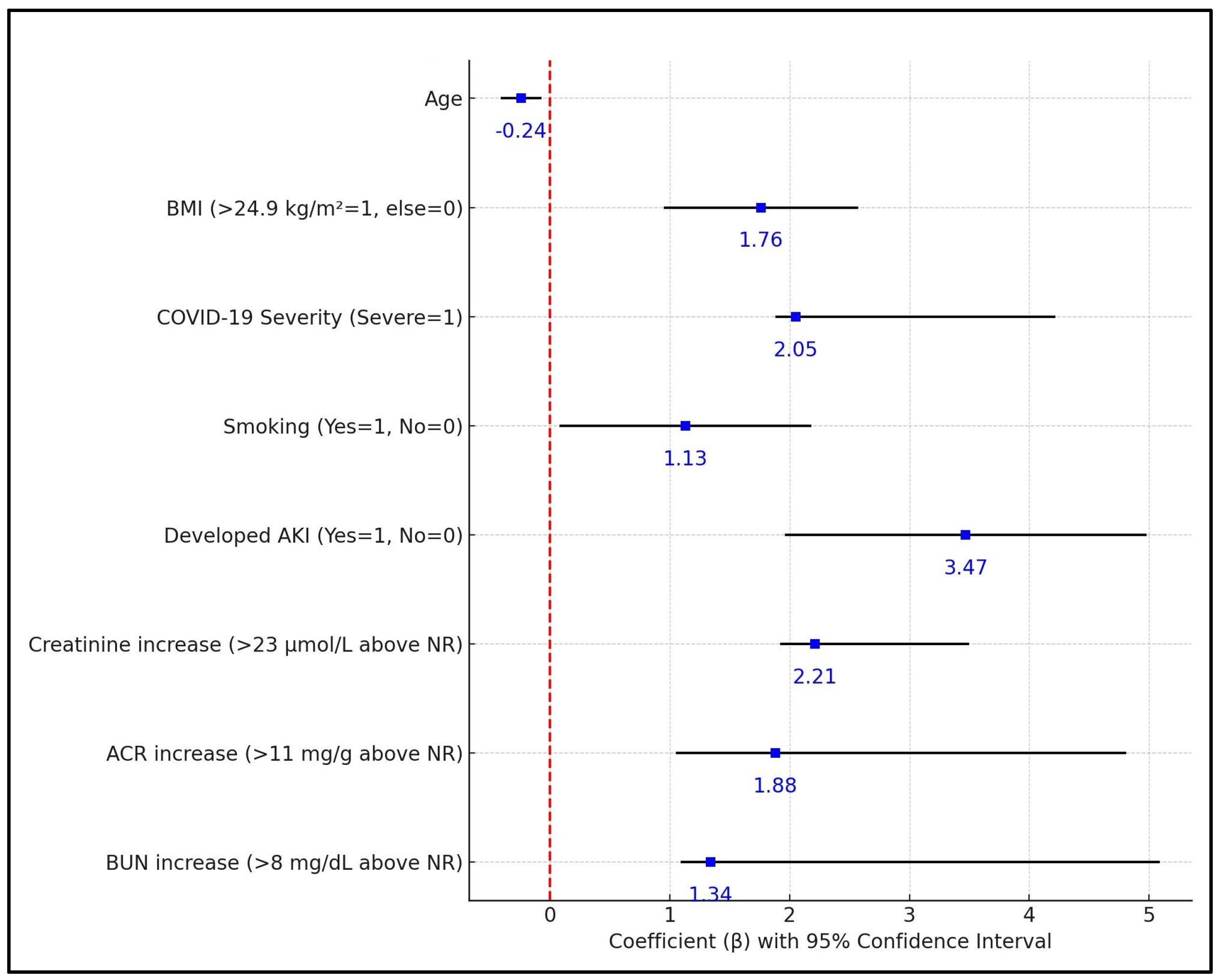
| Independent Variables | Coefficient (β) | Std. Error | p-Value | 95% CI |
|---|---|---|---|---|
| Age | −0.24 | 0.08 | 0.004 | (−0.40, −0.08) |
| COVID-19 Severity (Severe = 1) | 2.05 | 0.59 | 0.001 | (1.89, 4.21) |
| Developed AKI (Yes = 1, No = 0) | 3.47 | 0.76 | <0.001 | (1.97, 4.97) |
| Creatinine increase (>23 μmol/L above NR) | 2.21 | 0.65 | <0.001 | (1.93, 3.49) |
| ACR increase (>11 mg/g above NR) | 1.88 | 0.47 | <0.001 | (1.06, 4.80) |
| BUN increase (>8 mg/dL above NR) | 1.34 | 0.38 | 0.001 | (1.10, 5.08) |
Disclaimer/Publisher’s Note: The statements, opinions and data contained in all publications are solely those of the individual author(s) and contributor(s) and not of MDPI and/or the editor(s). MDPI and/or the editor(s) disclaim responsibility for any injury to people or property resulting from any ideas, methods, instructions or products referred to in the content. |
© 2024 by the authors. Licensee MDPI, Basel, Switzerland. This article is an open access article distributed under the terms and conditions of the Creative Commons Attribution (CC BY) license (https://creativecommons.org/licenses/by/4.0/).
Share and Cite
Boruga, M.; Septimiu-Radu, S.; Nandarge, P.S.; Elagez, A.; Doros, G.; Lazureanu, V.E.; Stoicescu, E.R.; Tanase, E.; Iacob, R.; Dumitrescu, A.; et al. Kidney Function Tests and Continuous eGFR Decrease at Six Months after SARS-CoV-2 Infection in Patients Clinically Diagnosed with Post-COVID Syndrome. Biomedicines 2024, 12, 950. https://0-doi-org.brum.beds.ac.uk/10.3390/biomedicines12050950
Boruga M, Septimiu-Radu S, Nandarge PS, Elagez A, Doros G, Lazureanu VE, Stoicescu ER, Tanase E, Iacob R, Dumitrescu A, et al. Kidney Function Tests and Continuous eGFR Decrease at Six Months after SARS-CoV-2 Infection in Patients Clinically Diagnosed with Post-COVID Syndrome. Biomedicines. 2024; 12(5):950. https://0-doi-org.brum.beds.ac.uk/10.3390/biomedicines12050950
Chicago/Turabian StyleBoruga, Madalina, Susa Septimiu-Radu, Prashant Sunil Nandarge, Ahmed Elagez, Gabriela Doros, Voichita Elena Lazureanu, Emil Robert Stoicescu, Elena Tanase, Roxana Iacob, Andreea Dumitrescu, and et al. 2024. "Kidney Function Tests and Continuous eGFR Decrease at Six Months after SARS-CoV-2 Infection in Patients Clinically Diagnosed with Post-COVID Syndrome" Biomedicines 12, no. 5: 950. https://0-doi-org.brum.beds.ac.uk/10.3390/biomedicines12050950