Communication

Detecting Class 1 Integrons and Their Variable Regions in Escherichia coli Whole-Genome Sequences Reported from Andean Community Countries

by
María Nicole Solis
1,
Karen Loaiza
2,
Lilibeth Torres-Elizalde
3,
Ivan Mina
4,
Miroslava Anna Šefcová
1 and
Marco Larrea-Álvarez
1,*
1
Facultad de Ciencias Médicas Enrique Ortega Moreira, Carrera de Medicina, Universidad Espíritu Santo, Samborondón 092301, Ecuador
2
Department of Bacteria, Parasites and Fungi, Statens Serum Institut, 2300 Copenhagen, Denmark
3
Graduate School Life Sciences and Health (GS LSH), Université Paris-Saclay, 91198 Gif-sur-Yvette, France
4
School of Biological Science and Engineering, Yachay-Tech University, Urcuquí 100650, Ecuador
*
Author to whom correspondence should be addressed.
Submission received: 28 March 2024 / Revised: 24 April 2024 / Accepted: 25 April 2024 / Published: 25 April 2024
(This article belongs to the Special Issue Genomic Analysis of Antimicrobial Drug-Resistant Bacteria)

Abstract

:
Various genetic elements, including integrons, are known to contribute to the development of antimicrobial resistance. Class 1 integrons have been identified in E. coli isolates and are associated with multidrug resistance in countries of the Andean Community. However, detailed information on the gene cassettes located on the variable regions of integrons is lacking. Here, we investigated the presence and diversity of class 1 integrons, using an in silico approach, in 2533 whole-genome sequences obtained from EnteroBase. IntFinder v1.0 revealed that almost one-third of isolates contained these platforms. Integron-bearing isolates were associated with environmental, food, human, and animal origins reported from all countries under scrutiny. Moreover, they were identified in clones known for their pathogenicity or multidrug resistance. Integrons carried cassettes associated with aminoglycoside (aadA), trimethoprim (dfrA), cephalosporin (blaOXA; blaDHA), and fluoroquinolone (aac(6′)-Ib-cr; qnrB) resistance. These platforms showed higher diversity and larger numbers than previously reported. Moreover, integrons carrying more than three cassettes in their variable regions were determined. Monitoring the prevalence and diversity of genetic elements is necessary for recognizing emergent patterns of resistance in pathogenic bacteria, especially in countries where various factors are recognized to favor the selection of resistant microorganisms.

1. Introduction

Escherichia coli is widely distributed in natural environments and is a common inhabitant of the gastrointestinal tract in animals, including humans. While it serves as an important commensal resident, it also poses a threat as a potential pathogen, capable of causing both intestinal and extra-intestinal diseases [1,2]. The rise of multidrug-resistant (MDR) E. coli can be attributed to prolonged antibiotic exposure, presenting substantial hurdles for public health systems [3]. This phenomenon has been observed globally, with prevalence rates influenced by geographical regions, populations, and countries [4,5]. In South America, several factors contribute to the dissemination of antimicrobial resistance. These include drug misuse in the community, the spread of genetic markers through the food chain, and environmental contamination from sewage disposal, not only from hospitals but also from industrial and urban sources [6]. In particular, strains of E. coli resistant to commonly used antibiotics have been documented in countries comprising the Andean Community—an intergovernmental organization consisting of Bolivia, Colombia, Ecuador, and Peru [7,8,9,10].
Genetic changes and the horizontal transmission of resistant traits are recognized as crucial factors driving the development of antimicrobial resistance. MDR phenotypes arise as bacteria acquire and disseminate resistance genes through horizontal transfer [11,12]. This process is facilitated by genetic elements such as gene cassettes/integrons, insertion sequences (IS), and transposons, which have the ability to move between and within DNA molecules. Additionally, plasmids and conjugative elements play a role in mobilizing genetic information between bacterial cells [13]. Integrons, in particular, are capable of integrating gene cassettes through natural recombination events, making them adequate platforms for gene expression [14]. They are associated with IS, transposons, and plasmids, which collectively facilitate the spread of resistance traits among bacterial populations [15,16]. A typical integron consists of a variable region and two conserved segments. These segments are known as the 5′ and 3′ conserved sequences (CS). The intI1 gene is situated within the 5′ CS and encodes a tyrosine recombinase responsible for integrating or excising genes at the attI site. Additionally, the intI1 gene contains the (Pc) promoter sequence, which drives the expression of gene cassettes located within the variable region [17,18]. Integrons are classified into five groups based on the sequence of the intI1 gene they carry, with class 1 integrons being particularly prevalent and clinically significant [15,16]. In contrast, the 3′ CS region may incorporate genes conferring resistance to sulphonamides (sul1) or quaternary ammonium compounds (qacEΔ1) [17]. These genes are frequently encountered in class 1 integrons, while class 2 integrons often harbor genes associated with transposition proteins (tns). Other classes of integrons have not been associated with gene cassettes [15]. The variable region of the integron, situated between these two segments, serves as a platform for the integration of various cassettes that confer resistance to multiple classes of antibiotics [15]. These gene cassettes typically consist of an open reading frame and a recombination site (attC), and they are integrated at the attI site and expressed from the Pc promoter. The level of transcription depends on the proximity of the cassette to the promoter, with genes located closer to it exhibiting higher expression levels [18]. Class 1 integrons have been linked to a range of gene cassettes conferring resistance to aminoglycosides, folate antagonists, β-lactams, quinolones, and other classes of antibiotics [16].
These integrons are frequently encountered in enteropathogenic bacteria [16]. Escherichia, Klebsiella, Salmonella, Shigella, and Yersinia are notable genera of the Enterobacteriaceae family that colonize the intestinal tract and can lead to intestinal, genitourinary, and bloodstream infections [19]. As mentioned earlier, the emergence of MDR E. coli has been linked to prolonged exposure to antibiotics. Indeed, class 1 integrons have been identified in E. coli isolates from various geographical regions [20]. The prevalence of these platforms and their associated cassettes is known to fluctuate over time, a phenomenon linked to selective antibiotic pressure. An increase in the prevalence of class 1 integrons in E. coli has been observed in South Korea and China [16,20]. Moreover, the incidence of class 1 integrons harboring multiple cassettes has increased among E. coli isolates, suggesting that they facilitate the acquisition of gene cassettes [16,20]. Therefore, the continuous monitoring of integrons is crucial, particularly for understanding the spread of drug resistance.
As mentioned previously, strains of MDR E. coli have been documented in countries forming the trade block called the Andean Community [7,8,9,10]. This organization promotes collaboration in industry, agriculture, social issues, and trade. The constant flow of people and resources increases the risk of the cross-transmission of microorganisms, particularly those resistant to multiple drugs. Integrons have been identified in MDR and pathogenic isolates of E. coli belonging to various clones reported in the countries under scrutiny. Several studies have documented the presence of various antibiotic-resistant genes, primarily dfrA and aadA, in the variable regions of these integrons [21,22,23,24], while others have solely documented the presence of the intI1 gene [25,26,27]. Class 1 integrons have been linked to an increase in MDR E. coli, attributed to the diversity of cassettes within their variable regions [20,28]. The limited information available on these cassettes among isolates from Andean countries may hinder our ability to study their dynamics and their relationship with resistance genes.
Bioinformatic tools have not only facilitated the identification of mobile genetic elements but have also helped establish crucial connections between resistance traits and pathogenic bacteria [29,30]. The objective of this study is to use IntFinder v1.0, a tool capable of detecting integrons in both raw reads and assembled genomes [31,32], to identify the association of integrons with resistance cassettes in isolates from countries within the Andean Community.

2. Results

2.1. Bacterial Isolate Dataset

The final dataset comprised 2533 whole-genome sequenced isolates of E. coli documented in countries of the Andean Community (Table S1, Supplementary Materials). The majority of isolates were reported in Ecuador (75%), followed by Peru (19%), Colombia (3%), Venezuela (2%), and Bolivia (1%). Nearly half of the isolates were sourced from human samples (47%), followed by those from animal samples (31%), the environment (20%), and food products (2%).

2.2. Integron Characterization by Country and Source

IntFinder v1.0 identified integrons in 29% of isolates, all classified as class 1 integrons (Table S2, Supplementary Materials). These integrons were distributed across all countries, with the highest prevalence observed in samples from Ecuador, followed by Peru, Colombia, Venezuela, and Bolivia. However, in terms of relative abundance, approximately 30% of isolates from Ecuador and Colombia tested positive for integrons, while they accounted for 20% in Peruvian samples. Less than 1% of isolates from Venezuela and Bolivia showed evidence of integrons (Figure 1A). These class 1 integrons were detected across various sources, including human, animal, environmental, and food-related samples. However, analysis of the relative abundance revealed that integrons were present in approximately 30% of environmental and animal samples, while 25% of human samples carried these sequences. Class 1 integrons were found in less than 20% of isolates derived from food or environmental samples (Figure 1B).

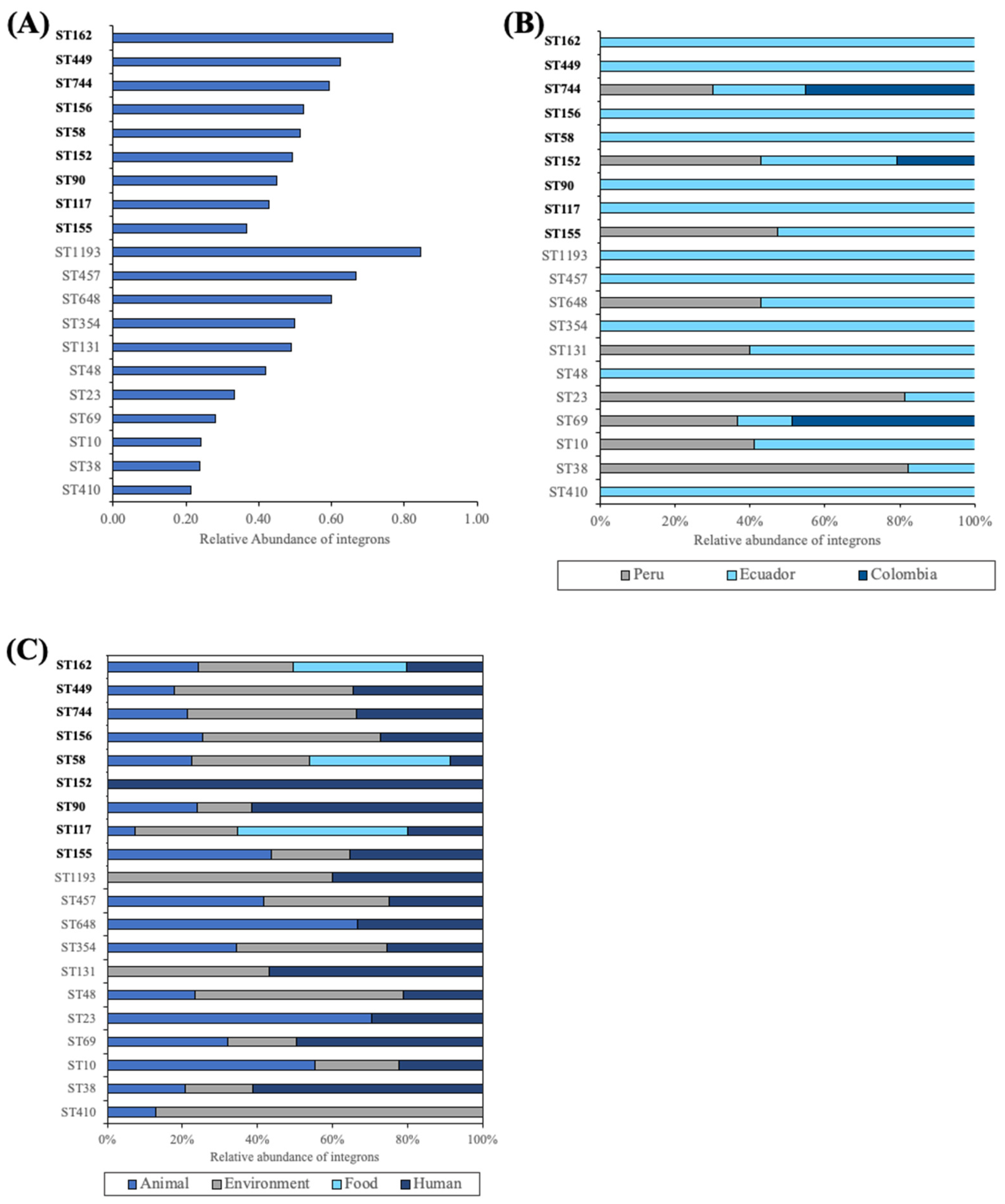
2.3. Integron Characterization by ST

Integron-positive isolates were associated with 149 different sequence types (STs). Figure 2A illustrates the prevalence of integrons among isolates associated with relevant pathogenic or MDR STs; the latter are highlighted in bold in the figure. In MDR clones, the highest occurrence of integrons was observed in isolates from ST162 (77%), followed by those associated with ST449 (63%) and ST744 (59%). Approximately half of the isolates belonging to ST156, ST58, and ST152 were positive for integrons, while in E. coli from ST90, ST117, and ST155, integrons were present in less than 50% of isolates. In pathogenic E. coli, integrons were most abundant in bacteria from ST1193 (85%), ST457 (67%), and ST648 (60%), whereas, in isolates from ST354 and ST131, integrons were present in half of them. They were also detected in 40% and 33% of E. coli associated with ST48 and ST23, respectively. In bacteria belonging to the remaining STs, these platforms were present in less than 30% of isolates.
Figure 2B,C depicts the aforementioned sequence types categorized by country and source, respectively. Integron-containing E. coli isolates belonging to STs known for multidrug resistance were predominantly reported in Ecuador, although bacteria from Peru were also associated with ST155, ST744, and ST152. E. coli from the latter two STs were also found in Colombian samples. Isolates from these STs mainly originated from environmental, human, and animal sources. ST117, ST58, and ST162 were also linked to food samples, while ST152 and ST156 were found in human samples. On the other hand, STs associated with pathogenic bacteria were predominantly documented in Ecuador, although some isolates belonging to ST648, ST131, ST23, ST69, ST10, and ST38 were also reported in Peru. E. coli from ST69 was also found in Colombia. Most of the STs were linked to animal, environmental, and human samples. Bacteria from ST1193 and ST131 originated from human and environmental samples, whereas those of ST648 and ST23 came from animal and human samples.

2.4. Integrons and Antibiotic Resistance Genes

Gene cassettes conferring resistance to aminoglycosides and dihydrofolate reductase inhibitors were the most prevalent, were detected across samples from all sources reported in all five countries, and were associated with all the aforementioned STs. Similarly, genes conferring resistance to phenicols were identified in isolates from Ecuador and Peru. Integrons carrying these genes were reported from various sources. Conversely, genes associated with β-lactam resistance were primarily detected in human isolates reported from Peru, Ecuador, and Colombia. Resistance markers for lincosamide and sulfonamide antibiotics were found in environmental and animal samples, with the latter also associated with genes conferring resistance to quinolones. These markers were also detected in human isolates, along with those associated with rifampicin resistance. All STs exhibited resistance markers for aminoglycosides and folate antagonists. Additionally, resistance to chloramphenicol was observed across most of them. Genes encoding β-lactam hydrolases were prevalent, particularly in ST152 and ST10. Among the ST156, ST744, and ST69 clones, there were also traits associated with quinolone resistance. Furthermore, rifampicin-resistant genes were detected in the latter two STs (Figure 3).
A total of 37 different integrons were identified by IntFinder v1.0. Resistance to aminoglycosides was mediated by adenylyl transferases encoded by seven different aadA alleles, while resistance to trimethoprim was associated with dihydrofolate reductases encoded by seven different dfrA alleles. Inactivation of aminoglycosides was also facilitated by drug-modifying enzymes, including adenylyl and acetyl transferases encoded by the ant(2′) and aac(3)-Vla genes, respectively. Additionally, resistance was linked to the aac(6′)-lb-cr gene, identified in certain integrons, encoding an acetyl transferase capable of inactivating both aminoglycoside and quinolone antibiotics. Quinolone resistance was also associated with the qnrB genes, which produce proteins that bind to topoisomerases and protect them from drugs. β-lactam inactivation was facilitated by hydrolases encoded by the blaOXA and blaDHA genes, while resistance to rifampicin was attributed to enzymes encoded by the arr genes, catalyzing the ADP-ribosylative inactivation of the antibiotic (Table 1).
Resistance to clindamycin was linked to nucleotidyl transferases encoded by the lnu(F) genes. Similarly, sequences associated with transferases that inactivate chloramphenicol (catB) were identified, although resistance to this antibiotic was mainly attributed to efflux pumps encoded by the cmlA1 genes. Additionally, sul3 genes were found, responsible for conferring resistance to sulfonamide antibiotics by encoding dihydropteroate synthases. Regarding the variable regions, approximately half of the integrons harbored an aadA cassette in the first position, while 30% contained a dfrA cassette. Among the remaining 20% of isolates, aac (8%), bla (6%), ant (3%), and lnu (3%) genes were identified as the first cassette. The majority of positive isolates contained integrons with either one of two cassettes in the variable regions. The predominant genes identified were aadA and dfrA, along with additional genes encoding β-lactamases, acetyl transferases, and nucleotidyl transferases. Integrons with three cassettes were detected in 16% of isolates, while those containing four cassettes represented 11% of the samples. Only a small fraction, less than 1% of isolates, exhibited a single integron with more than four cassettes in the variable region (Table 1).

3. Discussion

This study explores the presence and diversity of class 1 integrons among isolates documented in Ecuador, Colombia, Peru, Bolivia, and Venezuela, representing the Andean Community. Integrons were found in only 29% of isolates, displaying limited diversity in gene cassette content, albeit higher than previously reported. Despite this, the detected integrons contained genes conferring resistance to commonly used antibiotics, contributing to the emergence of multidrug-resistant phenotypes in E. coli. The use of bioinformatic tools has been instrumental in identifying mobile genetic elements carrying resistance genes and elucidating their relationship with pathogenic bacteria [29,30]. IntFinder v1.0, specifically, facilitates the detection of integrons using both raw reads and assembled genomes/contigs [31].
Integrons were found to be more common in isolates from Ecuador, Colombia, and Peru compared with those reported in Bolivia and Venezuela. Interestingly, integrons were equally distributed among animal, environmental, and human samples, but they were less frequently identified in those obtained from food samples. In Ecuador, integrons were detected in human, animal, environmental, and food sources, consistent with findings from previous studies [22,24,33,34]. In Peruvian isolates, integrons were detected in samples from both humans and animals, while Colombian isolates showed an association between these platforms and clinical sources. Indeed, integrons have been reported in E. coli from patients and farm animals [10,21,35], although there are limited data available on their presence in environmental samples. Clinical isolates from Bolivia and Venezuela were associated with integrons, although they have also been reported in environmental samples [36,37].
ST162 is recognized as a multidrug-resistant clone [38]. In Ecuadorian isolates derived from the environment, this sequence type has been linked to integrons carrying resistance markers for aminoglycosides and trimethoprim [23]. In fact, our analysis confirmed the presence of these genes within this ST, in addition to genes conferring resistance to chloramphenicol. Moreover, isolates associated with both human and animal samples were identified. Notably, ST162 has been documented in such samples in Ecuador, Bolivia, and Colombia, although references to integrons have not been previously noted [39,40,41]. Similarly, our findings reveal that ST155, isolated from human, animal, and environmental sources, harbored integrons containing genes conferring resistance to the specified antibiotics. ST155 is another well-established multidrug-resistant clone [42] observed in Peru, Ecuador, and Colombia; however, previous studies have not associated it with integrons [43,44,45]. Interestingly, in Bolivia, this sequence type is associated with the presence of the intI1 gene, although no references to gene cassettes within the variable region were identified [27]. ST58, ST744, ST90, and ST156 are recognized MDR clones [46,47,48,49] that have been documented in the area without previous references to integrons [27,40,50,51]. However, our investigation revealed that isolates belonging to these STs did indeed carry integrons containing resistance markers for aminoglycosides, trimethoprim, phenicols, and quinolones. Similarly, E. coli strains from the ST152, ST177, and ST449 clones also harbored integrons with the mentioned resistance markers. Although these clones are known for their multidrug resistance [52,53,54], they have not been previously documented in the area.
The majority of pathogenic STs harbored integrons carrying markers for aminoglycosides and trimethoprim. Specifically, ST131, ST1193, ST38, and ST457 were exclusively associated with these genes. These sequence types, along with ST10, ST48, ST410, and ST69, are well known for their involvement in extraintestinal infections [55,56,57,58,59,60]. Furthermore, the latter four also carried genes conferring resistance to chloramphenicol, while ST10 additionally contained markers for β-lactams. Integron-containing ST10 and ST410 have been documented in Ecuador and Bolivia [23,24,27], whereas the remaining STs have not been directly linked to these platforms. In Bolivia, ST48, ST410, and ST69 isolates were found to be positive for the intI1 gene and harbored many of the aforementioned resistance genes, although no references to the variable regions of integrons were identified [27]. These clones have also been reported in other countries, but the presence of integrons was not specifically assessed [10,45,51,61,62,63]. ST23, ST648, and ST354 have not been previously documented in the countries under study. However, these STs have been associated with extraintestinal infections [59,64,65]. Our analysis revealed that isolates belonging to these clones carried genes responsible for aminoglycosides, trimethoprim, and chloramphenicol resistance.
The most prevalent genes identified were those encoding various aminoglycoside adenylyl transferases (AadA) and dihydrofolate reductases (DfrA). These markers have previously been reported in E. coli associated with integrons in the scrutinized area, with specific alleles such as dfrA1, dfrA5, dfrA7, dfrA12, dfrA15, dfrA17, aadA1, aadA2, and aadA5 documented [21,22,36]. In addition to these known sequences, our findings revealed the presence of previously unreported genes, including aadA2b, aadA16, aadA17, aadA22, dfrA14, dfrA16, and dfrA27. Furthermore, other markers for aminoglycosides, such as those encoding acetyl and nucleotidyl transferases, were found. While these genes have been described in E. coli in the area [22], they have not been directly linked to integrons. Resistance to chloramphenicol was attributed to acetyl transferases (Cat) and efflux pumps (Cml1), predominantly identified in animal and human samples. Specifically, genes encoding CatB3 have been detected in clinical isolates from Peru [21]. The sequences identified in our study have been documented in clinical and animal sources from Bolivia and Ecuador, although a direct relationship with integrons was not assessed [27,33].
Our findings show that resistance to β-lactams was associated with hydrolases encoded by bla-OXA1 and blaDHA4, which were found in human and environmental isolates. A study reported the presence of intI1 and bla-OXA1 genes in both environmental and human isolates, although it was not specified whether bla-OXA1 was located in the variable region [27]. However, in Colombia, a class 1 integron carrying the blaVIM-4 gene, encoding for a metallo-β-lactamase, has been documented in clinical isolates [35]. Quinolone resistance was determined to be mediated by the aac(6′)-Ib-cr and qnrB4 genes. These markers have been documented in numerous studies in the area, particularly in association with clinical and environmental samples, yet they have not been directly related to integrons [22,25,27].
Among the identified sequences, a total of 27 different gene cassette complexes were detected, all of which contained resistance genes. Integrons are commonly recognized as genetic structures with a low cost [66]; specifically, the number of gene cassettes has a notable impact on their fitness. Integrons carrying a larger number of gene cassettes tend to incur higher costs, leading to their decreased prevalence. Consequently, integrons harboring fewer, less costly cassettes are more commonly observed [67,68]. In fact, over 70% of predicted integrons were found to have one or two cassettes, and less than 1% contained more than five resistance genes in the variable region. Moreover, in half of the integrons, the first position was occupied by either an aadA or a dfrA gene. These cassettes contain highly recombinogenic attC sites and are frequently detected in such positions in class 1 integrons due to their low cost [66]. Additionally, the relatively costly aac(6′)-lb gene was found at the first position in approximately 8% of integrons. It has been suggested that this cassette must be located near the promoter to achieve proper expression levels [66].
Our results revealed that integrons are more predominantly present and diverse in the studied area than previously reported in the literature. Additionally, they carry a greater number of genes than previously documented. It is worth noting that only simple integrons containing two cassettes in their variable region have been reported in E. coli. The present results highlight that larger integrons containing more than two cassettes are prevalent among relevant STs. These complex integrons may play an important role in expanding the array of resistant markers found in E. coli. They carry genes conferring resistance not only to antibiotics commonly used as first-line defenses against E. coli—such as fluoroquinolones, trimethoprim–sulfamethoxazole, and cephalosporins—but also to aminoglycosides, which are typically reserved for treating serious infections [69,70]. Resistance markers against chloramphenicol, clindamycin, and rifampicin were also detected. These drugs may serve as viable alternatives when other antibiotics are ineffective or deemed inappropriate [71,72,73]. Certainly, integrons are extensively distributed among E. coli strains documented across diverse geographical regions [20], and they are thought to play a crucial role in the emergence of multidrug-resistant phenotypes within Enterobacteriaceae [16]. These mosaic structures act as reservoirs of exchangeable cassettes, which are not only linked to drug resistance but also to virulence and pathogenicity [74].
The dataset utilized in this study was constructed using E. coli sequences sourced from EnteroBase, potentially introducing bias toward isolates derived from culturable and pathogenic bacteria. While it is expected that novel isolates will continue to be reported, the ones examined in this study serve as relevant examples. Moreover, the approach employed in this study relies on a database of well-described integrons, which only represents a partial population. However, despite these limitations, the findings presented here contribute to advancing our understanding of the relationship between integrons, resistance genes, and E. coli in the Andean region.

4. Materials and Methods

4.1. Selection of Dataset

A dataset of E. coli isolates (n = 2533) that were whole-genome-sequenced was compiled from EnteroBase (http://enterobase.warwick.ac.uk/, accessed on 22 March 2023), a platform dedicated to studying genomic variation in enterobacteria. The compilation of this dataset adhered to specific criteria: (i) inclusion of isolates reported from countries within the Andean Community, including Ecuador, Colombia, Bolivia, and Peru, with the addition of data from Venezuela up to 2006, as it was a former member; (ii) collection of samples spanning the period 1993 to 2023; and (iii) selection of non-repetitive whole genomes. Sequences in FASTA format were downloaded on 22 March 2023.

4.2. Multilocus Sequence Type Identification

The software MLST v2.19.0 developed by Torsten Seemann was employed to determine the sequence types (STs) using default settings (https://github.com/tseemann/mlst; accessed on 22 February 2023). The allele analysis scheme utilized for this purpose was from PubMLST.org [75]. The MLST scheme used was based on seven consensus genes, adk, fumC, gyrB, icd, mdh, purA, and recA.

4.3. IntFinder v1.0

IntFinder v1.0, developed by the Center for Genomic Epidemiology at the Technical University of Denmark (https://bitbucket.org/genomicepidemiology/intfinder/src/master/; accessed on 22 May 2023; software version: 2019-12-18; database version: 2019-11-29), was utilized to identify resistance integrons with modified parameters: threshold, 0.9, and min cov, 0.9. IntFinder v1.0 is accessible online (https://cge.food.dtu.dk/services/IntFinder-1.0/; accessed on 24 May 2023) and utilizes k-mer alignment for sequence detection, leveraging KMA v1.3.9, developed by the Center for Genomic Epidemiology at the Technical University of Denmark (https://bitbucket.org/genomicepidemiology/kma/src/master/, accessed on 24 May 2023) [76]. This alignment methodology enables detection based on an adjustable similarity threshold. Integron identification relies on detecting the presence of the integrase sequence, specifically, intI1. The integron database comprises data obtained from the public repository INTEGRALL (http://integrall.bio.ua.pt/; accessed on 24 May 2023) [77]. The database employs the unique numbers assigned by INTEGRALL to each integron (ln) [31,78]. Prediction of antimicrobial resistance genes in the integron database was carried out using the local standalone version of ResFinder v4.1 developed by the Center for Genomic Epidemiology at the Technical University of Denmark (https://bitbucket.org/genomicepidemiology/resfinder/src/master/; software version: 2019-01-29; database version: 2019-02-20, accessed on 24 May 2023) [79]. This analysis employed parameters requiring a minimum sequence identity of 90% and a minimum coverage of 60%.

5. Conclusions

In this study, we investigated the occurrence and diversity of class 1 integrons in isolates reported from countries of the Andean Community using an in silico approach. Utilizing IntFinder v1.0, we found that almost one-third of the isolates tested positive for integrons. Class 1 integrons were identified in environmental, food, human, and animal isolates belonging to various relevant clones known for their pathogenicity or multidrug resistance. Overall, the integrons carried cassettes conferring resistance to antibiotics used in E. coli infections, such as fluoroquinolones, trimethoprim, cephalosporins, and aminoglycosides. The integrons identified in this study exhibited a greater number and variety of cassettes compared with what has been previously reported in the literature. Furthermore, the majority of integrons observed carried only two or three cassettes in their variable regions. However, in certain cases, four or even six cassettes were identified. These large mosaic structures harbor markers conferring resistance to various antibiotic classes, thereby aiding bacteria in adapting to environmental stress. In South American countries, several factors are recognized as favoring the spread of antibiotic-resistant bacteria. Therefore, monitoring the presence and diversity of these platforms in the region appears imperative. In silico studies are not only useful for identifying genetic traits associated with mobile elements but also contribute to recognizing emergent patterns of resistance, particularly in species inhabiting the intestine and regularly exposed to antibiotic pressure.

Supplementary Materials

The following supporting information can be downloaded at https://0-www-mdpi-com.brum.beds.ac.uk/article/10.3390/antibiotics13050394/s1: Table S1. Dataset of whole-genome-sequenced E. coli isolates in Andean Community. Table S2. Integron-positive isolates.

Author Contributions

Conceptualization, M.N.S., K.L., M.A.Š., L.T.-E. and M.L.-Á.; methodology, M.N.S., K.L., L.T.-E., I.M. and M.L.-Á.; software, K.L.; validation, K.L.; formal analysis, M.N.S. and L.T.-E.; investigation, M.N.S., K.L., M.A.Š., L.T-E. and M.L.-Á.; data curation, L.T.-E.; writing—original draft preparation, M.L.-Á.; writing—review and editing, M.N.S., K.L., L.T.-E., I.M., M.A.Š. and M.L.-Á.; visualization, L.T.-E. and M.L.-Á.; supervision, K.L. and M.L.-Á.; project administration, M.L.-Á.; funding acquisition, M.L.-Á. All authors have read and agreed to the published version of the manuscript.

Funding

This study was supported by Universidad de Especialidades Espíritu Santo (UEES 2023-MED-003).

Institutional Review Board Statement

Not applicable.

Informed Consent Statement

Not applicable.

Data Availability Statement

The data are available upon request.

Conflicts of Interest

The authors declare no conflicts of interest.

References

  1. Leimbach, A.; Hacker, J.; Dobrindt, U. E. coli as an All-rounder: The Thin Line between Commensalism and Pathogenicity. In Between Pathogenicity and Commensalism: Current Topics in Microbiology and Immunology; Dobrindt, U., Hacker, J., Svanborg, C., Eds.; Springer: Heidelberg/Berlin, Germany, 2013; Volume 358, pp. 3–32. [Google Scholar]
  2. Conway, T.; Cohen, P.S. Commensal and pathogenic Escherichia coli metabolism in the gut. Metab. Bact. Pathog. 2015, 3, 343–362. [Google Scholar]
  3. Szmolka, A.; Nagy, B. Multidrug resistant commensal Escherichia coli in animals and its impact for public health. Front. Microbiol. 2013, 4, 258. [Google Scholar] [CrossRef]
  4. Johnston, B.D.; Thuras, P.; Porter, S.B.; Anacker, M.; VonBank, B.; Vagnone, P.S.; Witwer, M.; Castanheira, M.; Johnson, J.R. Global molecular epidemiology of carbapenem-resistant Escherichia coli (2002–2017). Eur. J. Clin. Microbiol. Infect. Dis. 2021; ahead of print. [Google Scholar]
  5. Erb, A.; Stürmer, T.; Marre, R.; Brenner, H. Prevalence of antibiotic resistance in Escherichia coli: Overview of geographical, temporal, and methodological variations. Eur. J. Clin. Microbiol. Infect. Dis. 2007, 26, 83–90. [Google Scholar] [CrossRef]
  6. Bonelli, R.R.; Moreira, B.M.; Picão, R.C. Antimicrobial resistance among Enterobacteriaceae in South America: History, current dissemination status, and associated socioeconomic factors. Drug Resist. Updates 2014, 17, 24–36. [Google Scholar] [CrossRef]
  7. Delgado-Blas, J.F.; Ovejero, C.M.; Abadia-Patiño, L.; Gonzalez-Zorn, B. Coexistence of mcr-1 and blaNDM-1 in Escherichia coli from Venezuela. Antimicrob. Agents Chemother. 2016, 60, 6356–6358. [Google Scholar] [CrossRef]
  8. Martinez, P.; Garzón, D.; Mattar, S. CTX-M-producing Escherichia coli and Klebsiella pneumoniae isolated from community-acquired urinary tract infections in Valledupar, Colombia. Braz. J. Infect. Dis. 2012, 16, 420–425. [Google Scholar] [CrossRef]
  9. Joffré, E.; Iñiguez Rojas, V. Molecular epidemiology of Enteroaggregative Escherichia coli (EAEC) isolates of hospitalized children from Bolivia reveals high heterogeneity and multidrug-resistance. Int. J. Mol. Sci. 2020, 21, 9543. [Google Scholar] [CrossRef]
  10. Espinoza, L.L.; Carhuaricra Huamán, D.; Rodríguez Cueva, C.; Durán Gonzales, C.; León, Y.I.; Silvestre Espejo, T.; Marcelo Monge, G.; Rosadio Alcántara, R.; Maturrano Hernández, L. Genomic analysis of multidrug-resistant Escherichia coli strains carrying the mcr-1 gene recovered from pigs in Lima, Peru. Comp. Immunol. Microbiol. Infect. Dis. 2023, 99, 102019. [Google Scholar] [CrossRef]
  11. Huddleston, J.R. Horizontal gene transfer in the human gastrointestinal tract: Potential spread of antibiotic resistance genes. Infect. Drug Resist. 2014, 7, 167–176. [Google Scholar] [CrossRef]
  12. Woodford, N.; Ellington, M.J. The Emergence of Antibiotic Resistance by Mutation. Clin. Microbiol. Infect. 2007, 13, 5–18. [Google Scholar] [CrossRef]
  13. Partridge, S.R.; Kwong, S.M.; Firth, N.; Jensen, S.O. Mobile genetic elements associated with antimicrobial resistance. Clin. Microbiol. Rev. 2018, 31, e00088-17. [Google Scholar] [CrossRef]
  14. Leverstein-van Hall, M.A.; Blok, H.E.M.; Donders, A.R.T.; Paauw, A.; Fluit, A.C.; Verhoef, J. Multidrug resistance among Enterobacteriaceae is strongly associated with the presence of integrons and is independent of species or isolate origin. J. Infect. Dis. 2003, 187, 251–259. [Google Scholar] [CrossRef]
  15. Deng, Y.; Bao, X.; Ji, L.; Chen, L.; Liu, J.; Miao, J.; Chen, D.; Bian, H.; Li, Y.; Yu, G. Resistance integrons: Class 1, 2 and 3 integrons. Ann. Clin. Microbiol. Antimicrob. 2015, 14, 45. [Google Scholar] [CrossRef]
  16. Kaushik, M.; Kumar, S.; Kapoor, R.K.; Virdi, J.S.; Gulati, P. Integrons in Enterobacteriaceae: Diversity, distribution and epidemiology. Int. J. Antimicrob. Agents 2018, 51, 167–176. [Google Scholar] [CrossRef]
  17. Canal, N.; Meneghetti, K.L.; de Almeida, C.P.; da Rosa Bastos, M.; Otton, L.M.; Corção, G. Characterization of the variable region in the class 1 integron of antimicrobial-resistant Escherichia coli isolated from surface water. Braz. J. Microbiol. 2016, 47, 337–344. [Google Scholar] [CrossRef]
  18. Luk-In, S.; Pulsrikarn, C.; Bangtrakulnonth, A.; Chatsuwan, T.; Kulwichit, W. Occurrence of a novel class 1 integron harboring qnrVC4 in Salmonella Rissen. Diagn. Microbiol. Infect. Dis. 2017, 88, 282–286. [Google Scholar] [CrossRef]
  19. Salimiyan Rizi, K.; Ghazvini, K.; Farsiani, H. Clinical and pathogenesis overview of Enterobacter infections. Rev. Clin. Med. 2020, 6, 146–154. [Google Scholar]
  20. Zhang, S.; Abbas, M.; Rehman, M.U.; Huang, Y.; Zhou, R.; Gong, S.; Yang, H.; Chen, S.; Wang, M.; Cheng, A. Dissemination of antibiotic resistance genes (ARGs) via integrons in Escherichia coli: A risk to human health. Environ. Pollut. 2020, 266, 115260. [Google Scholar] [CrossRef]
  21. Riveros, M.; Pons, M.J.; Durand, D.; Ochoa, T.J.; Ruiz, J. Class 1 and 2 integrons in Escherichia coli strains isolated from diarrhea and bacteremia in children less than 2 years of age from Peru. Am. J. Trop. Med. Hyg. 2023, 108, 181. [Google Scholar] [CrossRef]
  22. Chiluisa-Guacho, C.; Escobar-Perez, J.; Dutra-Asensi, M. First detection of the CTXM-15 producing Escherichia coli O25-ST131 pandemic clone in Ecuador. Pathogens 2018, 7, 42. [Google Scholar] [CrossRef]
  23. Ortega-Paredes, D.; Barba, P.; Mena-López, S.; Espinel, N.; Crespo, V.; Zurita, J. High quantities of multidrug-resistant Escherichia coli are present in the Machángara urban river in Quito, Ecuador. J. Water Health 2020, 18, 67–76. [Google Scholar] [CrossRef]
  24. Ortega-Paredes, D.; Barba, P.; Mena-López, S.; Espinel, N.; Zurita, J. Escherichia coli hyperepidemic clone ST410-A harboring blaCTX-M-15 isolated from fresh vegetables in a municipal market in Quito, Ecuador. Int. J. Food Microbiol. 2018, 280, 41–45. [Google Scholar] [CrossRef]
  25. Zhang, L.; Levy, K.; Trueba, G.; Cevallos, W.; Trostle, J.; Foxman, B.; Marrs, C.F.; Eisenberg, J.N.S. Effects of selection pressure and genetic association on the relationship between antibiotic resistance and virulence in Escherichia coli. Antimicrob. Agents Chemother. 2015, 59, 6733–6740. [Google Scholar] [CrossRef]
  26. Moser, K.A.; Zhang, L.; Spicknall, I.; Braykov, N.P.; Levy, K.; Marrs, C.F.; Foxman, B.; Trueba, G.; Cevallos, W.; Goldstick, J.; et al. The role of mobile genetic elements in the spread of antimicrobial-resistant Escherichia coli from chickens to humans in small-scale production poultry operations in rural Ecuador. Am. J. Epidemiol. 2018, 187, 558–567. [Google Scholar] [CrossRef]
  27. Calderon Toledo, C.; von Mentzer, A.; Agramont, J.; Thorell, K.; Zhou, Y.; Szabó, M.; Colque, P.; Kuhn, I.; Gutiérrez-Cortez, S.; Joffré, E. Circulation of enterotoxigenic Escherichia coli (ETEC) isolates expressing CS23 from the environment to clinical settings. mSystems 2023, 8, e00141-23. [Google Scholar] [CrossRef]
  28. Suhartono, S.; Savin, M.C.; Gbur, E.E. Transmissible plasmids and integrons shift Escherichia coli population toward larger multiple drug resistance numbers. Microb. Drug Resist. 2018, 24, 244–252. [Google Scholar] [CrossRef]
  29. Johansson, M.H.K.; Bortolaia, V.; Tansirichaiya, S.; Aarestrup, F.M.; Roberts, A.P.; Petersen, T.N. Detection of mobile genetic elements associated with antibiotic resistance in Salmonella enterica using a newly developed web tool: MobileElementFinder. J. Antimicrob. Chemother. 2021, 76, 101–109. [Google Scholar] [CrossRef]
  30. Singh, T.; Dar, S.A.; Singh, S.; Shekhar, C.; Wani, S.; Akhter, N.; Bashir, N.; Haque, S.; Ahmad, A.; Das, S. Integron mediated antimicrobial resistance in diarrheagenic Escherichia coli in children: In vitro and in silico analysis. Microb. Pathog. 2021, 150, 104680. [Google Scholar] [CrossRef]
  31. Torres-Elizalde, L.; Ortega-Paredes, D.; Loaiza, K.; Fernández-Moreira, E.; Larrea-Álvarez, M. In Silico detection of antimicrobial resistance integrons in Salmonella enterica isolates from countries of the Andean community. Antibiotics 2021, 10, 1388. [Google Scholar] [CrossRef]
  32. Srednik, M.E.; Morningstar-Shaw, B.R.; Hicks, J.A.; Tong, C.; Mackie, T.A.; Schlater, L.K. Whole-genome sequencing and phylogenetic analysis capture the emergence of a multi-drug resistant Salmonella enterica serovar Infantis clone from diagnostic animal samples in the United States. Front. Microbiol. 2023, 14, 1166908. [Google Scholar] [CrossRef]
  33. Salinas, L.; Cárdenas, P.; Johnson, T.J.; Vasco, K.; Graham, J.; Trueba, G. Diverse commensal Escherichia coli clones and plasmids disseminate antimicrobial resistance genes in domestic animals and children in a semirural community in Ecuador. mSphere 2019, 4, e00316-19. [Google Scholar] [CrossRef]
  34. Braykov, N.P.; Eisenberg, J.N.S.; Grossman, M.; Zhang, L.; Vasco, K.; Cevallos, W.; Muñoz, D.; Acevedo, A.; Moser, K.A.; Marrs, C.F.; et al. Antibiotic resistance in animal and environmental samples associated with small-scale poultry farming in northwestern Ecuador. mSphere 2016, 1, e00021-15. [Google Scholar] [CrossRef] [PubMed]
  35. Rada, A.M.; Correa, A.; Restrepo, E.; Capataz, C. Escherichia coli ST471 producing VIM-4 Metallo-β-Lactamase in Colombia. Microb. Drug Resist. 2022, 28, 288–292. [Google Scholar] [CrossRef] [PubMed]
  36. Guerrero, E.; Caraballo, L.; Takiff, H.; García, D.; Montiel, M. Phenotypic and genotypic study of antibiotic-resistant Escherichia coli isolates from a wastewater treatment plant in Zulia state, Venezuela. Investig. Clínica 2023, 64, 296–307. [Google Scholar] [CrossRef]
  37. Ginn, O.; Nichols, D.; Rocha-Melogno, L.; Bivins, A.; Berendes, D.; Soria, F.; Andrade, M.; Deshusses, M.A.; Bergin, M.; Brown, J. Antimicrobial resistance genes are enriched in aerosols near impacted urban surface waters in La Paz, Bolivia. Environ. Res. 2021, 194, 110730. [Google Scholar] [CrossRef] [PubMed]
  38. Hayashi, W.; Ohsaki, Y.; Taniguchi, Y.; Koide, S.; Kawamura, K.; Suzuki, M.; Kimura, K.; Wachino, J.-I.; Nagano, Y.; Arakawa, Y.; et al. High prevalence of blaCTX-M-14 among genetically diverse Escherichia coli recovered from retail raw chicken meat portions in Japan. Int. J. Food Microbiol. 2018, 284, 98–104. [Google Scholar] [CrossRef] [PubMed]
  39. Guzman-Otazo, J.; Gonzales-Siles, L.; Poma, V.; Bengtsson-Palme, J.; Thorell, K.; Flach, C.-F.; Iniguez, V.; Sjöling, Å. Diarrheal bacterial pathogens and multi-resistant enterobacteria in the Choqueyapu River in La Paz, Bolivia. PLoS ONE 2019, 14, e0210735. [Google Scholar] [CrossRef]
  40. Zurita, J.; Yánez, F.; Sevillano, G.; Ortega-Paredes, D.; Paz y Miño, A. Ready-to-eat street food: A potential source for dissemination of multidrug-resistant Escherichia coli epidemic clones in Quito, Ecuador. Lett. Appl. Microbiol. 2020, 70, 203–209. [Google Scholar] [CrossRef] [PubMed]
  41. Castellanos, L.R.; Donado-Godoy, P.; León, M.; Clavijo, V.; Arevalo, A.; Bernal, J.F.; Timmerman, A.J.; Mevius, D.J.; Wagenaar, J.A.; Hordijk, J. High heterogeneity of Escherichia coli sequence types harbouring ESBL/AmpC genes on IncI1 plasmids in the Colombian poultry chain. PLoS ONE 2017, 12, e0170777. [Google Scholar] [CrossRef]
  42. Liu, Z.; Wang, K.; Zhang, Y.; Xia, L.; Zhao, L.; Guo, C.; Liu, X.; Qin, L.; Hao, Z. High prevalence and diversity characteristics of blaNDM, mcr, and blaESBLs harboring multidrug-resistant Escherichia coli from chicken, pig, and cattle in China. Front. Cell. Infect. Microbiol. 2022, 11, 1364. [Google Scholar] [CrossRef]
  43. Murray, M.; Salvatierra, G.; Dávila-Barclay, A.; Ayzanoa, B.; Castillo-Vilcahuaman, C.; Huang, M.; Pajuelo, M.J.; Lescano, A.G.; Cabrera, L.; Calderón, M.; et al. Market chickens as a source of antibiotic-resistant Escherichia coli in a peri-urban community in Lima, Peru. Front. Microbiol. 2021, 12, 635871. [Google Scholar] [CrossRef] [PubMed]
  44. Calero-Cáceres, W.; Tadesse, D.; Jaramillo, K.; Villavicencio, X.; Mero, E.; Lalaleo, L.; Welsh, C.; Villacís, J.E.; Quentin, E.; Parra, H.; et al. Characterization of the genetic structure of mcr-1 gene among Escherichia coli isolates recovered from surface waters and sediments from Ecuador. Sci. Total Environ. 2022, 806, 150566. [Google Scholar] [CrossRef] [PubMed]
  45. De La Cadena, E.; Mojica, M.F.; Castillo, N.; Correa, A.; Appel, T.M.; García-Betancur, J.C.; Pallares, C.J.; Villegas, M.V. Genomic analysis of CTX-M-group-1-producing extraintestinal pathogenic E. coli (ExPEc) from patients with urinary tract infections (UTI) from Colombia. Antibiotics 2020, 9, 899. [Google Scholar] [CrossRef] [PubMed]
  46. Reid, C.J.; Cummins, M.L.; Börjesson, S.; Brouwer, M.S.M.; Hasman, H.; Hammerum, A.M.; Roer, L.; Hess, S.; Berendonk, T.; Nešporová, K.; et al. A role for ColV plasmids in the evolution of pathogenic Escherichia coli ST58. Nat. Commun. 2022, 13, 683. [Google Scholar] [CrossRef] [PubMed]
  47. Zingali, T.; Chapman, T.A.; Webster, J.; Roy Chowdhury, P.; Djordjevic, S.P. Genomic characterisation of a multiple drug resistant IncHI2 ST4 plasmid in Escherichia coli ST744 in Australia. Microorganisms 2020, 8, 896. [Google Scholar] [CrossRef] [PubMed]
  48. Tavares, R.D.S.; Tacão, M.; Ramalheira, E.; Ferreira, S.; Henriques, I. Report and comparative genomics of an NDM-5-producing Escherichia coli in a Portuguese hospital: Complex class 1 integrons as important players in bla NDM spread. Microorganisms 2022, 10, 2243. [Google Scholar] [CrossRef] [PubMed]
  49. Sartori, L.; Sellera, F.P.; Fuga, B.; Sano, E.; Monte, D.F.M.; Cardoso, B.; Côrtes, L.D.A.; Lincopan, N. Phylogenomic analysis of CTX-M-15-positive Escherichia coli from companion animal reveals intercontinental dissemination of ST90 within a One Health framework. Microb. Drug Resist. 2023, 29, 296–301. [Google Scholar] [CrossRef] [PubMed]
  50. Benavides, J.A.; Godreuil, S.; Opazo-Capurro, A.; Mahamat, O.O.; Falcon, N.; Oravcova, K.; Streicker, D.G.; Shiva, C. Long-term maintenance of multidrug-resistant Escherichia coli carried by vampire bats and shared with livestock in Peru. Sci. Total Environ. 2022, 810, 152045. [Google Scholar] [CrossRef] [PubMed]
  51. Zurita, J.; Sevillano, G.; Paz y Miño, A.; Haro, N.; Larrea-Álvarez, M.; Alcocer, I.; Ortega-Paredes, D. Dominance of ST131, B2, blaCTX-M-15, and papA-papC-kpsMII-uitA among ESBL Escherichia coli isolated from bloodstream infections in Quito, Ecuador: A 10-year surveillance study (2009–2019). J. Appl. Microbiol. 2023, 134, lxad269. [Google Scholar] [CrossRef]
  52. Lee, S.; An, J.-U.; Woo, J.; Song, H.; Yi, S.; Kim, W.-H.; Lee, J.-H.; Ryu, S.; Cho, S. Prevalence, characteristics, and clonal distribution of Escherichia coli carrying mobilized colistin resistance gene mcr-1.1 in swine farms and their differences according to swine production stages. Front. Microbiol. 2022, 13, 873856. [Google Scholar] [CrossRef]
  53. Zamudio, R.; Boerlin, P.; Beyrouthy, R.; Madec, J.-Y.; Schwarz, S.; Mulvey, M.R.; Zhanel, G.G.; Cormier, A.; Chalmers, G.; Bonnet, R.; et al. Dynamics of extended-spectrum cephalosporin resistance genes in Escherichia coli from Europe and North America. Nat. Commun. 2022, 13, 7490. [Google Scholar] [CrossRef] [PubMed]
  54. Izdebski, R.; Baraniak, A.; Fiett, J.; Adler, A.; Kazma, M.; Salomon, J.; Lawrence, C.; Rossini, A.; Salvia, A.; Samso, J.V.; et al. Clonal structure, extended-spectrum β-lactamases, and acquired AmpC-type cephalosporinases of Escherichia coli populations colonizing patients in rehabilitation centers in four countries. Antimicrob. Agents Chemother. 2013, 57, 309–316. [Google Scholar] [CrossRef] [PubMed]
  55. Platell, J.L.; Johnson, J.R.; Cobbold, R.N.; Trott, D.J. Multidrug-resistant extraintestinal pathogenic Escherichia coli of sequence type ST131 in animals and foods. Vet. Microbiol. 2011, 153, 99–108. [Google Scholar] [CrossRef] [PubMed]
  56. Kocsis, B.; Gulyás, D.; Szabó, D. Emergence and dissemination of extraintestinal pathogenic high-risk international clones of Escherichia coli. Life 2022, 12, 2077. [Google Scholar] [CrossRef] [PubMed]
  57. Li, Y.; Luo, Q.; Shi, X.; Lin, Y.; Qiu, Y.; Lv, D.; Jiang, Y.; Chen, Q.; Jiang, M.; Ma, H.; et al. Phenotypic and genotypic characterization of clinical Enterotoxigenic Escherichia coli isolates from Shenzhen, China. Foodborne Pathog. Dis. 2017, 14, 333–340. [Google Scholar] [CrossRef] [PubMed]
  58. Roer, L.; Overballe-Petersen, S.; Hansen, F.; Schønning, K.; Wang, M.; Røder, B.L.; Hansen, D.S.; Justesen, U.S.; Andersen, L.D.; Fulgsang-Damgaard, D.; et al. Escherichia coli sequence type 410 is causing new international high-risk clones. mSphere 2018, 3, e00337-18. [Google Scholar] [CrossRef] [PubMed]
  59. Manges, A.R. Escherichia coli causing bloodstream and other extraintestinal infections: Tracking the next pandemic. Lancet Infect. Dis. 2019, 19, 1269–1270. [Google Scholar] [CrossRef] [PubMed]
  60. Pitout, J.D.D.; Peirano, G.; Chen, L.; DeVinney, R.; Matsumura, Y. Escherichia coli ST1193: Following in the footsteps of E. coli ST131. Antimicrob. Agents Chemother. 2022, 66, e00511-22. [Google Scholar] [CrossRef] [PubMed]
  61. Amézquita-Montes, Z.; Tamborski, M.; Kopsombut, U.G.; Zhang, C.; Arzuza, O.S.; Gómez-Duarte, O.G. Genetic relatedness among Escherichia coli pathotypes isolated from food products for human consumption in Cartagena, Colombia. Foodborne Pathog. Dis. 2015, 12, 454–461. [Google Scholar] [CrossRef]
  62. Cuicapuza, D.; Loyola, S.; Velásquez, J.; Fernández, N.; Llanos, C.; Ruiz, J.; Tsukayama, P.; Tamariz, J. Molecular characterization of carbapenemase-producing Enterobacterales in a tertiary hospital in Lima, Peru. Microbiol. Spectr. 2024, 12, e02503-23. [Google Scholar] [CrossRef]
  63. Amato, H.K.; Loayza, F.; Salinas, L.; Paredes, D.; Garcia, D.; Sarzosa, S.; Saraiva-Garcia, C.; Johnson, T.J.; Pickering, A.J.; Riley, L.W.; et al. Risk factors for extended-spectrum beta-lactamase (ESBL)-producing E. coli carriage among children in a food animal-producing region of Ecuador: A repeated measures observational study. PLoS Med. 2023, 20, e1004299. [Google Scholar] [CrossRef] [PubMed]
  64. Schaufler, K.; Semmler, T.; Wieler, L.H.; Trott, D.J.; Pitout, J.; Peirano, G.; Bonnedahl, J.; Dolejska, M.; Literak, I.; Fuchs, S.; et al. Genomic and functional analysis of emerging virulent and multidrug-resistant Escherichia coli lineage sequence type 648. Antimicrob. Agents Chemother. 2019, 63, 10–1128. [Google Scholar] [CrossRef] [PubMed]
  65. Guo, S.; Wakeham, D.; Brouwers, H.J.M.; Cobbold, R.N.; Abraham, S.; Mollinger, J.L.; Johnson, J.R.; Chapman, T.A.; Gordon, D.M.; Barrs, V.R.; et al. Human-associated fluoroquinolone-resistant Escherichia coli clonal lineages, including ST354, isolated from canine feces and extraintestinal infections in Australia. Microbes Infect. 2015, 17, 266–274. [Google Scholar] [CrossRef] [PubMed]
  66. Lacotte, Y.; Ploy, M.C.; Raherison, S. Class 1 integrons are low-cost structures in Escherichia coli. ISME J. 2017, 11, 1535–1544. [Google Scholar] [CrossRef]
  67. Vasilakopoulou, A.; Psichogiou, M.; Tzouvelekis, L.; Tassios, P.T.; Kosmidis, C.; Petrikkos, G.; Charvalos, E.; Passiotou, M.; Avlami, A.; Daikos, G.L. Prevalence and characterization of class 1 integrons in Escherichia coli of poultry and human origin. Foodborne Pathog. Dis. 2009, 6, 1211–1218. [Google Scholar] [CrossRef] [PubMed]
  68. Kheiri, R.; Akhtari, L. Antimicrobial resistance and integron gene cassette arrays in commensal Escherichia coli from human and animal sources in IRI. Gut Pathog. 2016, 8, 40. [Google Scholar] [CrossRef] [PubMed]
  69. Ojdana, D.; Sieńko, A.; Sacha, P.; Majewski, P.; Wieczorek, P.; Wieczorek, A.; Tryniszewska, E. Genetic basis of enzymatic resistance of E. coli to aminoglycosides. Adv. Med. Sci. 2018, 63, 9–13. [Google Scholar] [CrossRef] [PubMed]
  70. Ahmed, M.N.; Vannoy, D.; Frederick, A.; Chang, S.; Lawler, E. First-line antimicrobial resistance patterns of Escherichia coli in children with urinary tract infection in emergency department and primary care clinics. Clin. Pediatr. 2016, 55, 19–28. [Google Scholar] [CrossRef] [PubMed]
  71. Bischoff, K.M.; White, D.G.; McDermott, P.F.; Zhao, S.; Gaines, S.; Maurer, J.J.; Nisbet, D.J. Characterization of chloramphenicol resistance in beta-hemolytic Escherichia coli associated with diarrhea in neonatal swine. J. Clin. Microbiol. 2002, 40, 389–394. [Google Scholar] [CrossRef]
  72. Boe, N.M.; Dellinger, E.P.; Minshew, B.H. Effect of clindamycin on growth and haemolysin production by Escherichia coli. J. Antimicrob. Chemother. 1983, 12, 105–116. [Google Scholar] [CrossRef]
  73. Weinstein, Z.B.; Zaman, M.H. Evolution of rifampin resistance in Escherichia coli and Mycobacterium smegmatis due to substandard drugs. Antimicrob. Agents Chemother. 2019, 63, 10–1128. [Google Scholar] [CrossRef] [PubMed]
  74. Ghaly, T.M.; Geoghegan, J.L.; Tetu, S.G.; Gillings, M.R. The peril and promise of integrons: Beyond antibiotic resistance. Trends Microbiol. 2020, 28, 455–464. [Google Scholar] [CrossRef] [PubMed]
  75. Clausen, P.T.L.C.; Zankari, E.; Aarestrup, F.M.; Lund, O. Benchmarking of methods for identification of antimicrobial resistance genes in bacterial whole genome data. J. Antimicrob. Chemother. 2016, 71, 2484–2488. [Google Scholar] [CrossRef] [PubMed]
  76. Clausen, P.T.L.C.; Aarestrup, F.M.; Lund, O. Rapid and precise alignment of raw reads against redundant databases with KMA. BMC Bioinform. 2018, 19, 307. [Google Scholar] [CrossRef] [PubMed]
  77. Moura, A.; Soares, M.; Pereira, C.; Leitão, N.; Henriques, I.; Correia, A. Integrall: A database and search engine for integrons, integrases and gene cassettes. Bioinformatics 2009, 25, 1096–1098. [Google Scholar] [CrossRef]
  78. Loaiza, K. IntFinder Development and Validation. Available online: https://github.com/kalilamali/Integrons (accessed on 25 September 2021).
  79. Koczura, R.; Mokracka, J.; Jabłońska, L.; Gozdecka, E.; Kubek, M.; Kaznowski, A. Antimicrobial resistance of integron-harboring Escherichia coli isolates from clinical samples, wastewater treatment plant and river water. Sci. Total Environ. 2012, 414, 680–685. [Google Scholar] [CrossRef]
Figure 1. Abundance of integrons detected in E. coli isolates classified by (A) country and (B) source.
Antibiotics 13 00394 g001
Figure 2. Abundance of integrons detected in E. coli isolates according to sequence type (A), organized by (B) country and (C) source. MDR STs are highlighted in bold.
Antibiotics 13 00394 g002
Figure 3. Integron-associated resistance genes classified by (A) country, (B) source of isolation, and (C) sequence type.
Antibiotics 13 00394 g003
Table 1. Resistance markers located in the variable regions of class 1 integrons detected in E. coli isolates reported in countries of the Andean Community.
IntegronGene Cassettes in the Variable RegionAntimicrobial Resistance PatternFrequency (%)Accession Number
In1741aadA1, cmlA1, aadA2, dfrA12, aadA17, lnu(F)AG, CHL, FA, LIN0.4CP042600
In37aac(6′)-Ib-cr, blaOXA-1, catB3, arr-3AG, CIP, PEN-CP, CHL, RIF0.1AY259086
In1021aac(6′)-Ib-cr, arr-3, dfrA27, aadA16AG, CIP, RIF, FA0.1KF921558
In1001aac(6′)-Ib3, aac(6′)-Ib-cr, catB3, dfrA1AG, CIP, CHL, FA0.5KF921553
In1598aadA16, dfrA27, arr-3, aac(6′)-Ib-crAG, FA, RIF, CIP0.4MG196293
In1558dfrA12, aadA2, cmlA1, aadA1FA, AG, CHL8.5CP031549
In640dfrA12, aadA2, cmlA1, aadA1FA, AG, CHL1.4FM244708
In1632aadA1, cmlA1, aadA2bAG, CHL2.2CP034788
In1671aadA1, cmlA1, aadA2bAG, CHL0.3CP036168
In1405aadA22, lnu(F), sul3AG, LIN, SUL0.9CP021843
In1004aadA2b, cmlA1, aadA1AG, CHL10.0KF921558
In1153aadA2b, cmlA1, aadA1AG, CHL1.7CP010575
In1179aadA2b, cmlA1, aadA1AG, CHL0.3CP011644
In1621blaDHA-1, qnrB4, dfrA17PEN-CP, CIP, FA0.4MK048477
In1058blaOXA-4, aadA2, cmlA1PEN-CP, AG, CHL0.4KJ463833
In1262aadA2, dfrA12AG, FA0.3KX710093
In322aadA1, blaOXA-1AG, PEN-CP8.6AM991977
In1265aadA1, dfrA1AG, FA7.4CP011540
In1077aadA1, aac(3)-VIaAG2.3CP009409
In1637aadA2, dfrA12AG, FA0.1LN830952
In1756aadA2, dfrA12AG, FA0.1CP042894
In1438aadA2, dfrA12AG, FA14.3CP022692
In406aadA2, dfrA12AG, FA0.1AP012055
In1546aadA5, dfrA17AG, FA17.2CP031110
In294ant(2″)-Ia, aadA2bAG0.1AJ971341
In1412dfrA12, aadA2AG, FA0.4CP019647
In1181dfrA17, aadA5AG, FA0.1CP006642
In1449dfrA17, aadA5AG, FA0.3CP023145
In1450dfrA17, aadA5AG, FA2.7CM008265
In1363lnu(F), aadA17LIN, AG0.5CP019443
In1612dfrA5FA3.3CP034201
In862aadA1AG0.8CP011540
In530aadA1AG3.9AM055748
In18dfrA1FA0.1X17478
In191dfrA14FA8.1HF545433
In1210dfrA16FA0.3KT884517
In1205dfrA17FA1.2CP012626
AG: aminoglycosides; CHL: chloramphenicol; FA: folate antagonist; LIN: lincosamide; PEN: penicillin; CP: cephalosporin; RIF: rifampicin; SUL: sulfonamide.
Disclaimer/Publisher’s Note: The statements, opinions and data contained in all publications are solely those of the individual author(s) and contributor(s) and not of MDPI and/or the editor(s). MDPI and/or the editor(s) disclaim responsibility for any injury to people or property resulting from any ideas, methods, instructions or products referred to in the content.

