Article

Chemical Characterization and Effect of a Lactobacilli-Postbiotic on Streptococcus mutans Biofilm In Vitro

by
Guilherme Bandeira Santana
1,
Patrick Veras Quelemes
1,
Enedina Rodrigues da Silva Neta
2,
Sidney Gonçalo de Lima
2 and
Gláuber Campos Vale
1,*
1
Postgraduate Program in Dentistry, Federal University of Piauí, Teresina 64049-550, Brazil
2
Organic Geochemistry Laboratory, Postgraduate Program in Chemistry, Federal University of Piauí, Teresina 64049-550, Brazil
*
Author to whom correspondence should be addressed.
Submission received: 13 March 2024 / Revised: 10 April 2024 / Accepted: 16 April 2024 / Published: 23 April 2024
(This article belongs to the Special Issue A Contemporary Look at Oral Microbe Management)

Abstract

:
Postbiotic is the term used to define the soluble factors, metabolic products, or byproducts released by live probiotic bacteria or after its lysis. The objective of this study was to carry out the chemical characterization of the postbiotic of Lacticaseibacillus rhamnosus LR-32 and to evaluate its in vitro effect on the development of the Streptococcus mutans biofilm. After the cultivation of the probiotic strain, the postbiotic was extracted by centrifuging the culture and filtering the supernatant. This postbiotic was characterized by using gas chromatography coupled with mass spectrometry (GC–MS), and then it was used to determine the growth inhibition of S. mutans in its planktonic form; additionally, its effects on the following parameters in 48 h biofilm were evaluated: viable bacteria, dry weight, and gene expression of glucosyltransferases and VicR gene. The control group consisted of the biofilm without any treatment. A paired t-test was performed for statistical analysis, with the p-value set at 5%. Seventeen compounds of various chemical classes were identified in the postbiotic, including sugars, amino acids, vitamins, and acids. The treatment with the postbiotic led to an inhibition of the growth of S. mutans in its planktonic form, as well as a decrease in the number of viable bacteria, reduction in dry weight, and a negative regulation of the gene expression of gtfB, gtfC, gtfD, and vicR in its biofilm state, compared with the nontreated group (p < 0.05). The postbiotic of L. rhamnosus impaired the development of S. mutans biofilm.

1. Introduction

Probiotics are live microorganisms that, in ideal quantities, can provide health benefits due to their antibacterial, protective, nutritional, and immunomodulatory functions [1]. Lacticaseibacillus rhamnosus, a facultative anaerobic and Gram-positive probiotic bacterium [2], has been frequently and effectively studied in the prevention and treatment of various gastrointestinal diseases [3]. Specifically, the L. rhamnosus LR-32 strain is considered safe for human consumption and has shown an in vitro inhibitory effect against pathogens such as Staphylococcus aureus and Escherichia coli, being able to modulate immune function [4]. Furthermore, L. rhamnosus Lr-32 was able to reduce H. pylori adhesion and inflammation caused by H. pylori infection in human gastric adenocarcinoma cells [5].
Several studies presented positive results regarding oral health after the addition of probiotics to treat periodontal diseases [6,7,8,9,10] in addition to reducing plaque index and gingival index [11]. The positive effect of probiotics on Streptococcus mutans, one of the main microorganisms involved in the development of dental caries, is also proven both for the reduction and inhibition of these bacteria [12]. Furthermore, L. rhamnosus LR-32 presented an effect on the control of candidiasis, since its incorporation as an additive in cheese decreased the levels of Candida albicans colonization in denture wearers [13]. Also, Lactobacillus supernatant exhibited effects against Candida/bacteria adhesion and biofilm formation on silicone [14].
In turn, postbiotic is the term used to define soluble factors, products, or metabolic byproducts released by live bacteria or released after bacterial lysis [15,16]. Such products are excreted into the cell-free supernatant of the bacterial suspension during its growth and can provide health benefits through a direct or indirect mechanism [17]. Postbiotics have an advantage over probiotics since they optimize their beneficial effects, bypass the technical challenge of colonization efficiency, and keep microorganisms viable and stable in the product at high doses, since they are influenced by external factors, such as temperature and pH [18]. Additionally, postbiotics can be added to toothpaste, chewing gum, natto, chips, popcorn, and lollipops [19].
Lactobacillus-derived postbiotics have been shown to inhibit biofilms of several organisms, including pathogens such as C. albicans [20], S. aureus [21], and Porphyromonas gingivalis [22]. This effect may be related to the chemical composition of the postbiotic used [18]. Particularly, the postbiotic strain L. rhamsnosus LR-32 had a positive effect on periodontopathogenic bacteria, reducing the expression of inflammatory genes in gingival epithelial cells, and presented potential for antibacterial activity in periodontal tissues [10]. However, there are no studies showing the chemical composition and effect of this strain of postbiotic for S. mutans bacteria and biofilms.
Thus, this work aimed to carry out the chemical characterization of the postbiotic L. rhamnosus LR-32 and evaluate its effect on the development of the S. mutans biofilm.

2. Materials and Methods

2.1. Obtaining the Postbiotic (Conditioned Culture Medium)

The postbiotic (conditioned culture medium) was obtained from the probiotic strain L. rhamnosus LR-32 (Danisco, Madison, WI, USA) as previously described [10]. Briefly, the strain was initially cultivated using MRS Lactobacilli agar in 5% CO2 (microaerophilic condition) at 37 °C for 48 h. The colonies were cultured in MRS broth at 37 °C for 18 h and then centrifuged at 5000× g for 10 min at 4 °C. The supernatant was transferred to a new tube and filtered through a polyvinylidene fluoride (PVDF) filter with a pore size of 0.22 μm (Millipore Co., Billerica, MA, USA).
The postbiotic was cultivated in brain heart infusion (BHI) agar to prove its sterility. The product was packaged in a 2 mL tube and frozen in a −20 °C freezer for the antimicrobial tests.
Subsequently, the obtained postbiotic was freeze-dried using a benchtop freeze dryer for 48 h (LS300; Terroni, São Paulo, Brazil), with freezing temperature: −40 °C, pump pressure: 100 mTorr and shelf temperature: −60 °C. The samples were then placed in a desiccator containing phosphorus pentoxide for desiccation and subsequent chemical analysis.

2.2. Gas Chromatography Coupled with Mass Spectrometry (GC–MS)

2.2.1. Analysis Method

Gas chromatography coupled with mass spectrometry (GC–MS) analysis was con-ducted using a chromatograph (GC–MS-QP2010 SE, AOC-5000 autoinjector from SHIMADZU) under the following conditions: injector temperature set at 290 °C, initial oven temperature of 60 °C, with a heating ramp of 4 °C/min up to 315 °C, maintaining this temperature for 25 min, a split ratio of 1:10, interface temperature of 320 °C, and source temperature of 230 °C. The mass range was from 57 to 700 Daltons, with electron impact ionization set at 70 eV. A DB5-MS UI column (Agilent, Santa Clara, CA, USA), measuring 30 m × 0.25 mm and featuring an internal film thickness of 0.25 µm, was utilized for chromatographic separation of components. The stationary phase consisted of diphenyl dimethylpolysiloxane, and helium was the carrier gas. Identification and quantification of the constituents were achieved by comparing retention indices, interpreting fragmentation patterns observed in the mass spectra, cross-referencing with the analysis system’s database (considering only spectra with a similarity index equal to or greater than 90%), and consulting relevant data from the literature (Wiley Registry/NIST Mass Spectral Library 2023).

2.2.2. Silylation Method

Before the derivatization process, the samples were placed in a container containing phosphorus pentoxide for 24 h. Approximately 5.0 mg of sample was used in a 5 mL round-bottomed flask, cleaned with dichloromethane, containing a magnetic bar for derivatization. Then, with the aid of a glass syringe, pyridine and BSTFA (N,O-bis(trimethylsilyl)trifluoroacetamide) were added in a 1:1 (v/v) ratio, and the acidic fraction was added, obeying the proportion of 1 mg of fraction for each 50 µL of BSTFA and 50 µL of pyridine. The derivatization flask was placed in a sand bath system coupled with a thermometer under a heating plate, maintaining the temperature between 50 °C and 70 °C for 1 h and 30 min. After silylation, the samples were filtered directly into the injection vial with a PTFE syringe filter (0.22 µm, 13 mm) (Allcrom, São Paulo, Brazil). After filtration, the volume was made up to 1.0 mL with ethyl acetate.

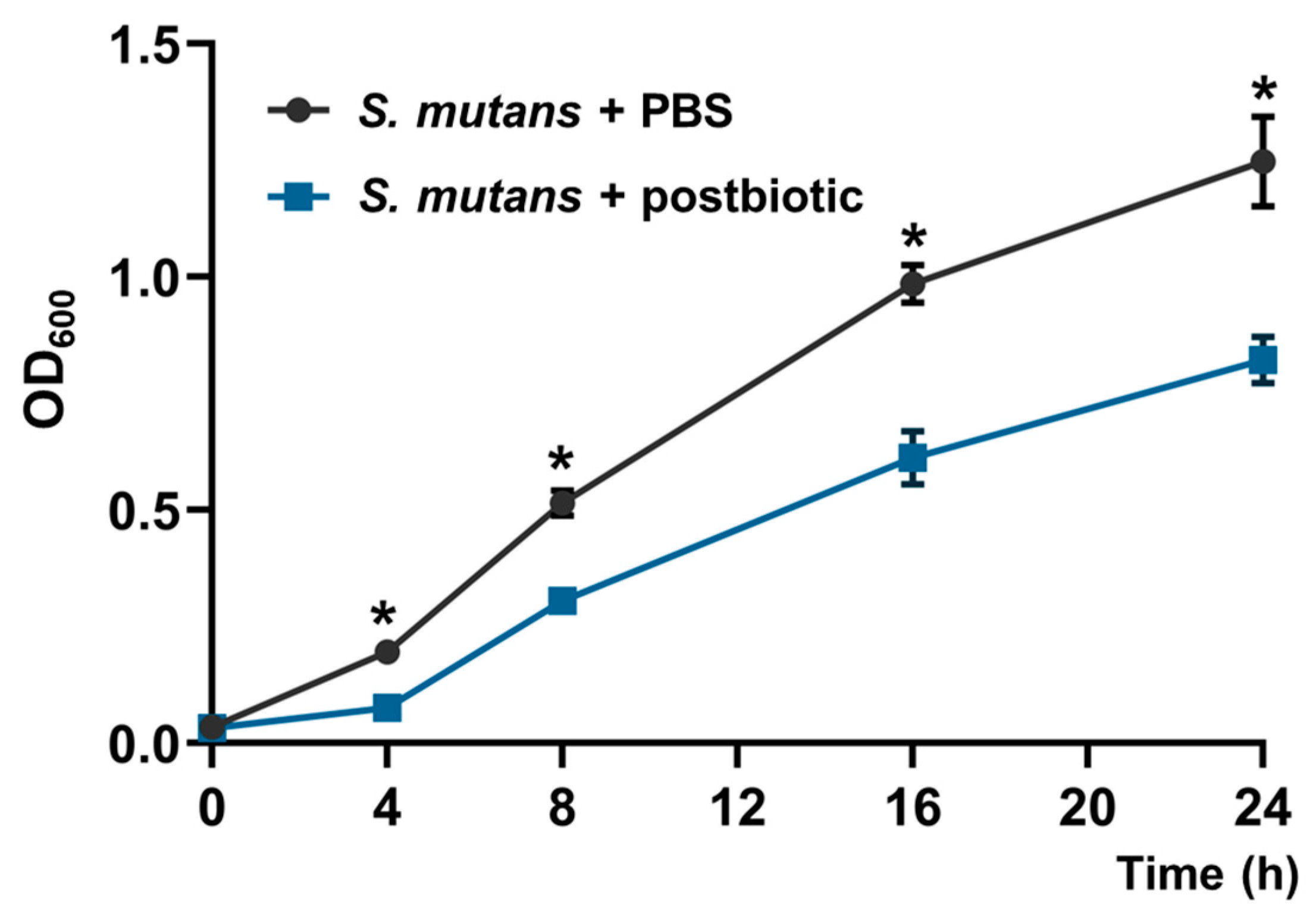
2.3. Effect on Planktonic Cells of S. mutans

Aliquots (250 µL) of L. rhamnosus LR-32 conditioned medium (prepared as described in item 2.1) or PBS and 250 µL of 108 CFU/mL of S. mutans UA130 (ATCC 700611) were inoculated into 5 mL of BHI broth with 0.12% glucose. Then, bacterial growth was monitored at time zero and in regular intervals within 24 h, in triplicate, determining the optical density at 600 nm in a spectrophotometer.

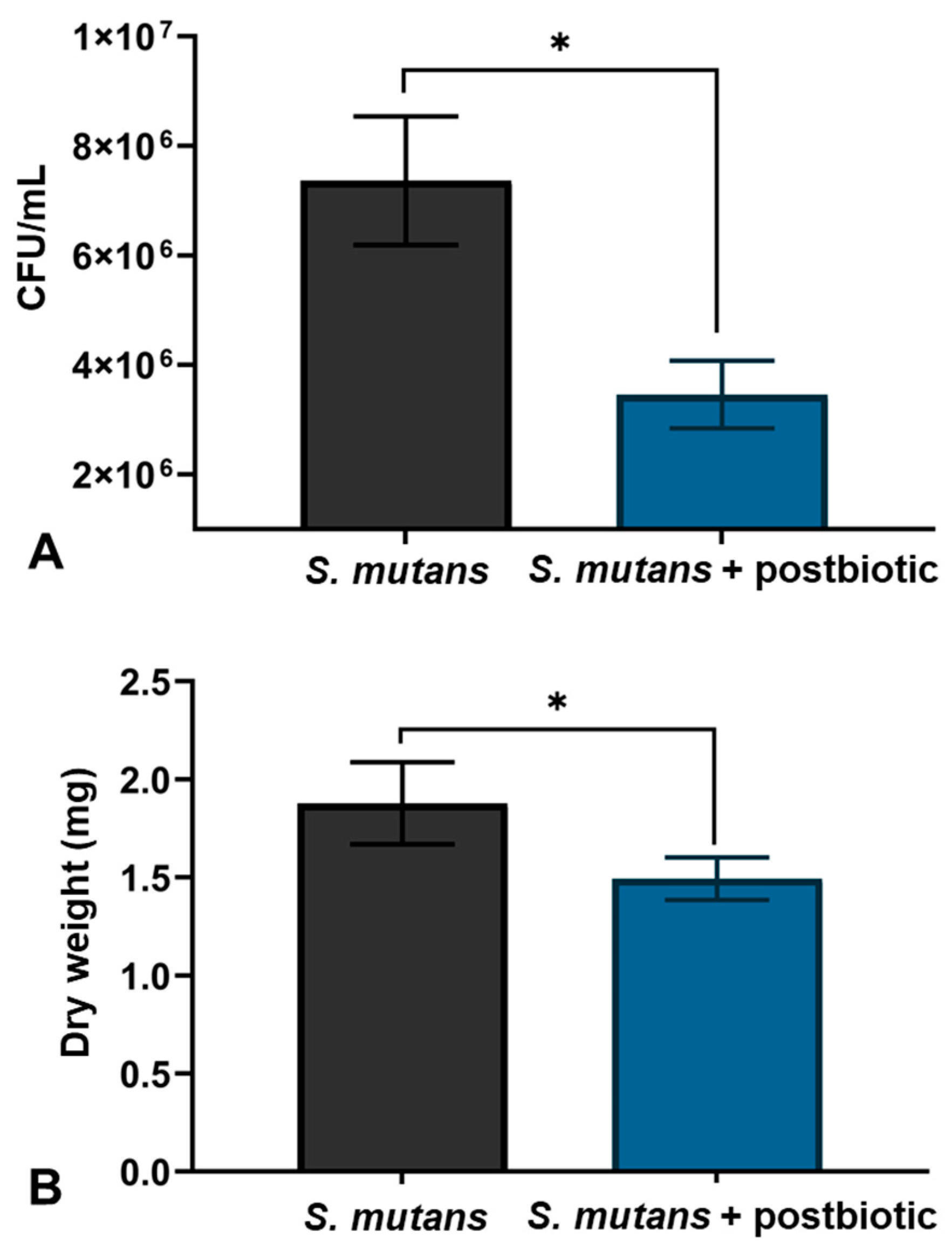
2.4. Effect on Biofilm of S. mutans

S. mutans UA130 was cultured in tubes containing BHI broth plus 1% sucrose for 18 h. After this period, the suspension was vortexed, and 2 mL (approximately 1 × 10⁶ CFU/mL) was dispensed in 24-well plates on hydroxyapatite (HA) disks (2.93 cm2; Clarkson Chromatography Products, Inc., South Williamsport, PA, USA) for 8 h, for initial bacterial adhesion. Previously, the disks were incubated with clarified saliva and filtered on a 0.22 μm polyvinylidene fluoride (PVDF) filter for 1 h at 37 °C in 24-well plates.
Then, the discs were gently washed with sterile 0.9% NaCl saline solution and the culture medium BHI broth plus 1% sucrose, containing the conditioned medium of L. rhamnosus LR-32 (prepared as described in item 2.1) at a dilution of 1:4 (culture medium: conditioned medium), was added to the S. mutans biofilms adhered to the discs. Untreated biofilms were considered as the control group. The plates were incubated in microaerophilic condition at 37 °C for 24 h.
At the end of the experiment, the biofilms were gently washed with saline solution, and collected to determine viable bacteria, dry weight, and gene expression by RT-qPCR. The experiment was carried out in triplicate, and within each experiment, four biofilm samples were used for each of the analyses, described as follows.

2.4.1. Determination of Live Bacteria Count

S. mutans biofilm samples were added with saline solution, at a proportion of 1 mL/mg of wet weight, homogenized in a vortex tube shaker and sonicated at 40 W, 5% amplitude, 6 pulses of 10 s each. From this suspension, dilutions with saline solution were made in the proportion of 10−1 to 10−5 times and inoculated on the BHI agar, to count total bacteria. Colony-forming units (CFUs) were counted, and the results were expressed as CFU/mg of wet biofilm.

2.4.2. Determination of Dry Weight of Biofilms

Biofilm samples were dried in a desiccator containing phosphorus pentoxide for 24 h, and the dry weight of the biofilm was determined in microtubes previously weighed on a precision analytical balance.

2.4.3. Effect of Postbiotics on Gene Expression of S. mutans

Glucan production by S. mutans was analyzed indirectly using gtfs expression comparison. Furthermore, the expression of the vicR gene (vic, for virulence control) was determined. Biofilms were washed three times with PBS. Total RNA was then isolated with a TRIzol® Max Bacterial RNA Isolation Kit (Invitrogen Life Tech, Carlsbad, CA, USA) according to the manufacturer’s instructions. The cDNA was synthesized by mixing total RNA (1 mg) and Maxime™ RT Premix (random primer; INTON, Gyeonggi, Republic of Korea) in a 20 μL reaction volume and by incubating the mixture at 45 °C for 1 h. The cDNAs were mixed with 10 μL of SYBR PremixExTaq and 0.4 μM of each primer pair in 20 μL final volume and subjected to 40 cycles of PCR (95 °C for 15 s, 60 °C for 15 s, and 72 °C for 33 s) with real-time PCR model StepOne Plus (Thermo Fisher, Foster City, CA, USA). PCR products were investigated for each amplification product using an amplification dissociation curve. The 16S rRNA gene (housekeeping gene) was used as a reference to normalize expression levels and to quantify changes in gtfs and VicR expression between samples treated with postbiotics and untreated samples. The primer sequences for real-time RT-PCR are described in Table 1.

2.5. Statistical Analysis

Experiments were carried out in triplicate with four replications within each experiment. The data generated were analyzed by one-way analysis of variance (ANOVA) and Tukey’s post hoc test using the Graphpad Prism 9.02 program (Graphpad, La Jolla, CA, USA). The results were considered significant for p-values lower than 0.05.

3. Results

3.1. Chemical Characterization

The chromatographic profile of the L. rhamnosus LR-32 postbiotic is presented in Figure 1. The majority of species in the sample are silanol (03), citric acid (10), and β-D-glucopyranose (13), thus exerting a greater influence on the characteristics of the postbiotic. In addition to these, other compounds present were α-D-galactopyranose, lactose, and maltose, showing the great abundance of sugars in the sample.
Table 2 presents the chemical composition of L. rhamnosus LR-32 postbiotic, silylated in BSTFA and analyzed by using GC–MS. In the chromatogram, a total of 17 (seventeen) compounds were identified, from various chemical classes, including 5 (five) sugars, 2 (two) amino acids, 1 (one) vitamin, and 3 (three) acids, among others.

3.2. Effect of the Postbiotics on S. mutans

Figure 2 shows the effect of L. rhamnosus LR-32 postbiotic on the growth of S. mutans determined by optical density (600 nm) over 24 h. When analyzing the growth of planktonic bacteria, an attenuation was observed in comparison to the control group containing PBS, showing statistical differences in the periods of 4 h, 8 h, 16 h, and 24 h.
Figure 3A shows the count of viable bacteria in S. mutans biofilms formed over 48 h (CFU/mL). There was less bacterial colonization in formed biofilms in which the postbiotic was added, showing a significant difference when compared to the control group (untreated biofilms).
Figure 3B shows the dry weight of S. mutans biofilms formed over 48 h of treatment and after being dried for 24 h in a desiccator with phosphorus pentoxide (mg). The dry weight of the biofilms that were treated with the conditioned medium was lower, with a statistically significant difference in comparison to the control group.
Figure 4 shows the relative gene expression (mean ± SD of foldchange) of the gtfB, gtfC, gtfD, and VicR genes of S. mutans by RT-PCR. Lower gene expression can be observed in all genes analyzed with a statistically significant difference in the groups treated with the postbiotic when compared to the control group.

4. Discussion

This study determined the composition of a postbiotic derived from L. rhamnosus LR-32 and its effect on the development of S. mutans biofilms in vitro. The results indicate a reduction in the development of these biofilms by analyzing the following parameters: viability, dry weight, and expression of genes associated with the virulence of that cariogenic bacterium. This result may be due to the presence of exo-metabolites in the analyzed postbiotic, such as acids and antimicrobial products like hydrogen peroxide [25]. When observing the majority of compounds (Table 2), there is a certain abundance of organic acids, peptides, and amino acids, corroborating the studies by Hossain et al. [26] and Moradi et al. [18], where the same classes of compounds were found.
It is important to mention that there may still be other types of compounds; furthermore, the loss or modification of metabolites when going through purification and/or detection processes cannot be ruled out [26]. It is known that L. rhamnosus LR-32 can produce hydrogen peroxide [22]; however, in the freeze-drying process of the conditioned medium, materials such as hydrogen peroxide and oxygen metabolites lose their antimicrobial activity [27]. When comparing the chemical profile of the postbiotic studied with other postbiotics like the ones derived from Lactobacillus spp., a diversity of profiles is observed, which can be explained by the type of bacteria, the analysis methods (GC–MS or HPLC), and the nature of the conditioned medium [18].
In the present study, the presence of Silanol (17.98%) in the postbiotic was observed, which may be related to the reduction in CFU of S. mutans (Figure 3), since, similarly, a reduction in CFUs of E. coli (Gram-negative) and S. aureus (Gram-positive) was observed in a study that analyzed the surface potential of biomaterials with silanol groups to prevent the formation of bacterial biofilm [28]. Furthermore, regarding biofilm formation prevention, that study demonstrated that silanol on the surface of the biomaterial showed an effect only on the S. aureus biofilm, suggesting an effect only on Gram-positive bacteria, such as S. mutans.
The presence of citric acid, another abundant compound in the postbiotic evaluated, may also be related to the reduction in the number of viable bacteria and is considered a safe disinfectant. A study conducted evaluated the effectiveness of citric acid and sodium hypochlorite as disinfectants against the bacteria Mycoplasma bovis; this study demonstrated that a concentration of 0.5% citric acid significantly reduces the infectivity of such bacteria [29]. Moreover, the antimicrobial effect of using citric acid, which occurs through membrane damage and loss of viability, at concentrations of 1% and 10% at pH 9.5 on E. coli and K. aerogenes was shown in another study [30].
The decreased growth of S. mutans in this study may also have been caused by organic acids produced by L. rhamnosus present in the conditioned medium. Lactic acid bacteria can produce a series of growth inhibitors, such as organic acids, bacteriocins, and hydrogen peroxide, which have an effect mainly against Gram-positive pathogens [31]. Additionally, probiotic bacteria can inhibit the growth of S. mutans, especially when looking at the effect of Lactobacillus paracasei, L. plantarum, and L. rhamnosus, which present the best results against this microorganism [32]. It was also observed that culture supernatants of L. rhamnosus GG, L. plantarum 299V, L. reuteri PTA 5289, and SD2112 reduced the viability of S. mutans [33], and the antimicrobial activity depended on the acidity level of the lactobacilli culture, which may explain the results of our study since the postbiotic used was not neutralized.
The dry weight of biofilms was reduced when exposed to the postbiotic, which can be highlighted by the lower number of viable bacteria, but also by the decrease in the polysaccharide matrix of the S. mutans biofilm. That matrix is composed of extracellular polysaccharides that are produced from the fragmentation of the sucrose molecule by enzymes—glucosyltransferases—produced by the cariogenic bacteria. These enzymes are responsible for the production of glucans from the glucose molecule originating from sucrose, which is responsible for producing acids and making the biofilm more cariogenic [34]. The decrease observed in the polysaccharide matrix, resulting from the decrease in the production of insoluble glucans due to the conditioned medium, was previously described [23] and corroborates with decreases observed in the present study.
The ability of S. mutans to produce extracellular glucans from sucrose using glucosyltransferases (gtfs) is considered a virulence factor of this bacterium [35]. For that species, there are three well-described glucosyltransferases. The gtfB gene product provides cellular aggregation and cohesion of different bacterial strains, enabling bacteria to produce glucans. The gtfC is the gene most related to the acquired pellicle and is the one with the greatest capacity for adhesion to hydroxyapatite among the three gtfs. Glucosyltransferase-D generates water-soluble glucans that can be metabolized or stored by bacterial strains. In addition to these, the vicRK gene exerts a self-protective function in response to external stress, being related to the signal transduction system; this gene also modulates gene expression in response to acid tolerance and oxidative stress [24].
As shown in Figure 4, a lower gene expression can be observed in all analyzed genes (gtfB, gtfC, gtfD, and vicR) when RT-PCR was performed, showing a negative regulation when compared to the control group. A similar result was observed in a study where the biosurfactant produced by the analyzed postbiotic significantly decreased the gene expression of gtfB and gtfC in S. mutans biofilms [35]. Similarly, bacterial lysates (BLs), another type of postbiotic, of Lactobacillus plantarum and L. rhamnosus GG downregulated gtfB, gtfC, and gtfD by S. mutans, suggesting that BLs reduce the synthesis of extracellular polysaccharide and thereby reduce S. mutans biofilm, but did not eradicate the established S. mutans biofilm [36].
This research has the limitations of an in vitro study, and the results must be interpreted with caution since the biofilm formed in the oral cavity is much more complex than the single-specie model used in the present research. Moreover, although postbiotics are considered superior to probiotics because of their good acid–base and thermal stability, their ease of storage and use, and high safety [19], further research should be carried out with other types of postbiotics (peptidoglycans, lipopolysaccharides, and pili) to test the safety and effectiveness of these formulations in the oral cavity.

5. Conclusions

The postbiotic from L. rhamnosus LR-32, characterized by 17 diverse chemical compounds, primarily citric acid, silanol, and β-D-glucopyranose, exhibited notable inhibitory effects on S. mutans growth in both planktonic and biofilm states in vitro. This intervention reduced biofilm dry weight and downregulated essential genes (gtfB, gtfC, gtfD, vicR). These findings underscore the postbiotic’s potential in countering S. mutans and its biofilm development, offering promising implications for future oral health strategies of antimicrobial management.

Author Contributions

Conceptualization, G.B.S. and G.C.V.; methodology, G.B.S., G.C.V. and S.G.d.L.; formal analysis, G.B.S., G.C.V., P.V.Q. and S.G.d.L.; investigation, G.B.S., E.R.d.S.N. and G.C.V.; resources, G.C.V.; data curation, G.B.S., G.C.V. and P.V.Q.; writing—original draft preparation, G.B.S., P.V.Q. and G.C.V.; writing—review and editing, G.B.S., P.V.Q., G.C.V. and S.G.d.L.; supervision, G.C.V.; project administration, G.C.V.; funding acquisition, G.C.V. All authors have read and agreed to the published version of the manuscript.

Funding

This research was funded by CNPq (process nº421385/2023-5) and FAPEPI Ed. 08/2018 PRONEM.

Data Availability Statement

The raw data supporting the conclusions of this article will be made available by the authors on request.

Conflicts of Interest

The authors declare no conflicts of interest.

References

  1. Hill, C.; Guarner, F.; Reid, G.; Gibson, G.R.; Merenstein, D.J.; Pot, B.; Morelli, L.; Canani, R.B.; Flint, H.J.; Salminen, S.; et al. Expert consensus document. The International Scientific Association for Probiotics and Prebiotics consensus statement on the scope and appropriate use of the term probiotic. Nat. Rev. Gastroenterol. Hepatol. 2014, 11, 506–514. [Google Scholar] [CrossRef]
  2. Cicenia, A.; Scirocco, A.; Carabotti, M.; Pallotta, L.; Marignani, M.; Severi, C. Postbiotic activities of lactobacilli-derived factors. J. Clin. Gastroenterol. 2014, 48 (Suppl. S1), S18–S22. [Google Scholar] [CrossRef] [PubMed]
  3. Fayol-Messaoudi, D.; Berger, C.N.; Coconnier-Polter, M.H.; Liévin-Le Moal, V.; Servin, A.L. pH-, Lactic acid-, and non-lactic acid-dependent activities of probiotic Lactobacilli against Salmonella enterica Serovar Typhimurium. Appl. Environ. Microbiol. 2005, 71, 6008–6013. [Google Scholar] [CrossRef] [PubMed]
  4. Memorandum, D.T. Lactobacillus rhamnosus, 2008; Volume 32.
  5. Shen, S.; Ren, F.; Qin, H.; Bukhari, I.; Yang, J.; Gao, D.; Ouwehand, A.C.; Lehtinen, M.J.; Zheng, P.; Mi, Y. Lactobacillus acidophilus NCFM and Lactiplantibacillus plantarum Lp-115 inhibit Helicobacter pylori colonization and gastric inflammation in a murine model. Front. Cell Infect. Microbiol. 2023, 13, 1196084. [Google Scholar] [CrossRef] [PubMed]
  6. Alanzi, A.; Honkala, S.; Honkala, E.; Varghese, A.; Tolvanen, M.; Söderling, E. Effect of Lactobacillus rhamnosus and Bifidobacterium lactis on gingival health, dental plaque, and periodontopathogens in adolescents: A randomised placebocontrolled clinical trial. Benef. Microbes 2018, 9, 593–602. [Google Scholar] [CrossRef] [PubMed]
  7. Cagetti, M.G.; Mastroberardino, S.; Milia, E.; Cocco, F.; Lingström, P.; Campus, G. The use of probiotic strains in caries prevention: A systematic review. Nutrients 2013, 5, 2530–2550. [Google Scholar] [CrossRef] [PubMed]
  8. Keller, M.K.; Hasslöf, P.; Stecksén-Blicks, C.; Twetman, S. Co-aggregation and growth inhibition of probiotic lactobacilli and clinical isolates of mutans streptococci: An in vitro study. Acta Odontol. Scand. 2011, 69, 263–268. [Google Scholar] [CrossRef] [PubMed]
  9. Luo, S.C.; Wei, S.M.; Luo, X.T.; Yang, Q.Q.; Wong, K.H.; Cheung, P.C.K.; Zhang, B.B. How probiotics, prebiotics, synbiotics, and postbiotics prevent dental caries: An oral microbiota perspective. NPJ Biofilms Microbiomes 2024, 10, 14. [Google Scholar] [CrossRef] [PubMed]
  10. Vale, G.C.; Mayer, M.P.A. Effect of probiotic Lactobacillus rhamnosus by-products on gingival epithelial cells challenged with Porphyromonas gingivalis. Arch. Oral. Biol. 2021, 128, 105174. [Google Scholar] [CrossRef]
  11. Laleman, I.; Detailleur, V.; Slot, D.E.; Slomka, V.; Quirynen, M.; Teughels, W. Probiotics reduce mutans streptococci counts in humans: A systematic review and meta-analysis. Clin. Oral. Investig. 2014, 18, 1539–1552. [Google Scholar] [CrossRef]
  12. Liévin-Le Moal, V.; Servin, A.L. Anti-infective activities of lactobacillus strains in the human intestinal microbiota: From probiotics to gastrointestinal anti-infectious biotherapeutic agents. Clin. Microbiol. Rev. 2014, 27, 167–199. [Google Scholar] [CrossRef] [PubMed]
  13. Miyazima, T.Y.; Ishikawa, K.H.; Mayer, M.; Saad, S.; Nakamae, A. Cheese supplemented with probiotics reduced the Candida levels in denture wearers-RCT. Oral. Dis. 2017, 23, 919–925. [Google Scholar] [CrossRef] [PubMed]
  14. Tan, Y.; Leonhard, M.; Moser, D.; Schneider-Stickler, B. Inhibition activity of Lactobacilli supernatant against fungal-bacterial multispecies biofilms on silicone. Microb. Pathog. 2017, 113, 197–201. [Google Scholar] [CrossRef] [PubMed]
  15. Aguilar-Toalá, J.E.; Hall, F.G.; Urbizo-Reyes, U.C.; Garcia, H.S.; Vallejo-Cordoba, B.; González-Córdova, A.F.; Hernández-Mendoza, A.; Liceaga, A.M. In Silico Prediction and In Vitro Assessment of Multifunctional Properties of Postbiotics Obtained From Two Probiotic Bacteria. Probiotics Antimicrob. Proteins 2020, 12, 608–622. [Google Scholar] [CrossRef] [PubMed]
  16. Liang, B.; Xing, D. The Current and Future Perspectives of Postbiotics. Probiotics Antimicro. Prot. 2023, 15, 1626–1643. [Google Scholar] [CrossRef] [PubMed]
  17. Ishikawa, K.H.; Bueno, M.R.; Kawamoto, D.; Simionato, M.R.L.; Mayer, M.P.A. Lactobacilli postbiotics reduce biofilm formation and alter transcription of virulence genes of Aggregatibacter actinomycetemcomitans. Mol. Oral. Microbiol. 2021, 36, 92–102. [Google Scholar] [CrossRef] [PubMed]
  18. Moradi, M.; Mardani, K.; Tajik, H. Characterization and application of postbiotics of Lactobacillus spp. on Listeria monocytogenes in vitro and in food models. Lebensm.-Wiss. Technol. 2019, 111, 457–464. [Google Scholar] [CrossRef]
  19. Salminen, S.; Collado, M.C.; Endo, A.; Hill, C.; Lebeer, S.; Quigley, E.M.M.; Sanders, M.E.; Shamir, R.; Swann, J.R.; Szajewska, H.; et al. The International Scientific Association of Probiotics and Prebiotics (ISAPP) consensus statement on the definition and scope of postbiotics. Nat. Rev. Gastroenterol. Hepatol. 2021, 18, 649–667. [Google Scholar] [CrossRef] [PubMed]
  20. Matsubara, V.H.; Wang, Y.; Bandara, H.M.H.N.; Mayer, M.P.A.; Samaranayake, L.P. Probiotic lactobacilli inhibit early stages of Candida albicans biofilm development by reducing their growth, cell adhesion, and filamentation. Appl. Microbiol. Biotech. 2016, 100, 6415–6426. [Google Scholar] [CrossRef]
  21. Koohestani, M.; Moradi, M.; Tajik, H.; Badali, A. Effects of cellfree supernatant of Lactobacillus acidophilus LA5 and Lactobacillus casei 431 against planktonic form and biofilm of Staphylococcus aureus. Vet. Res. Forum 2018, 9, 301–306. [Google Scholar]
  22. Ishikawa, K.H.; Mita, D.; Kawamoto, D.; Nicoli, J.R.; Albuquerque-Souza, E.; Lorenzetti, M.R.; Mayer, M.P.A. Probiotics alter biofilm formation and the transcription of Porphyromonas gingivalis virulence-associated genes. J. Oral. Microbiol. 2020, 20, 1805553. [Google Scholar] [CrossRef] [PubMed]
  23. Lee, S.; Kim, Y. A comparative study of the effect of probiotics on cariogenic biofilm model for preventing dental caries. Arch. Microbiol. 2014, 196, 601–609. [Google Scholar] [CrossRef] [PubMed]
  24. Wu, J.; Fan, Y.; Wang, X.; Jiang, X.; Zou, J.; Huang, R. Effects of the natural compound, oxyresveratrol, on the growth of Streptococcus mutans, and on biofilm formation, acid production, and virulence gene expression. Eur. J. Oral. Sci. 2020, 128, 18–26. [Google Scholar] [CrossRef] [PubMed]
  25. Surendran, N.M.; Amalaradjou, M.A.; Venkitanarayanan, K. Antivirulence Properties of Probiotics in Combating Microbial Pathogenesis. Adv. Appl. Microbiol. 2017, 98, 1–29. [Google Scholar]
  26. Hossain, M.I.; Mizan, F.R.; Roy, P.K.; Nahar, S.; Toushik, S.H.; Ashrafudoulla, M.; Jahid, I.K.; Lee, J.; Ha, S. Listeria monocytogenes biofilm inhibition on food contact surfaces by application of postbiotics from Lactobacillus curvatus B.67 and Lactobacillus plantarum M.2. Food Res. Int. 2021, 148, 110595. [Google Scholar] [CrossRef] [PubMed]
  27. Rodríguez, J.M.; Martínez, M.I.; Suárez, A.M.; Martínez, J.M.; Hernández, P.E. Research note: Unsuitability of the MRS medium for the screening of hydrogen peroxide-producing lactic acid bacteria. Lett. Appl. Microbiol. 1997, 25, 73–74. [Google Scholar] [CrossRef] [PubMed]
  28. Rodrigues, A.I.; Franco, A.R.; Rodrigues, F.J.; Leonor, I.B.; Reis, R.L. Unveiling the effect of three-dimensional bioactive fibre mesh scaffolds functionalized with silanol groups on bacteria growth. J. Biomed. Mater. Res. A 2016, 104, 2189–2199. [Google Scholar] [CrossRef]
  29. Mahdizadeh, S.; Sawford, K.; van Andel, M.; Browning, G.F. Efficacy of citric acid and sodium hypochlorite as disinfectants against Mycoplasma bovis. Vet. Microbiol. 2020, 243, 108630. [Google Scholar] [CrossRef]
  30. Burel, C.; Kala, A.; Purevdorj-Gage, L. Impact of pH on citric acid antimicrobial activity against Gram-negative bacteria. Lett. Appl. Microbiol. 2021, 72, 332–340. [Google Scholar] [CrossRef]
  31. Diop, M.B.; Alvarez, V.B.; Guiro, A.T.; Thonart, P. Efficiency of Neutralized Antibacterial Culture Supernatant from Bacteriocinogenic Lactic Acid Bacteria Supplemented With Salt in Control of Microorganisms Present in Senegalese Artisanally Handled Fish by Immersion Preservative Technology During Guedj Seafood Processing at 10 °C and 30 °C. J. Food Microbiol. Saf. Hyg. 2016, 1, 1. [Google Scholar] [CrossRef]
  32. Lin, X.; Chen, X.; Chen, Y.; Jiang, W.; Chen, H. The effect of five probiotic lactobacilli strains on the growth and biofilm formation of Streptococcus mutans. Oral. Dis. 2015, 21, 128–134. [Google Scholar] [CrossRef] [PubMed]
  33. Söderling, E.M.; Marttinen, A.M.; Haukioja, A.L. Probiotic lactobacilli interfere with Streptococcus mutans biofilm formation in vitro. Curr. Microbiol. 2011, 62, 618–622. [Google Scholar] [CrossRef] [PubMed]
  34. Bowen, W.H.; Koo, H. Biology of Streptococcus mutans—Derived glucosyltransferases: Role in extracellular matrix formation of cariogenic biofilms. Caries Res. 2011, 45, 69–86. [Google Scholar] [CrossRef] [PubMed]
  35. Tahmourespour, A.; Salehi, R.; Kermanshahi, R.K. Lactobacillus Acidophilus-Derived Biosurfactant Effect on GTFB and GTFC Expression Level in Streptococcus mutans Biofilm Cells. Braz. J. Microbiol. 2011, 42, 330–339. [Google Scholar] [CrossRef]
  36. Kim, M.Y.; Hyun, I.K.; An, S.; Kim, D.; Kim, K.H.; Kang, S.S. In vitro anti-inflammatory and antibiofilm activities of bacterial lysates from lactobacilli against oral pathogenic bacteria. Food Funct. 2022, 13, 12755–12765. [Google Scholar] [CrossRef]
Figure 1. Chromatographic profile of L. rhamnosus LR-32 postbiotic that was silylated in BSTFA and analyzed by GC–MS.
Microorganisms 12 00843 g001
Figure 2. Effect of L. rhamnosus LR-32 postbiotic on the growth of S. mutans determined by optical density (OD at 600 nm); * p < 0.05.
Microorganisms 12 00843 g002
Figure 3. (A) Count of viable bacteria in S. mutans biofilms (CFU/mL) formed with and without the postbiotic L. rhamnosus LR-32. (B) Dry weight of S. mutans biofilms formed (mg) with and without the postbiotic L. rhamnosus LR-32. The vertical bars indicate the standard deviation; * p < 0.05.
Microorganisms 12 00843 g003
Figure 4. Relative gene expression (mean ± SD of foldchange) of the gtfB, gtfC, gtfD, and VicR genes of S. mutans with and without treatment with the postbiotic of L. rhamnosus LR-32 by RT-PCR; * p < 0.05.
Microorganisms 12 00843 g004
Table 1. Sequences of the primers used in the study.
GeneDescriptionPrimer Sequence (5′-3′)Reference
gtfBProduction of water-insoluble glucan5′-AGC AAT GCA GCC AAT CTA CAA AT-3′
5′-ACG AAC TTT GCC GTT ATT GTC A-3′
[23]
gtfCProduction of water-soluble and insoluble glucan5′-CTC AAC CAA CCG CCA CTG TT-3′
5′-GGT TAA CGT CAA TAG AAT CTG TAG TAG C-3′
[23]
gtfDProduction of water-soluble glucan5′-CAC AGG CAA AAG CTG AAT TAC A-3′
5′-GAT GGC CGC TAA GTC AAC AG-3′
[23]
vicRResponse Regulator5′-TGA CACGA TTA CAG CCT TTG ATG-3′
5′-CGT CTA GTT CTG GTA ACA TTA AGT CCA ATA-3′
[24]
16S rRNAInternal standard normalizer5′-AGT CTG AGT 5′-GAA TAA AAG GCT A-3′
5′-GTT AGC TCC GGC ACT AAG CC-3′
[23]
Table 2. Chemical composition of L. rhamnosus LR-32 postbiotic that was silylated in BSTFA and analyzed by GC–MS.
RTSimilarityConstituentMolecular FormulaAbsolute
Area
Relative
Area (%)
110.11996Trimethylsilyl Ester of L-valineC11H27NO2Si22,565,5041.59%
215.08294Pentanoic acid C5H10O2712,9000.44%
316.33694Silanol, Phosphoric acid tri-TMSC9H27O4PSi328,993,17817.98%
416.492943,7-Dioxa-2,8-disilanonaneC9H24O2Si23,325,7452.06%
517.05695Trimethylsilyl ester of O-trimethylsilyl-L-ThreonineC10H25NO3Si22,381,2191.48%
617.82195Butanedioic acidC4H6O41,288,8290.80%
724.22894L-Proline, Pyroglutamic acid di-TMSC5H7NO34,212,7012.61%
824.97895Phenylalanine 1TMSC12H19NO2Si2,606,5981.62%
931.26790Phosphoric acid, α.-GlycerC3H9O6P428,8720.27%
1032.76887Citric acidC6H8O731,491,76819.53%
1133.37290Glucofuranoside, methyl 2,3,5,6-tetrakis-O-(trimethylsilyl)C19H46O6Si46,408,4263.98%
1234.59196α-D-GalactopyranoseC6H12O621,217,49013.16%
1336.84496β-D-GlucopyranoseC6H12O628,402,98317.62%
1439.25591Myo-inositolC6H12O61,018,4040.63%
1541.64890Tryptophan 2TMSC17H28N2O2Si2724,7740.45%
1650.70594Lactose-OctarmsC36H86O11Si812,470,9227.74%
1752.99492Maltose-OctarmsC36H86O11Si812,963,5188.04%
Disclaimer/Publisher’s Note: The statements, opinions and data contained in all publications are solely those of the individual author(s) and contributor(s) and not of MDPI and/or the editor(s). MDPI and/or the editor(s) disclaim responsibility for any injury to people or property resulting from any ideas, methods, instructions or products referred to in the content.

