Article

Association of Human Papilloma Virus, Cytomegalovirus, and Epstein–Barr Virus with Breast Cancer in Jordanian Women

1
Department of Microbiology, Pathology, and Forensic Medicine, Faculty of Medicine, The Hashemite University, Zarqa 13133, Jordan
2
Department of Anatomy, Physiology & Biochemistry, Faculty of Medicine, The Hashemite University, Zarqa 13133, Jordan
3
Department of Pharmacology and Public Health, Faculty of Medicine, The Hashemite University, Zarqa 13133, Jordan
4
Department of Pathology and Microbiology, Faculty of Medicine, Jordan University of Science and Technology, Irbid 22110, Jordan
5
Department of Pharmacology and Toxicology, College of Pharmacy, King Saud University, Riyadh 11421, Saudi Arabia
*
Authors to whom correspondence should be addressed.
Submission received: 22 March 2024 / Revised: 9 April 2024 / Accepted: 23 April 2024 / Published: 25 April 2024
(This article belongs to the Section Infectious Disease)

Abstract

:
Background and Objectives: The investigation of oncogenic viruses and their potential association with breast cancer (BC) remains an intriguing area of study. The current work aims to assess evidence of three specific viruses, human papillomavirus (HPV), cytomegalovirus (CMV), and Epstein–Barr virus (EBV) in BC samples and to explore their relationship with relevant clinicopathological variables. Materials and Methods: The analysis involved BC samples from 110 Jordanian female patients diagnosed with BC and breast tissue samples from 30 control patients with no evidence of breast malignancy, investigated using real-time PCR. The findings were then correlated with various clinico-pathological characteristics of BC. Results: HPV was detected in 27 (24.5%), CMV in 15 (13.6%), and EBV in 18 (16.4%) BC patients. None of the control samples was positive for HPV or CMV while EBV was detected in only one (3.3%) sample. While (HPV/EBV), (HPV/CMV), and (EBV/CMV) co-infections were reported in 1.8%, 2.7%, and 5.5%, respectively, coinfection with the three viruses (HPV/CMV/EBV) was not reported in our cohort. A statistically significant association was observed between HPV status and age (p = 0.047), and between clinical stage and CMV infection (p = 0.015). Conclusions: Our findings indicate the presence or co-presence of HPV, CMV, and EBV in the BC subpopulation, suggesting a potential role in its development and/or progression. Further investigation is required to elucidate the underlying mechanisms that account for the exact role of oncoviruses in breast carcinogenesis.

1. Introduction

According to the World Health Organization (WHO), breast cancer (BC) was the most common cancer on the global level and the leading cause of female cancer deaths in 2020 [1]. In the Arab world, Jordan is among the top ten countries in BC incidence and death rates [2], with BC being the most diagnosed female malignancy (38.4%) [3]. Subsequently, a thorough understanding of BC’s etiology, molecular pathways, risk factors, and health outcomes is essential.
The development of BC is multifactorial, with a set of proposed contributing elements that can increase its risk including age, smoking, alcohol intake, genetic predisposition, and environmental factors, particularly viral infections [4]. It is hypothesized that several types of viruses can infect breast epithelial cells and become dormant, causing what is called a “latent infection”. These types of infections contribute to cancer development through multiple molecular pathways. For example, a virus can inhibit apoptosis in the infected cell [5], it can integrate its DNA in an oncogenic region of the cell’s DNA [6], activate telomerase, which contributes to cellular immortality [7], or induce mitosis in fully differentiated cells leading to their uncontrollable division [8]. Moreover, the effect of many oncogenic viruses is propagated through affecting anti-oncogenic proteins and interfering with the function of cell cycle regulators such as p53, RAS, retinoblastoma protein (Rb), and many others [8,9]. In addition, viruses can contribute to cancer development through invading the immune system and interfering with the elimination of viral particles and virus-infected cells [10,11].
Several viruses fall into this category including the Epstein–Barr virus (EBV) [12], the human papilloma virus (HPV) [13], the cytomegalovirus (CMV) [14], and others [15,16]. These viruses are linked to various cancers, including neuroblastoma, B cell lymphomas, and cervical and oropharyngeal cancers [17,18,19,20]. The association between being infected with these viruses and the occurrence of BC is still a matter of active research with no conclusive evidence. Worldwide, chronic infections (both viral and bacterial) are the cause of a fifth of cancer cases, and in developing countries, they are responsible for 20% of cancer-related deaths [21]. Several studies in our region showed a positive correlation between viral infections and BC in Iran, Pakistan, Syria, and Sudan [22,23,24,25], while other studies have rejected this causal relationship [26,27], indicating that a role for viral infections and the pathogenesis of BC is still unresolved.
In Jordan, only a single published study has evaluated the association between BC and viral infections such as EBV and HPV [28]. The aim of this study was to further investigate evidence of HPV, EBV, and CMV in BC samples obtained from a Jordanian female subpopulation.

2. Materials and Methods

2.1. Sample Collection

A retrospective screening of the medical records of King Abdullah University Hospital (KAUH) patients, diagnosed with BC between 2018 and 2022, was performed. The eligibility criteria for patient inclusion were as follows: patients with a histologically confirmed diagnosis of BC, aged between 20–80 years, who had not undergone radiotherapy or chemotherapy prior to tumor excision, and their samples tested positive for GAPDH (housekeeping gene) by PCR.
The samples comprised 120 formalin-fixed paraffin-embedded (FFPE) tissue blocks of BC along with 30 samples of non-malignant breast lesions as controls. Of those 120 BC samples, 10 samples were excluded from the analysis due to the unavailability of their molecular subtyping data. The samples were obtained from KAUH, Irbid, Jordan. All research activities within this project were carried out in accordance with ethical approval granted by the Hashemite University Institutional Review Board (IRB) (No. 7/14/2020/2021) and KAUH IRB (No. 42/151/2022).

2.2. DNA Extraction

FFPE breast tissue blocks were obtained from the KAUH Pathology Department for each patient. A 20 μm thick section was prepared from each block, and genomic DNA extraction was performed using the QIAamp DNA FFPE Tissue Kit (Qiagen, Hilden, Germany) following the manufacturer’s protocol. Briefly, the FFPE samples were deparaffinized through sequential washes with warmed (100%) xylene and (100%) ethanol to remove residual xylene. Following this, the samples were lysed using 20 μL proteinase K and ATL lysis buffer (Roche, Munich, Germany) and incubated for two hours at 56 °C initially, followed by an additional hour at 90 °C to reverse formalin crosslinking. Subsequently, 4 μL of RNAse (100 mg/mL) was added, and the mixture was incubated at room temperature for 2 min. Next, AL buffer and ethanol were added, mixed thoroughly, and the lysate containing DNA was transferred to the MinElute column for several washing steps. Finally, DNA was eluted in 200 μL ATE buffer and stored at −20 °C for subsequent experiments. The concentration of DNA was measured using a Qubit 3.0 Fluorometer (Thermo Fisher Scientific, Waltham, MA, USA) following the manufacturer’s instructions.

2.3. HPV Detection in Breast Cancer Samples

HPV detection and genotyping were conducted using the REALQUALITY RQ-Multi HPV detection kit (AB ANALITICA, Padua, Italy), as previously described [17]. This kit enables the identification of 28 HPV genotypes, including 14 high-risk (16, 18, 31, 33, 35, 39, 45, 51, 52, 56, 58, 59, 66, 68), 6 potential high-risk (26, 53, 67, 70, 73, 82), and 8 low-risk (6, 11, 40, 42, 43, 44, 55, 83) types. It amplifies a short sequence within the E6 and E7 genes of the HPV genome, individually detecting HPV 16 and HPV 18 genotypes, while other genotypes are detected as pools. Additionally, human beta-globin serves as an internal control. The kit includes both negative and positive controls, with the positive control containing plasmid DNA fragments of HPV 16, 18, and 33 for high-risk HPV, and fragments of HPV 6, 26, and 40 genotypes for low-risk HPV.
RT-PCR amplification was conducted using a Bioer RT-PCR thermocycler (China). The PCR mixture had a total volume of 25 μL, consisting of 22.5 μL of master mix and 2.5 μL of template DNA (3–15 ng/µL), positive control, or negative control. Thermocycling conditions included an initial step of 2 min at 50 °C for UNG activation, followed by initial denaturation at 95 °C for 10 min, then 45 cycles at 95 °C for 15 s, 60 °C for 60 s, and 72 °C for 60 s. Data were interpreted according to the manufacturers’ guidelines. Briefly, for HPV-negative samples, an amplification signal of the internal control (beta globin) with a Ct value ≤ 34 was regarded as appropriate, while values exceeding 34 were considered inadequate. Conversely, internal control values exceeding 34 were considered acceptable for HPV-positive samples, as per the manufacturer’s guidelines. Regarding viral target detection, any amplification signal demonstrating a proper sigmoid curve shape with a Ct value ≤ 40 was classified as positive.

2.4. EBV and CMV Detection in Breast Cancer Samples

To initially assess the quality of the extracted DNA samples, all samples underwent PCR using GAPDH primers. The GAPDH primers used were as follows: forward primer (GAPDH-F): 5′ GAGTCAACGGATTTGGTCGT, reverse primer (GAPDH-R): 5′ TTGATTTTGGAGGGATCTCG, generating a PCR product of 237 bp. Additionally, for the detection of human cytomegalovirus (HCMV), primers conserved for the GB region were utilized. The forward primer sequence was 5′-TCTGGGAAGCCTCGGAACG, and the reverse primer sequence was 5′-GAAACGCGCGGCAATCGG. To screen BC samples for the prevalence of the EBV-EBNA 2 gene, specific primers EBNA2F: 5′-AGGACAGCCGTTGCCCTAGTG and EBNA2R: 5′-TAGCGGACAAGCCGAATACCCT targeting the EBV-EBNA2 gene were utilized.
Initially, a conventional PCR was conducted to determine the optimal annealing temperature for each primer. Subsequently, the QuantiTect SYBR® Green PCR Kit (Cat. No. 204143, QIAGEN) was employed to amplify the target DNA for each sample using the aforementioned primers. In the conventional PCR protocol, each reaction included 12.5 µL of Taq DNA polymerase, 2 µL of template DNA, 8.5 µL of nuclease-free water, and 1 µL each for the forward and reverse primers per sample. Positive and negative controls were included in every run. The resulting PCR products were then loaded onto a 2% agarose gel, and the DNA bands were visualized and documented using a gel documentation system. After completion of the run, a UV light device was utilized to visualize the distinct DNA bands.
During the reaction setup, both the samples and all reagents from the QuantiTect SYBR® Green PCR Kit were kept on ice. Using filter tips, 12.5 μL of QuantiTect SYBR Green PCR Master Mix, 8.5 μL of RNase-free water, 2 µL of template DNA, and 1 µL each of the forward and reverse primers per sample were accurately added to the PCR tube, resulting in a total volume of 25 µL. Following this, the tubes underwent centrifugation at maximum speed and were then placed in the PCR device. The temperatures for the three PCR stages were set as follows: 95 °C for 15 s, 56 °C for 30 s, and 72 °C for 30 s. These stages were repeated for 40 cycles.

2.5. Statistical Analysis

Statistical analysis was conducted utilizing SPSS version 22. Variations in proportions were assessed using the Chi-square test. A significance level of p < 0.05 was chosen to determine statistical significance.

3. Results

3.1. Clinicopathological Features within the Study Population

The clinicopathological characteristics of the patient’s population, including estrogen receptor (ER), progesterone receptor (PR), and human epidermal growth factor receptor 2 (HER2), are summarized in Table 1. The final analysis comprised 110 tumor samples (10 samples were excluded from the analysis due to the unavailability of their molecular subtyping data), with the mean age of 55.7 years, and a standard deviation (SD) of ±11.6. There was a slightly larger number of patients under 56 years of age (n = 56, 51%) than patients older than 56 years of age (n = 54, 49%). Molecular subtyping of BC revealed that 66% were luminal A (ER+ and/or PR+ with HER2−), 19% were luminal B (ER+ and/or PR+ with HER2+), 6% were HER2+ positive only (ER and PR both negative), and 9% were triple-negative BC (TNBC). Additionally, clinical staging of the sample cohort showed that 44% were classified as stage I, 16% as stage II, 14% as stage III, and 26% as stage IV. Lymphovascular invasion was identified in over half of the samples (52%; 57/110), while lymph node involvement by cancerous cells was observed in 67% (74/110) of cases. These clinicopathological characteristics were selected because BC is characterized by its heterogeneity, encompassing various histological subtypes with unique clinicopathological profiles. The prognosis for these special histological types varies significantly, indicating profound heterogeneity within the disease. Importantly, the histological subtype serves as an independent prognostic indicator, emphasizing its crucial role in predicting outcomes for BC patients.

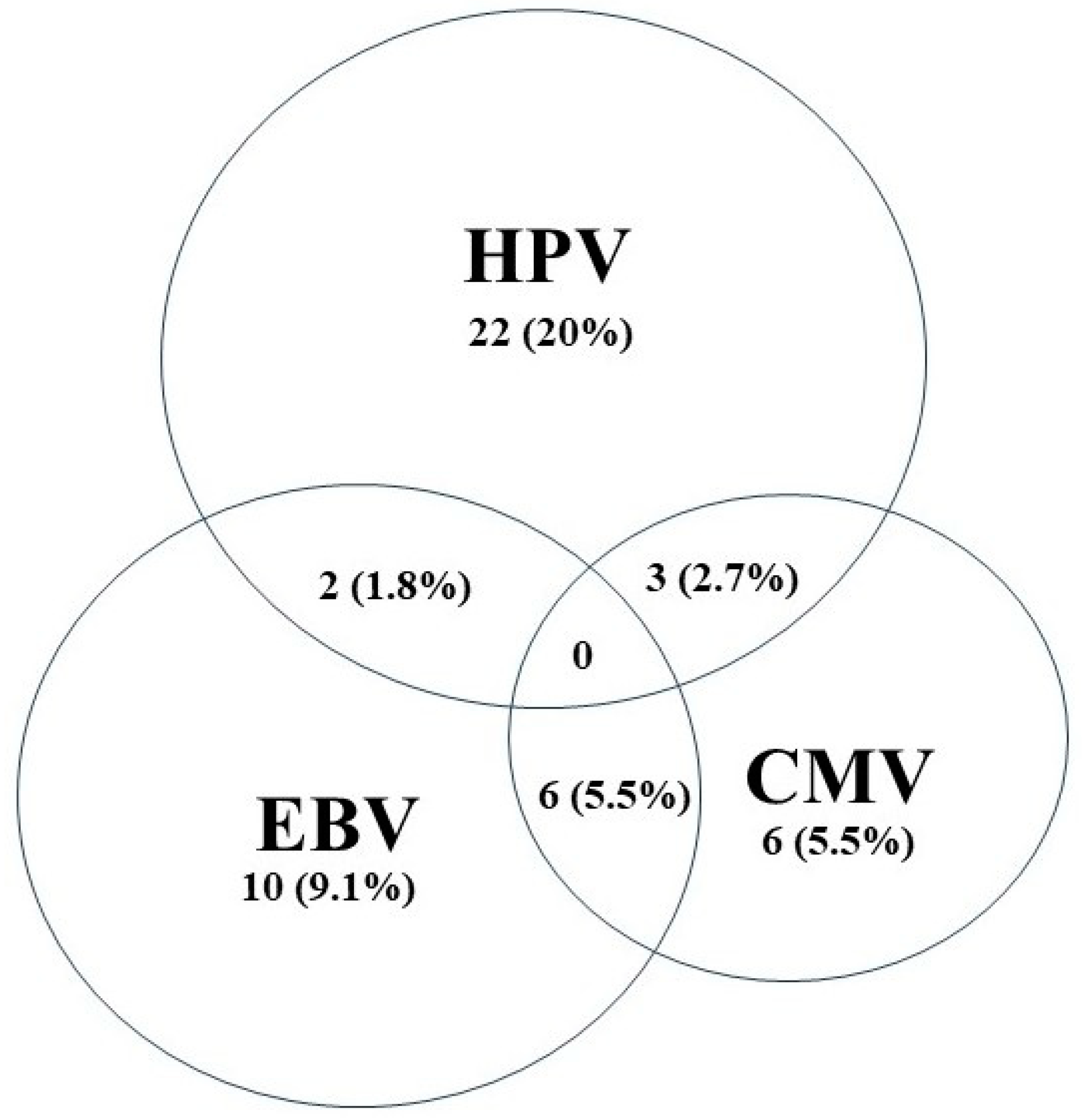
3.2. Detection of High-Risk HPV Subtypes, EBV, and CMV by RT-PCR

Our data show that 27 out of 110 samples tested positive for high-risk HPVs (HR-HPVs), accounting for 24.5% of the total sample number (Table 1). Among those, the most frequently detected HR-HPV subtype was HPV16, which was present in 23 (85.2%) cases, followed by HPV18 (n = 3, 11.1%), and other genotypes (one of the following HR-HPV genotypes 31, 33, 35, 39, 45, 51, 52, 56, 58, 59, 66, 68) seen in 3 (11.1%) samples. HPV double infection (HPV16 and others) was seen in two samples (7.4%) (Table 1).
On the other hand, we observed that 18 samples (16.4%) tested positive for EBV and 15 samples (13.6%) tested positive for CMV. More notably, our data show that HR-HPVs and EBV coinfection occurred in 1.8% (n = 2), HR-HPVs and CMV coinfection occurred in 2.7% (n = 3), and EBV and CMV coinfection occurred in 5.5% (n = 6) of the investigated BC cases (Figure 1).
Lastly, none of the normal breast samples showed positivity for HR-HPVs or CMV, with only one sample being positive for EBV. These variations among normal breast samples were statistically insignificant (p > 0.05).

3.3. Clinicopathological Association with HR-HPV, CMV, and EBV Positivity

An analytical association was conducted between each viral infection and the clinicopathological characteristics of the 110 tumor samples (Table 1). Among individuals aged ≤56 years, HPV was the most frequently observed, with 18 positive cases compared to 7 for CMV and 8 for EBV. Conversely, within the age group >56 years, an almost similar number of positive cases were found for each viral marker, with 9 for HPV, 8 for CMV, and 10 for EBV.
Among luminal A tumors, 20 were positive for HPV, which was the highest among the three screened viruses, compared to 10 and 11 positive cases for CMV and EBV, respectively. Similar findings were observed in luminal B cases, where HPV positivity was highest with four cases compared to two cases for each CMV and EBV. Among HER2+ cases, two were positive for HPV, while only one sample was positive for CMV or EBV. Notably, only one case of TNBC was positive for HPV, while two cases were positive for CMV and four for EBV.
Regarding clinical stage, 10 stage I cases exhibited positive HPV compared to 11 and 9 for CMV and EBV, respectively. In stage II samples, six were HPV positive, two for EBV, and no positive cases were observed for CMV. Among stage III tumors, there were four samples with positive HPV and three cases positive for CMV or EBV. In stage IV samples, HPV positivity was observed in seven cases, while only one case was positive for CMV and four for EBV.
HPV positivity was reported in 15 cases with lymphovascular invasion, whereas only 4 and 7 cases were reported positive for CMV and EBV, respectively. In contrast, among lymphovascular-invasion-free samples positivity rates were 12, 11, and 11 cases for HPV, CMV, and EBV, respectively. Among the subset where lymph nodes were involved, there were 18 cases positive for HPV, 7 for CMV, and 12 for EBV. Conversely, in cases that lacked lymph nodes involvement, nine were positive for HPV, seven for CMV, and five for EBV.
Out of the 110 tumor samples analyzed, 14 patients had a history of smoking. Among these, four patients tested positive for HPV, four patients tested positive for CMV, while only one patient tested positive for EBV. Conversely, among the 96 patients with no previous smoking history, 23, 11, and 17 tested positive for HPV, CMV, and EBV, respectively (Table 1).
Additionally, we investigated the relationship between family history and the presence of HPV, CMV, and EBV. We observed that the majority of HPV-positive cases (22 out of 27) had a positive family history of breast cancer. Conversely, most of the CMV-positive cases (17 out of 21) and EBV-positive cases (11 out of 18) were found in patients with no previous family history of breast cancer (Table 1).
Regarding the correlation between carcinoma in situ (CIS) and viral infections, we found that most cases were seen in patients lacking all three viral infections: 56 out of 83 in HPV-negative cases, 69 out of 95 for CMV-negative cases, and 70 out of 92 in EBV-negative cases. For tumor size correlation, 16 out of 27 HPV-positive cases, 7 out of 15 CMV-positive cases, and 11 out of 18 EBV-positive cases had tumors with dimensions exceeding 3 cm (Table 1).
Finally, we explored the relationship between HPV, CMV, EBV, and distant metastasis. The majority of samples positive for viral infections were observed among patients without distant metastasis (Table 1).
It is notable that a statistically significant association (p < 0.05) was observed between HPV status and age (p = 0.047), indicating that most HR-HPV infections (66.6%) occurred among younger aged patients. Clinical staging and CMV infection were also statistically significant (p = 0.015), highlighting that most CMV infections (73.3%) occurred in patients diagnosed with stage I BC (Table 1). Despite lacking statistical significance, it was noted that most cases of HR-HPV, CMV, and EBV infections were recorded in patients with grade II BC. CIS and HPV infection showed a statistically significant correlation indicating that CIS in BC is not necessarily linked to HPV infection or arises as a consequence of it, as observed in cervical cancer.

4. Discussion

This study represents the first investigation into the presence/co-presence of HR-HPVs, CMV, and EBV in human BC and its association with the immunohistochemical subtyping within a Jordanian subpopulation. Our findings indicate that HR-HPVs were detected in 24.5% of BC samples, whereas none of the normal breast samples tested positive for HR-HPVs. Previous studies have reported the presence of HR-HPVs in BC patients worldwide, including in samples from the Middle East and North Africa (MENA) region, with prevalence ranging from 21% to 65% [28,29,30,31]. Specifically, our results align with several reports from Egypt, Iran, and Jordan, where high rates of HR-HPVs (22.2%, 25.9%, and 21%, respectively) were observed [28,32,33]. However, it is noteworthy that in the Jordanian population, HPV prevalence seems to largely vary (range: 31–75%) in different types of cancers, including those closely associated with its infection such as oropharyngeal and cervical cancers, with HPV 16 being the most prominent [17,34]. HPV 16 genotype was the most predominant genotype in BC among Jordanian women in our study, similar to the Iraqi and Egyptian population [32,35]. HPV 18 genotype was reported as the most common in BC patients in Iran [29,33], and HPV 52 genotype among the Qatari and Lebanese populations [30,31]. Interestingly, the sole study examining the prevalence of BC among Jordanian women, conducted in 2020 by Al Hamad et al., reported HPV 18 as the most prevalent genotype in their studied subpopulation [28]. However, our findings are more consistent with all previous studies conducted in Jordan, which have collectively identified HPV 16 as the most predominant genotype among patients with cervical and oropharyngeal cancers [17,18,36,37,38].
Our findings are consistent with HPV genotypes that are commonly found in cancers worldwide where most cases are caused by genotypes 16 and 18. Likewise, a Canadian study revealed HPV 16 genotype as the most frequent in BC among Canadian women, whereas BC samples from the Qatari population did not detect the presence of HPV 16 [31,39]. Additionally, PCR analysis on BC samples from Syria detected HPV 33 genotype as the most prevalent subtype, followed by HPV genotypes 35, 18, and 16 [40]. Overall, the variation in HPV prevalence and genotype distribution can be attributed to different geographical distribution, sample size, and methodological differences. Among our cohort, only one patient infected with HPV was diagnosed with colon cancer and mucoepidermoid carcinoma of the parotid gland. Despite multiple studies showing relation between cervical lesions and the presence of HPV in BC, none of the patients with HPV infection in our study were diagnosed with cervical or oropharyngeal cancers [13].
Studies investigating the presence of CMV in BC patients in the MENA region are scarce. Worldwide, the detection of CMV among BC patients ranged from 0–100%. In the MENA region six studies were found: three in Iran, two in Egypt, and one in Iraq. In these studies, CMV was detected in 6.3–54% of the BC tissue compared to 0–28% in controls [41,42,43,44,45]. In our study, CMV was detected in 13.6% of the patients, which aligns with previous studies [42,43,44,46]. It is noteworthy that a New Zealand study revealed that CMV was completely absent in BC tissue samples from New Zealand women [47], while a study in China has shown that all BC cases were positive for CMV [48]. The differences in CMV distribution can be attributed to age, different socioeconomic status, hygiene practices, sexual behaviors, immune status, healthcare infrastructure, and geographical variations [49,50,51,52]. Table 2 summarizes the prevalence of CMV in BC tissue and controls among patients in the designated countries.
EBV presence is detected in approximately 30–50% of cancer cases globally. Studies from Turkey and Syria have reported EBV positivity rates of 58% and 52%, respectively, in BC cases, while reports from Qatar, Tunisia, Lebanon, and Egypt revealed EBV presence in 49%, 44%, 40%, and 37.5% of BC samples, respectively [23,30,31,53,54,55]. Additionally, EBV presence in BC tissue has been reported in Iran and Iraq in 27%, and 22.5%, respectively [56,57]. In our study, we found that EBV is present in approximately 16.4% of BC cases in Jordanian women. However, the mechanism by which EBV infects mammary epithelial cells is still not fully understood. A study by Hu et al. demonstrated that EBV infects mammary epithelial cells expressing CD21 and promotes the growth of early mammary epithelial cells with a stem cell phenotype [58]. EBV infection alters gene expression and stimulates oncogenic signaling via c-MET [58]. Moreover, EBV infection, along with activated RAS, triggers BC development; however, EBV is not essential once malignant transformation has occurred [58]. Our data suggest that the prevalence of EBV in BC tissues in Jordan is lower than its prevalence in the MENA and the world.
Table 2. Prevalence of CMV in BC tissue and controls among patients in the indicated countries.
CountryYearCMV Positive in BCCMV Positive in ControlsReference
New Zealand20150%3%Richardson et al. [47]
Egypt201818%5%El Shazly et al. [43]
China2018100%---Cui et al. [48]
Iraq20186.3%5%Kadhim et al. [56]
Iran201954%28%Sepahvand et al. [45]
Iran 202133%12%Fard et al. [41]
Iran202116.3%2%Nakhaie et al. [42]
Canada202218.4%10%Yang et al. [46]
Egypt202413.8%0%Elnegery et al. [44]
Previous studies indicate that the co-occurrence of HR-HPVs and EBV contributes to the onset and advancement of various cancers, including BC [59,60]. Our findings indicate that only 1.8% of BC cases in Jordanian women are co-infected with both high-risk HPVs and EBV. This contrasts with studies conducted in Qatar, Syria, and Lebanon, where high-risk HPVs and EBV were co-present in 47%, 32%, and 29% of BC samples, respectively [30,31,61]. Additionally, coinfection of high-risk HPVs and CMV was observed in 2.7% of cases, and EBV and CMV coinfection in 5.5% of BC cases. These results support the potential cooperative role of HR-HPVs and EBV oncoproteins in initiating and/or advancing various subtypes of human BC, as reported previously [31,54,62,63].

5. Conclusions

In conclusion, our findings highlight the predominance of HR-HPV infection among a Jordanian female subpopulation, and the co-existence of HR-HPV, CMV, and EBV coinfections suggesting an association with tumor grade and stage. This investigation proposes that an HPV vaccine could potentially be utilized to prevent the development and progression of specific subtypes of BC, yet the oncogenic role of HPV in BC requires further analysis. Moreover, future research involving larger cohorts in Jordan and the MENA region to validate the co-presence and HPV subtypes in BC is welcomed.

Author Contributions

Conceptualization, A.I.K. and T.S.; resources and data curation, A.I.K., N.H., S.A.S., M.A. and H.A.-M. (Hadeel Al-Momani), methodology, H.A.-M. (Hadeel Al-Momani), N.H., A.S. and U.S., writing—original draft preparation, A.I.K., U.S., S.A.S., M.R.A. and T.S.; writing—review and editing, A.I.K., T.S., S.A.S., N.H., H.A.-M. (Hadeel Al-Momani), M.A. and H.A.-M. (Hafez Al-Momani); supervision, A.I.K.; project administration, A.I.K. and N.H. All authors have read and agreed to the published version of the manuscript.

Funding

Work in Ashraf Khasawneh laboratory is supported by a grant provided by the Deanship of Scientific Research, The Hashemite University (grant no. 745/56/2022).

Institutional Review Board Statement

The study was conducted in accordance with the Declaration of Helsinki and approved by the Institutional Review Board (IRB) of The Hashemite University (No. 7/14/2020/2021, date of approval: 29 August 2021) and KAUH (No. 42/151/2022, date of approval: 12 September 2022).

Informed Consent Statement

Obtaining informed consents for this work was waived by both IRB protocols since all the samples used for this work were surplus (archived) breast tumor tissue samples, and that patients undergoing surgery or biopsy collection provide informed consent to donate any excess tissue (i.e., beyond that needed for diagnostic purposes).

Data Availability Statement

The data presented in this study are available from corresponding authors upon request.

Acknowledgments

All authors are highly thankful to the Researchers Supporting Project number (RSPD2024R786), King Saud University, Riyadh, Saudi Arabia. We would also like to thank Princess Iman Center for Research and Laboratory Sciences (JRMS) for providing the CMV- and EBV-positive controls. We would like to express our gratitude to Gordon Zubrod for his meticulous language and grammatical review of the manuscript.

Conflicts of Interest

The authors declare no conflicts of interest.

References

  1. Breast Cancer—IARC. Available online: https://www.iarc.who.int/cancer-type/breast-cancer/ (accessed on 7 March 2024).
  2. Jawad Hashim, M.; Al-Shamsi, F.A.; Al-Marzooqi, N.A.; Al-Qasemi, S.S.; Mokdad, A.H.; Khan, G. Burden of Breast Cancer in the Arab World: Findings from Global Burden of Disease, 2016. J. Epidemiol. Glob. Health 2018, 8, 54–58. [Google Scholar] [CrossRef]
  3. Jordan Cancer Registry 2023—Google Search. Available online: https://www.google.com/search?sca_esv=ed313d50f24d50b8&q=Jordan+cancer+registry+2023&sa=X&ved=2ahUKEwi15e_J5uKEAxWYUaQEHQznBXwQ1QJ6BAhJEAE&biw=1536&bih=730&dpr=1.25 (accessed on 7 March 2024).
  4. Winters, S.; Martin, C.; Murphy, D.; Shokar, N.K. Breast Cancer Epidemiology, Prevention, and Screening. Prog. Mol. Biol. Transl. Sci. 2017, 151, 1–32. [Google Scholar] [CrossRef]
  5. Zamaraev, A.V.; Zhivotovsky, B.; Kopeina, G.S. Viral Infections: Negative Regulators of Apoptosis and Oncogenic Factors. Biochemistry 2020, 85, 1191–1201. [Google Scholar] [CrossRef]
  6. Williams, V.M.; Filippova, M.; Soto, U.; Duerksen-Hughes, P.J. HPV-DNA Integration and Carcinogenesis: Putative Roles for Inflammation and Oxidative Stress. Future Virol. 2011, 6, 45–57. [Google Scholar] [CrossRef]
  7. Benites-Zapata, V.A.; Ulloque-Badaracco, J.R.; Alarcón-Braga, E.A.; Fernández-Alonso, A.M.; López-Baena, M.T.; Pérez-López, F.R. Telomerase Activity and Telomere Length in Women with Breast Cancer or without Malignancy: A Systematic Review and Meta-Analysis. Maturitas 2024, 180, 107882. [Google Scholar] [CrossRef]
  8. White, M.K.; Pagano, J.S.; Khalili, K. Viruses and Human Cancers: A Long Road of Discovery of Molecular Paradigms. Clin. Microbiol. Rev. 2014, 27, 463–481. [Google Scholar] [CrossRef]
  9. Saleh, T.; Khasawneh, A.I.; Himsawi, N.; Abu-Raideh, J.; Ejeilat, V.; Elshazly, A.M.; Gewirtz, D.A. Senolytic Therapy: A Potential Approach for the Elimination of Oncogene-Induced Senescent HPV-Positive Cells. Int. J. Mol. Sci. 2022, 23, 15512. [Google Scholar] [CrossRef]
  10. AK, G.; AB, R. The Invisible Enemy—How Human Papillomaviruses Avoid Recognition and Clearance by the Host Immune System. Open Virol. J. 2012, 6, 249–256. [Google Scholar] [CrossRef]
  11. Middeldorp, J.M.; Pegtel, D.M. Multiple Roles of LMP1 in Epstein-Barr Virus Induced Immune Escape. Semin. Cancer Biol. 2008, 18, 388–396. [Google Scholar] [CrossRef] [PubMed]
  12. Arias-Calvachi, C.; Blanco, R.; Calaf, G.M.; Aguayo, F. Epstein-Barr Virus Association with Breast Cancer: Evidence and Perspectives. Biology 2022, 11, 799. [Google Scholar] [CrossRef]
  13. Kudela, E.; Kudelova, E.; Kozubík, E.; Rokos, T.; Pribulova, T.; Holubekova, V.; Biringer, K. HPV-Associated Breast Cancer: Myth or Fact? Pathogens 2022, 11, 1510. [Google Scholar] [CrossRef]
  14. Geisler, J.; Touma, J.; Rahbar, A.; Söderberg-Nauclér, C.; Vetvik, K. A Review of the Potential Role of Human Cytomegalovirus (HCMV) Infections in Breast Cancer Carcinogenesis and Abnormal Immunity. Cancers 2019, 11, 1842. [Google Scholar] [CrossRef] [PubMed]
  15. Gao, A.; Kouznetsova, V.L.; Tsigelny, I.F. Bovine Leukemia Virus Relation to Human Breast Cancer: Meta-Analysis. Microb. Pathog. 2020, 149, 104417. [Google Scholar] [CrossRef]
  16. Lawson, J.S.; Glenn, W.K. Mouse Mammary Tumour Virus (MMTV) in Human Breast Cancer—The Value of Bradford Hill Criteria. Viruses 2022, 14, 721. [Google Scholar] [CrossRef]
  17. Khasawneh, A.I.; Himsawi, N.; Abu-Raideh, J.; Salameh, M.; Abdullah, N.; Khasawneh, R.; Saleh, T. Prevalence of Human Papillomavirus Associated with Head and Neck Squamous Cell Carcinoma in Jordanian Patients. Open Microbiol. J. 2020, 14, 57–64. [Google Scholar] [CrossRef]
  18. Khasawneh, A.I.; Asali, F.F.; Kilani, R.M.; Abu-Raideh, J.A.; Himsawi, N.M.; Salameh, M.A.; Al Ghabbiesh, G.H.; Saleh, T. Prevalence and Genotype Distribution of Human Papillomavirus among a Subpopulation of Jordanian Women. Int. J. Women’s Health Reprod. Sci. 2021, 9, 17–23. [Google Scholar] [CrossRef]
  19. Crombie, J.L.; LaCasce, A.S. Epstein Barr Virus Associated B-Cell Lymphomas and Iatrogenic Lymphoproliferative Disorders. Front. Oncol. 2019, 9, 436516. [Google Scholar] [CrossRef]
  20. Rovigatti, U.; Papaioannou, G.; Mammas, I.N.; Koutsaftiki, C.; Papatheodoropoulou, A.; Theodoridou, M.; Spandidos, D.A. Neuroblastoma in Childhood and Its Potential Viral Involvement: A Webinar by the Paediatric Virology Study Group. World Acad. Sci. J. 2022, 4, 16. [Google Scholar] [CrossRef]
  21. van Elsland, D.; Neefjes, J. Bacterial Infections and Cancer. EMBO Rep. 2018, 19, e46632. [Google Scholar] [CrossRef]
  22. Yahia, Z.A.; Adam, A.A.M.; Elgizouli, M.; Hussein, A.; Masri, M.A.; Kamal, M.; Mohamed, H.S.; Alzaki, K.; Elhassan, A.M.; Hamad, K.; et al. Epstein Barr Virus: A Prime Candidate of Breast Cancer Aetiology in Sudanese Patients. Infect. Agents Cancer 2014, 9, 9. [Google Scholar] [CrossRef]
  23. Aboulkassim, T.; Yasmeen, A.; Akil, N.; Batist, G.; Moustafa Al, A.E. Incidence of Epstein–Barr Virus in Syrian Women with Breast Cancer: A Tissue Microarray Study. Hum. Vaccin. Immunother. 2015, 11, 951. [Google Scholar] [CrossRef] [PubMed]
  24. Mofrad, M.G.; Kazeminezhad, B.; Faghihloo, E. Prevalence of Epstein-Barr Virus (EBV) in Iranian Breast Carcinoma Patients. Asian Pac. J. Cancer Prev. 2020, 21, 133–137. [Google Scholar] [CrossRef] [PubMed]
  25. Afzal, S.; Fiaz, K.; Noor, A.; Sindhu, A.S.; Hanif, A.; Bibi, A.; Asad, M.; Nawaz, S.; Zafar, S.; Ayub, S.; et al. Interrelated Oncogenic Viruses and Breast Cancer. Front. Mol. Biosci. 2022, 9, 781111. [Google Scholar] [CrossRef] [PubMed]
  26. Kadivar, M.; Monabati, A.; Joulaee, A.; Hosseini, N. Epstein-Barr Virus and Breast Cancer: Lack of Evidence for an Association in Iranian Women. Pathol. Oncol. Res. 2011, 17, 489–492. [Google Scholar] [CrossRef] [PubMed]
  27. Aguayo, F.; Khan, N.; Koriyama, C.; González, C.; Ampuero, S.; Padilla, O.; Solís, L.; Eizuru, Y.; Corvalán, A.; Akiba, S. Human Papillomavirus and Epstein-Barr Virus Infections in Breast Cancer from Chile. Infect. Agents Cancer 2011, 6, 7. [Google Scholar] [CrossRef] [PubMed]
  28. Al Hamad, M.; Matalka, I.; Al Zoubi, M.S.; Armogida, I.; Khasawneh, R.; Al-Husaini, M.; Sughayer, M.; Jaradat, S.; Al-Nasser, A.D.; Mazzanti, C.M. Human Mammary Tumor Virus, Human Papilloma Virus, and Epstein-Barr Infection Are Associated with Sporadic Breast Cancer. Breast Cancer 2020, 14, 1178223420976388. [Google Scholar] [CrossRef] [PubMed]
  29. Khodabandehlou, N.; Mostafaei, S.; Etemadi, A.; Ghasemi, A.; Payandeh, M.; Hadifar, S.; Hossein Norooznezhad, A.; Kazemnejad, A.; Moghoofei, M. Human Papilloma Virus and Breast Cancer: The Role of Inflammation and Viral Expressed Proteins. BMC Cancer 2019, 19, 61. [Google Scholar] [CrossRef] [PubMed]
  30. Nagi, K.; Gupta, I.; Jurdi, N.; Jabeen, A.; Yasmeen, A.; Batist, G.; Vranic, S.; Al-Moustafa, A.E. High-Risk Human Papillomaviruses and Epstein–Barr Virus in Breast Cancer in Lebanese Women and Their Association with Tumor Grade: A Molecular and Tissue Microarray Study. Cancer Cell Int. 2021, 21, 308. [Google Scholar] [CrossRef] [PubMed]
  31. Gupta, I.; Jabeen, A.; Al-Sarraf, R.; Farghaly, H.; Vranic, S.; Sultan, A.A.; Al Moustafa, A.E.; Al-Thawadi, H. The Co-Presence of High-Risk Human Papillomaviruses and Epstein-Barr Virus Is Linked with Tumor Grade and Stage in Qatari Women with Breast Cancer. Hum. Vaccin. Immunother. 2021, 17, 982. [Google Scholar] [CrossRef] [PubMed]
  32. El-Sheikh, N.; Mousa, N.O.; Tawfeik, A.M.; Saleh, A.M.; Elshikh, I.; Deyab, M.; Ragheb, F.; Moneer, M.M.; Kawashti, A.; Osman, A.; et al. Assessment of Human Papillomavirus Infection and Risk Factors in Egyptian Women with Breast Cancer. Breast Cancer 2021, 15, 1178223421996279. [Google Scholar] [CrossRef]
  33. Sigaroodi, A.; Nadji, S.A.; Naghshvar, F.; Nategh, R.; Emami, H.; Velayati, A.A. Human Papillomavirus Is Associated with Breast Cancer in the North Part of Iran. Sci. World J. 2012, 2012, 837191. [Google Scholar] [CrossRef] [PubMed]
  34. Sughayer, M.A.; Abdelhadi, M.; Abdeen, G.; Otay, L.; Dayeh, T. Human Papillomavirus Genotypes in Invasive Cervical Cancer in Jordan. Int. J. Gynecol. Obstet. 2010, 108, 74–75. [Google Scholar] [CrossRef] [PubMed]
  35. Ali, S.H.M.; Al-Alwan, N.A.S.; Al-Alwany, S.H.M. Detection and Genotyping of Human Papillomavirus in Breast Cancer Tissues from Iraqi Patients. East. Mediterr. Health J. 2014, 20, 372–377. [Google Scholar] [CrossRef] [PubMed]
  36. Bishtawi, M.; Saleh, H.; Khadra, M. Prevalence of HPV types in jordanian women with abnormal pap smear igcs19-0625 153 alarming rise in cervical cytological abnormalities in women living in jordan. Int. J. Gynecol. Cancer 2019, 29, A70. [Google Scholar] [CrossRef]
  37. Al-Qudah, M.A.; Al-Shaikh, A.F.; Haddad, H.K.; Elhassan, M.A.; Elhassan, O.B.; Dababneh, M.N.; Zaitoun, A.W.; Al Ghamdi, N.S.; Al-Najjar, B.Y. Prevalence and Detection of Sexually Transmitted Cases of Laryngeal Carcinoma Papilloma Virus · Cyclin-Dependent Kinase Inhibitor P16 · Cyclin B · Cyclin E · PCR. Head Neck Pathol. 2020, 14, 909–914. [Google Scholar] [CrossRef]
  38. Freij, M.A.; Saleh, H.H.; Farsakh, H.A.A.; Khadra, M.M.; Ijmail, A.A.; Rahal, B.O.; Waldali, M.H.; Najeeb, N.S.; Tahtamouni, L.H. Type-Specific Prevalence of Human Papillomavirus among Women with Abnormal Cytology in Jordan. Eur. J. Gynaecol. Oncol. 2017, 38, 901–904. [Google Scholar] [CrossRef]
  39. Yasmeen, A.; Bismar, T.A.; Kandouz, M.; Foulkes, W.D.; Desprez, P.-Y.; Al Moustafa, A.-E. E6/E7 of HPV Type 16 Promotes Cell Invasion and Metastasis of Human Breast Cancer Cells. Cell Cycle 2007, 6, 2038–2042. [Google Scholar] [CrossRef] [PubMed]
  40. Akil, N.; Yasmeen, A.; Kassab, A.; Ghabreau, L.; Darnel, A.D.; Al Moustafa, A.-E. High-Risk Human Papillomavirus Infections in Breast Cancer in Syrian Women and Their Association with Id-1 Expression: A Tissue Microarray Study. Br. J. Cancer 2008, 99, 404–407. [Google Scholar] [CrossRef]
  41. Fard, Z.T.; Khayamzadeh, M.; Zarei, M. Molecular Detection of Human Cytomegalovirus in Breast Cancer of Iranian Women Using Real-Time PCR. Int. J. Cancer Manag. 2021, 14, 115720. [Google Scholar] [CrossRef]
  42. Nakhaie, M.; Charostad, J.; Ghaderi-Zefrehi, H.; Arabzadeh, S.A.M.; Pourhosseini, F.; Makvandi, M. Prevalence of Human Cytomegalovirus in Breast Cancer: A Systematic Review. Asian Pac. J. Cancer Biol. 2021, 6, 161–169. [Google Scholar] [CrossRef]
  43. El Shazly, D.F.; Bahnassey, A.A.; Omar, O.S.; Elsayed, E.T.; Al-Hindawi, A.; El-Desouky, E.; Youssef, H.; Zekri, A.R.N. Detection of Human Cytomegalovirus in Malignant and Benign Breast Tumors in Egyptian Women. Clin. Breast Cancer 2018, 18, e629–e642. [Google Scholar] [CrossRef] [PubMed]
  44. Elnegery, A.A.; Abou Elela, M.A.; Mohamed, S.T.; Ragab, O.F.A.; Yussif, S.M.I.; Abou El-Khier, N.T. Human Cytomegalovirus and COX2 Expression among Women with Breast Tumors. Egypt. J. Med. Microbiol. 2024, 33, 73–82. [Google Scholar] [CrossRef]
  45. Sepahvand, P.; Makvandi, M.; Samarbafzadeh, A.; Talaei-Zadeh, A.; Ranjbari, N.; Nisi, N.; Azaran, A.; Jalilian, S.; Pirmoradi, R.; Makvandi, K.; et al. Human Cytomegalovirus DNA among Women with Breast Cancer. Asian Pac. J. Cancer Prev. 2019, 20, 2275. [Google Scholar] [CrossRef] [PubMed]
  46. Yang, Z.; Tang, X.; Hasing, M.E.; Pang, X.; Ghosh, S.; McMullen, T.P.W.; Brindley, D.N.; Hemmings, D.G. Human Cytomegalovirus Seropositivity and Viral DNA in Breast Tumors Are Associated with Poor Patient Prognosis. Cancers 2022, 14, 1148. [Google Scholar] [CrossRef] [PubMed]
  47. Richardson, A.K.; Currie, M.J.; Robinson, B.A.; Morrin, H.; Phung, Y.; Pearson, J.F.; Anderson, T.P.; Potter, J.D.; Walker, L.C. Cytomegalovirus and Epstein-Barr Virus in Breast Cancer. PLoS ONE 2015, 10, e0118989. [Google Scholar] [CrossRef] [PubMed]
  48. Cui, J.; Wang, Q.; Wang, H.B.; Wang, B.; Li, L. Protein and DNA Evidences of HCMV Infection in Primary Breast Cancer Tissues and Metastatic Sentinel Lymph Nodes. Cancer Biomark. 2018, 21, 769–780. [Google Scholar] [CrossRef] [PubMed]
  49. Richardson, A.K.; Walker, L.C.; Cox, B.; Rollag, H.; Robinson, B.A.; Morrin, H.; Pearson, J.F.; Potter, J.D.; Paterson, M.; Surcel, H.M.; et al. Breast Cancer and Cytomegalovirus. Clin. Transl. Oncol. 2020, 22, 585–602. [Google Scholar] [CrossRef] [PubMed]
  50. El Baba, R.; Herbein, G. Immune Landscape of CMV Infection in Cancer Patients: From “Canonical” Diseases Toward Virus-Elicited Oncomodulation. Front. Immunol. 2021, 12, 730765. [Google Scholar] [CrossRef] [PubMed]
  51. Balegamire, S.J.; McClymont, E.; Croteau, A.; Dodin, P.; Gantt, S.; Besharati, A.A.; Renaud, C.; Mâsse, B.; Boucoiran, I. Prevalence, Incidence, and Risk Factors Associated with Cytomegalovirus Infection in Healthcare and Childcare Worker: A Systematic Review and Meta-Analysis. Syst. Rev. 2022, 11, 131. [Google Scholar] [CrossRef]
  52. Lachmann, R.; Loenenbach, A.; Waterboer, T.; Brenner, N.; Pawlita, M.; Michel, A.; Thamm, M.; Poethko-Müller, C.; Wichmann, O.; Wiese-Posselt, M. Cytomegalovirus (CMV) Seroprevalence in the Adult Population of Germany. PLoS ONE 2018, 13, e0200267. [Google Scholar] [CrossRef]
  53. Kalkan, A.; Ozdarendeli, A.; Bulut, Y.; Yekeler, H.; Cobanoglu, B.; Doymaz, M.Z. Investigation of Epstein-Barr Virus DNA in Formalin-Fixed and Paraffin- Embedded Breast Cancer Tissues. Med. Princ. Pract. 2005, 14, 268–271. [Google Scholar] [CrossRef] [PubMed]
  54. Metwally, S.A.; Abo-Shadi, M.A.; Abdel Fattah, N.F.; Barakat, A.B.; Rabee, O.A.; Osman, A.M.; Helal, A.M.; Hashem, T.; Moneer, M.M.; Chehadeh, W.; et al. Presence of Hpv, Ebv and Hmtv Viruses among Egyptian Breast Cancer Women: Molecular Detection and Clinical Relevance. Infect. Drug Resist. 2021, 14, 2327–2339. [Google Scholar] [CrossRef] [PubMed]
  55. Mekrazi, S.; Kallel, I.; Jamai, D.; Yengui, M.; Khabir, A.; Gdoura, R. Epstein-Barr Virus in Breast Carcinoma and in Triple Negative Cases Impact on Clinical Outcomes. Pathol. Res. Pract. 2023, 245, 154484. [Google Scholar] [CrossRef] [PubMed]
  56. Shadood, H.K.A.L.; Atiya, S.A.-A.; Kardar, G.A.; Shadood, H.K.A.L.; Atiya, S.A.-A.; Kardar, G.A. Correlation of Breast Cancer with the Epstein Bar Virus and Human Cytomegalovirus Frequency and the Expression of Estrogen Receptor-Beta and IL-6 Receptor in Iraqi Women. Nat. Sci. 2018, 10, 182–192. [Google Scholar] [CrossRef]
  57. Farahmand, M.; Monavari, S.H.; Shoja, Z.; Ghaffari, H.; Tavakoli, M.; Tavakoli, A. Epstein–Barr Virus and Risk of Breast Cancer: A Systematic Review and Meta-Analysis. Future Oncol. 2019, 15, 2873–2885. [Google Scholar] [CrossRef] [PubMed]
  58. Hu, H.; Luo, M.-L.; Desmedt, C.; Nabavi, S.; Yadegarynia, S.; Hong, A.; Konstantinopoulos, P.A.; Gabrielson, E.; Hines-Boykin, R.; Pihan, G.; et al. Epstein-Barr Virus Infection of Mammary Epithelial Cells Promotes Malignant Transformation. EBioMedicine 2016, 9, 148–160. [Google Scholar] [CrossRef] [PubMed]
  59. Tempera, I.; Lieberman, P.M. Oncogenic Viruses as Entropic Drivers of Cancer Evolution. Front. Virol. 2021, 1, 753366. [Google Scholar] [CrossRef] [PubMed]
  60. Kannampuzha, S.; Gopalakrishnan, A.V.; Padinharayil, H.; Alappat, R.R.; Anilkumar, K.V.; George, A.; Dey, A.; Vellingiri, B.; Madhyastha, H.; Ganesan, R.; et al. Onco-Pathogen Mediated Cancer Progression and Associated Signaling Pathways in Cancer Development. Pathogens 2023, 12, 770. [Google Scholar] [CrossRef] [PubMed]
  61. Al Moustafa, A.-E.; Al-Antary, N.; Aboulkassim, T.; Akil, N.; Batist, G.; Yasmeen, A. Co-Prevalence of Epstein-Barr Virus and High-Risk Human Papillomaviruses in Syrian Women with Breast Cancer. Hum. Vaccin. Immunother. 2016, 12, 1936–1939. [Google Scholar] [CrossRef]
  62. Al-Thawadi, H.; Gupta, I.; Jabeen, A.; Skenderi, F.; Aboulkassim, T.; Yasmeen, A.; Malki, M.I.; Batist, G.; Vranic, S.; Al Moustafa, A.E. Co-Presence of Human Papillomaviruses and Epstein-Barr Virus Is Linked with Advanced Tumor Stage: A Tissue Microarray Study in Head and Neck Cancer Patients. Cancer Cell Int. 2020, 20, 361. [Google Scholar] [CrossRef]
  63. Fernandes, Q.; Gupta, I.; Vranic, S.; Al Moustafa, A.E. Human Papillomaviruses and Epstein–Barr Virus Interactions in Colorectal Cancer: A Brief Review. Pathogens 2020, 9, 300. [Google Scholar] [CrossRef] [PubMed]
Figure 1. Venn diagram depicting single and multiple infections with HPV, CMV, and EBV in Jordanian breast cancer samples (n = 110).
Medicina 60 00699 g001
Table 1. Clinicopathological association of HR-HPV, CMV, and EBV status among malignant breast samples (n = 110). HR-HPV: high-risk human papilloma virus; CMV: cytomegalovirus; EBV: Epstein–Barr virus; ER: estrogen receptor; PR: progesterone receptor; HER2: human epidermal growth factor receptor 2, LV: lymphovascular invasion; LN: lymph node involvement; CIS: carcinoma in situ; +: positive; −: negative. Bolded p values indicate statistical significance (p < 0.05).
HR-HPVCMVEBV
+p Value+p Value+p Value
Age≤5618380.0477490.7868480.612
>569458461044
Luminal AER+/PR+, HER2−20530.59510630.78311620.166
Luminal BER+/PR+, HER2+417219219
HER+ ER−/PR−, HER2+241515
TNBCER−/PR−, HER2−192846
StageStage I10380.72611370.0159390.841
Stage II612018216
Stage III411312312
Stage IV722128425
GradeGrade 10100.110370.147280.798
Grade 21842951951
Grade 3931337733
LVPresent15420.6664530.0517500.304
Not identified124111421142
LN Positive18560.4547670.13112621.000
Negative922724526
Smoking historyYes4100.7434100.0981130.459
No2373 1185 1779
Family historyPositive22640.2383110.5117130.057
Negative317 1775 1175
CISPositive2560.01112690.75511700.242
Negative2527 326 722
Tumor size>3 cm16390.3767781.00011440.440
≤3 cm1144 847 748
Disclaimer/Publisher’s Note: The statements, opinions and data contained in all publications are solely those of the individual author(s) and contributor(s) and not of MDPI and/or the editor(s). MDPI and/or the editor(s) disclaim responsibility for any injury to people or property resulting from any ideas, methods, instructions or products referred to in the content.

