
Physicochemical Properties of Inorganic and Hybrid Hydroxyapatite-Based Granules Modified with Citric Acid or Polyethylene Glycol

 , , ,
, , ,  , and
, and
Abstract
:1. Introduction
2. Results and Discussion
2.1. Phase Composition
2.2. Granules’ Chemical Composition
2.3. Specific Surface Area
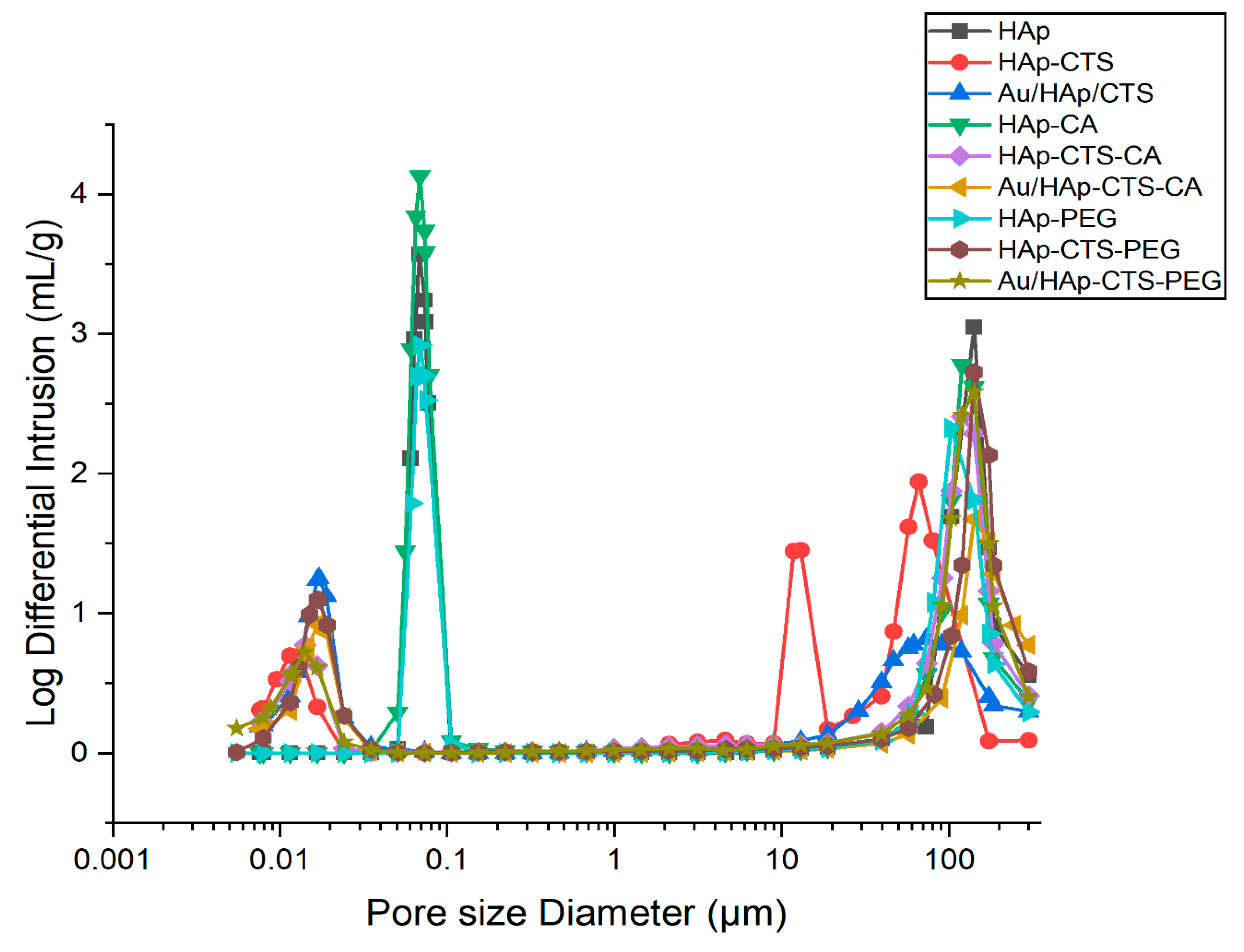
2.4. Open Porosity
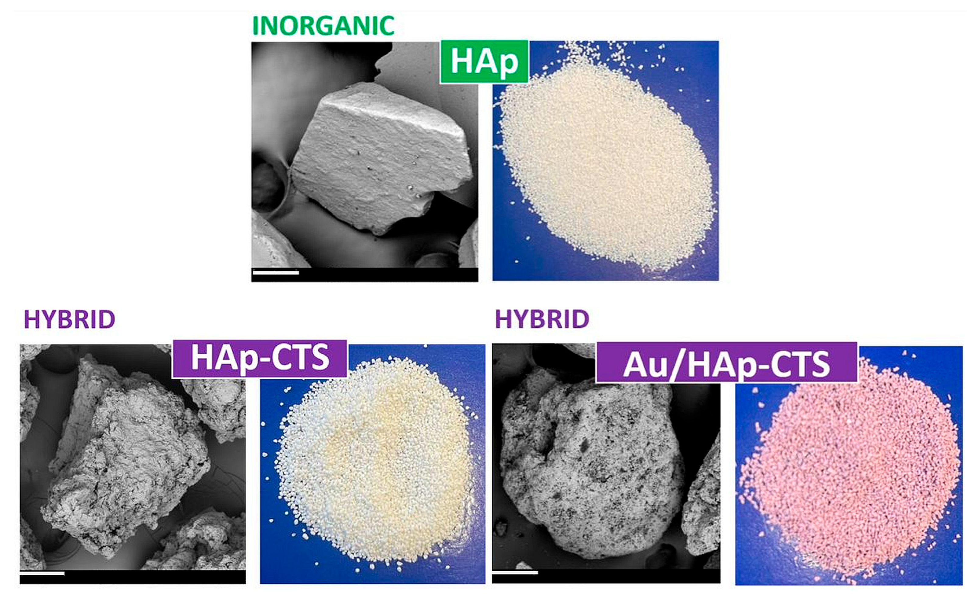
2.5. Microstructure
2.6. Biomicroconcretes’ Compressive Strength
3. Materials and Methods
3.1. Materials
3.1.1. Inorganic HAp Granules
3.1.2. Hybrid HAp-CTS Granules
3.1.3. Hybrid Au-HAp-CTS Granules
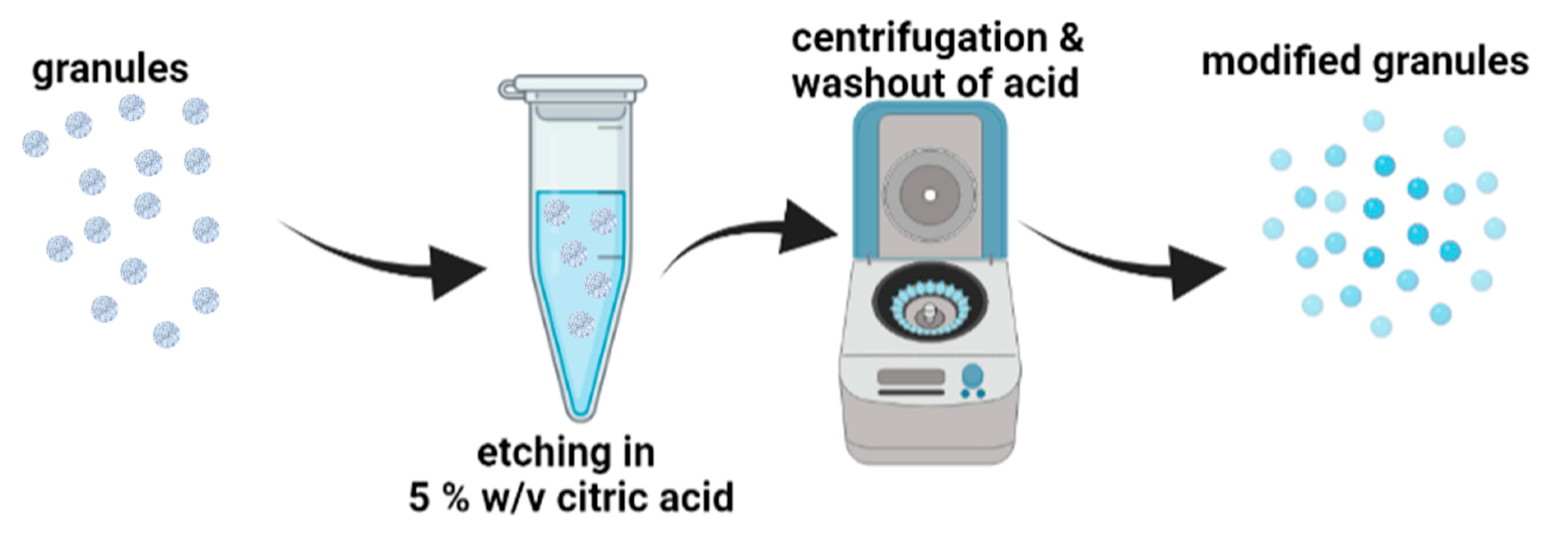
3.1.4. Modification of the Granules with Citric Acid
3.1.5. Modification of the Granules with Polyethylene Glycol
3.1.6. Granules/Tricalcium Phosphate-Based Biomicroconcretes
3.2. Methods
3.2.1. Phase Composition
3.2.2. Specific Surface Area
3.2.3. Open Porosity
3.2.4. Microstructure
3.2.5. FT-IR Analysis
3.2.6. Compressive Strength Measurements
4. Conclusions
Supplementary Materials
Author Contributions
Funding
Institutional Review Board Statement
Informed Consent Statement
Data Availability Statement
Acknowledgments
Conflicts of Interest
References
- We, B. A new calcium phosphate water setting cement. Cem. Res. Prog. 1986, 352–379. [Google Scholar]
- Takeyama, H.; Maruta, M.; Sato, T.; Kajimoto, N.; Fujii, E.; Matsuura, T.; Tsuru, K. Fabrication of bioresorbable hydroxyapatite bone grafts through the setting reaction of calcium phosphate cement. Dent. Mater. J. 2022, 41, 882–888. [Google Scholar] [CrossRef] [PubMed]
- Eliaz, N.; Metoki, N. Calcium phosphate bioceramics: A review of their history, structure, properties, coating technologies and biomedical applications. Materials 2017, 10, 334. [Google Scholar] [CrossRef] [PubMed]
- Schröter, L.; Kaiser, F.; Stein, S.; Gbureck, U.; Ignatius, A. Biological and mechanical performance and degradation characteristics of calcium phosphate cements in large animals and humans. Acta Biomater. 2020, 117, 1–20. [Google Scholar] [CrossRef] [PubMed]
- Czechowska, J.; Cichoń, E.; Belcarz, A.; Ślósarczyk, A.; Zima, A. Effect of gold nanoparticles and silicon on the bioactivity and antibacterial properties of hydroxyapatite/chitosan/tricalcium phosphate-based biomicroconcretes. Materials 2021, 14, 3854. [Google Scholar] [CrossRef] [PubMed]
- Ślósarczyk, A.; Czechowska, J.; Cichoń, E.; Zima, A. New hybrid bioactive composites for bone substitution. Processes 2020, 8, 335. [Google Scholar] [CrossRef]
- Sarkar, S.K.; Lee, B.Y.; Padalhin, A.R.; Sarker, A.; Carpena, N.; Kim, B.; Paul, K.; Choi, H.J.; Bae, S.H.; Lee, B.T. Brushite-based calcium phosphate cement with multichannel hydroxyapatite granule loading for improved bone regeneration. J. Biomater. Appl. 2016, 30, 823–837. [Google Scholar] [CrossRef] [PubMed]
- Fuchs, A.; Kreczy, D.; Brückner, T.; Gbureck, U.; Stahlhut, P.; Bengel, M.; Hoess, A.; Nies, B.; Bator, J.; Klammert, U.; et al. Bone regeneration capacity of newly developed spherical magnesium phosphate cement granules. Clin. Oral Investig. 2021, 1–15. [Google Scholar] [CrossRef] [PubMed]
- Zima, A. Hydroxyapatite-chitosan based bioactive hybrid biomaterials with improved mechanical strength. Spectrochim. Acta Part A Mol. Biomol. Spectrosc. 2018, 193, 175–184. [Google Scholar] [CrossRef]
- Czechowska, J. Self-assembling, hybrid hydroxyapatite-methylcellulose granules, modified with nano-silver. Mater. Lett. 2021, 300, 130156. [Google Scholar] [CrossRef]
- Dziadek, M.; Zima, A.; Cichoń, E.; Czechowska, J.; Ślósarczyk, A. Biomicroconcretes based on the hybrid HAp/CTS granules, α-TCP and pectins as a novel injectable bone substitutes. Mater. Lett. 2020, 265, 127457. [Google Scholar] [CrossRef]
- Martău, G.A.; Mihai, M.; Vodnar, D.C. The use of chitosan, alginate, and pectin in the biomedical and food sector—Biocompatibility, bioadhesiveness, and biodegradability. Polymers 2019, 11, 1837. [Google Scholar] [CrossRef] [PubMed]
- Garg, U.; Chauhan, S.; Nagaich, U.; Jain, N. Current advances in chitosan nanoparticles based drug delivery and targeting. Adv. Pharm. Bull. 2019, 9, 195. [Google Scholar] [CrossRef] [PubMed]
- Pańtak, P.; Cichoń, E.; Czechowska, J.; Zima, A. Influence of natural polysaccharides on properties of the biomicroconcrete-type bioceramics. Materials 2021, 14, 7496. [Google Scholar] [CrossRef] [PubMed]
- Lee, H.J.; Kim, B.; Padalhin, A.R.; Lee, B.T. Incorporation of chitosan-alginate complex into injectable calcium phosphate cement system as a bone graft material. Mater. Sci. Eng. C 2019, 94, 385–392. [Google Scholar] [CrossRef] [PubMed]
- Czechowska, J.; Zima, A.; Ślósarczyk, A. Comparative study on physicochemical properties of alpha-TCP/calcium sulphate dihydrate biomicroconcretes containing chitosan, sodium alginate or methylcellulose. Acta Bioeng. Biomech. 2020, 22, 47–56. [Google Scholar] [CrossRef] [PubMed]
- Skibiński, S.; Czechowska, J.P.; Cichoń, E.; Seta, M.; Gondek, A.; Cudnoch-Jędrzejewska, A.; Zima, A. Study on βTCP/P (3HB) Scaffolds—Physicochemical Properties and Biological Performance in Low Oxygen Concentration. Int. J. Mol. Sci. 2022, 23, 11587. [Google Scholar] [CrossRef] [PubMed]
- Nakonieczny, D.S.; Slíva, A.; Paszenda, Z.; Hundáková, M.; Kratošová, G.; Holešová, S.; Simha Martynková, G. Simple approach to medical grade alumina and zirconia ceramics surface alteration via acid etching treatment. Crystals 2021, 11, 1232. [Google Scholar] [CrossRef]
- Kazimierczak, P.; Przekora, A. Osteoconductive and osteoinductive surface modifications of biomaterials for bone regeneration: A concise review. Coatings 2020, 10, 971. [Google Scholar] [CrossRef]
- Kowalczyk, P.; Wojasiński, M.; Jaroszewicz, J.; Kopeć, K.; Ciach, T. Controlled formation of highly porous polylactic acid-calcium phosphate granules with defined structure. Biomater. Adv. 2023, 144, 213195. [Google Scholar] [CrossRef] [PubMed]
- Siniscalco, D.; Dutreilh-Colas, M.; Hjezi, Z.; Cornette, J.; El Felss, N.; Champion, E.; Damia, C. Functionalization of hydroxyapatite ceramics: Raman mapping investigation of silanization. Ceramics 2019, 2, 372–384. [Google Scholar] [CrossRef]
- Aneb, K.; Oudadesse, H.; Khireddine, H.; Lefeuvre, B.; Merdrignac-Conanec, O.; Tessier, F.; Lucas, A. Study of the effect of ordered porosity and surface silanization on in vitro bioactivity of sol-gel-derived bioactive glasses. Mater. Today Commun. 2023, 34, 104992. [Google Scholar] [CrossRef]
- de los Ángeles Ramírez, M.; Bindini, E.; Moretti, P.; Illia, G.J.S.; Amenitsch, H.; Andreozzi, P.; Moya, S.E. Impact of PEGylation on the degradation and pore organization in mesoporous silica nanoparticles: A study of the inner mesoporous structure in physiologically relevant ionic conditions. Colloids Surf. B Biointerfaces 2022, 219, 112797. [Google Scholar]
- Ali, M.; Mujtaba-ul-Hassan, S.; Ahmad, J.; Khurshid, A.; Shahzad, F.; Iqbal, Z.; Waheed, K. Fabrication of PEGylated Porous Alumina Whiskers (PAW) for drug delivery applications. Mater. Lett. 2019, 241, 23–26. [Google Scholar] [CrossRef]
- Kumar, P.; Saini, M.; Kumar, V.; Dehiya, B.S.; Sindhu, A.; Fouad, H.; Ahmad, N.; Mahmood, A.; Hashem, M. Polyethylene glycol (PEG) modified porous Ca5 (PO4) 2SiO4 bioceramics: Struc-tural, morphologic and bioactivity analysis. Coatings 2020, 10, 538. [Google Scholar] [CrossRef]
- Gyawali, D.; Nair, P.; Zhang, Y.; Tran, R.T.; Zhang, C.; Samchukov, M.; Yang, J. Citric acid-derived in situ crosslinkable biodegradable polymers for cell delivery. Biomaterials 2010, 31, 9092–9105. [Google Scholar] [CrossRef]
- Reena, R.; Sindhu, R.; Balakumaran, P.A.; Pandey, A.; Awasthi, M.K.; Binod, P. Insight into citric acid: A versatile organic acid. Fuel 2022, 327, 125181. [Google Scholar] [CrossRef]
- Salihu, R.; Abd Razak, S.I.; Zawawi, N.A.; Kadir, M.R.A.; Ismail, N.I.; Jusoh, N.; Nayan, N.H.M. Citric acid: A green cross-linker of biomaterials for biomedical applications. Eur. Polym. J. 2021, 146, 110271. [Google Scholar] [CrossRef]
- Tran, R.T.; Yang, J.; Ameer, G.A. Citrate-based biomaterials and their applications in regenerative engineering. Annu. Rev. Mater. Res. 2015, 45, 277–310. [Google Scholar] [CrossRef] [PubMed]
- Barbour, M.E.; Parker, D.M.; Allen, G.C.; Jandt, K.D. Enamel dissolution in citric acid as a function of calcium and phosphate concentrations and degree of saturation with respect to hydroxyapatite. Eur. J. Oral Sci. 2003, 111, 428–433. [Google Scholar] [CrossRef] [PubMed]
- Ibrahim, M.; Ramadan, E.; Elsadek, N.E.; Emam, S.E.; Shimizu, T.; Ando, H.; Ishida, T. Polyethylene glycol (PEG): The nature, immunogenicity, and role in the hypersensitivity of PEGylated products. J. Control. Release 2022, 351, 215–230. [Google Scholar] [CrossRef]
- Assis, C.M.D.; Vercik, L.C.D.O.; Santos, M.L.D.; Fook, M.V.L.; Guastaldi, A.C. Comparison of crystallinity between natural hydroxyapatite and synthetic cp-Ti/HA coatings. Mater. Res. 2005, 8, 207–211. [Google Scholar] [CrossRef]
- Panda, P.K.; Yang, J.M.; Chang, Y.H.; Su, W.W. Modification of different molecular weights of chitosan by p-Coumaric acid: Preparation, characterization and effect of molecular weight on its water solubility and antioxidant property. Int. J. Biol. Macromol. 2019, 136, 661–667. [Google Scholar] [CrossRef] [PubMed]
- Pashameah, R.A.; Ibrahium, H.A.; Awwad, N.S.; Farea, M.O.; Ahmed, H.A.; El-Morsy, M.A.; Menazea, A.A. Modification and development of the optical, structural, thermal and electrical characterization of Chitosan incorporated with Au/Bi2O3/Mo NPs fabricated by laser ablation. J. Inorg. Organomet. Polym. Mater. 2022, 32, 2729–2736. [Google Scholar] [CrossRef]
- Vijayan, A.A.S.; Kumar, G.S.V. PEG grafted chitosan scaffold for dual growth factor delivery for enhanced wound healing. Sci. Rep. 2019, 9, 19165. [Google Scholar] [CrossRef] [PubMed]
- Rydén, L.; Omar, O.; Johansson, A.; Jimbo, R.; Palmquist, A.; Thomsen, P. Inflammatory cell response to ultra-thin amorphous and crystalline hydroxyapatite surfaces. J. Mater. Sci. Mater. Med. 2017, 28, 9. [Google Scholar] [CrossRef]
- Gawish, S.M.; Abo El-Ola, S.M.; Ramadan, A.M.; Abou El-Kheir, A.A. Citric acid used as a crosslinking agent for the grafting of chitosan onto woolen fabric. J. Appl. Polym. Sci. 2012, 123, 3345–3353. [Google Scholar] [CrossRef]
- Khouri, J. Chitosan Edible Films Crosslinked by Citric Acid. 2019. Available online: http://hdl.handle.net/10012/14877 (accessed on 1 July 2023).
- Lazaro-Carrillo, A.; Filice, M.; Guillén, M.J.; Amaro, R.; Viñambres, M.; Tabero, A.; Marciello, M. Tailor-made PEG coated iron oxide nanoparticles as contrast agents for long lasting magnetic resonance molecular imaging of solid cancers. Mater. Sci. Eng. C 2020, 107, 110262. [Google Scholar] [CrossRef] [PubMed]
- Jegatheeswaran, S.; Sundrarajan, M. PEGylation of novel hydroxyapatite/PEG/Ag nanocomposite particles to improve its antibacterial efficacy. Mater. Sci. Eng. C 2015, 51, 174–181. [Google Scholar] [CrossRef] [PubMed]
- Feng, D.; Feng, Y.; Li, P.; Zang, Y.; Wang, C.; Zhang, X. Modified mesoporous silica filled with PEG as a shape-stabilized phase change materials for improved thermal energy storage performance. Microporous Mesoporous Mater. 2020, 292, 109756. [Google Scholar] [CrossRef]
- Vallet-Regí, M.; Balas, F.; Arcos, D. Mesoporous materials for drug delivery. Angew. Chem. Int. Ed. 2007, 46, 7548–7558. [Google Scholar] [CrossRef] [PubMed]
- Misra, D.N. Interaction of citric acid with hydroxyapatite: Surface exchange of ions and precipitation of calcium citrate. J. Dent. Res. 1996, 75, 1418–1425. [Google Scholar] [CrossRef] [PubMed]
- Li, S.H.; de Wijn, J.R.; Layrolle, P.; de Groot, K. Novel method to manufacture porous hydroxyapatite by dual-phase mixing. J. Am. Ceram. Soc. 2003, 86, 65–72. [Google Scholar] [CrossRef]
- Bégin, A.; Van Calsteren, M.R. Antimicrobial films produced from chitosan. Int. J. Biol. Macromol. 1999, 26, 63–67. [Google Scholar] [CrossRef] [PubMed]
- Khouri, J.; Penlidis, A.; Moresoli, C. Viscoelastic properties of crosslinked chitosan films. Processes 2019, 7, 157. [Google Scholar] [CrossRef]
- Utomo, J.; Noerjannah, L.I.; Rohmah, N.Z. Physical and mechanical properties of hydroxyapatite/polyethylene glycol nanocomposites. Mater. Today Proc. 2021, 44, 3263–3267. [Google Scholar]
- Nazarian, A.; Von Stechow, D.; Zurakowski, D.; Müller, R.; Snyder, B.D. Bone volume fraction explains the variation in strength and stiffness of cancellous bone affected by metastatic cancer and osteoporosis. Calcif. Tissue Int. 2008, 83, 368–379. [Google Scholar] [CrossRef]
- Kopperdahl, D.L.; Keaveny, T.M. Yield strain behavior of trabecular bone. J. Biomech. 1998, 31, 601–608. [Google Scholar] [CrossRef] [PubMed]
- Chung, E.J.; Sugimoto, M.J.; Ameer, G.A. The role of hydroxyapatite in citric acid-based nanocomposites: Surface characteristics, degradation, and osteogenicity in vitro. Acta Biomater. 2011, 7, 4057–4063. [Google Scholar] [CrossRef] [PubMed]
- Wang, S.; Xu, C.; Yu, S.; Wu, X.; Jie, Z.; Dai, H. Citric acid enhances the physical properties, cytocompatibility and osteogenesis of magnesium calcium phosphate cement. J. Mech. Behav. Biomed. Mater. 2019, 94, 42–50. [Google Scholar] [CrossRef] [PubMed]
- Rhodes, A.; Sandhu, S.S.; Onis, S.J. Surface modification of biomaterials by covalent binding of poly (ethylene glycol)(PEG). Surf. Modif. Biomater. 2011, 39–55. [Google Scholar] [CrossRef]
- Shen, C.C.; Hsu, S.H.; Chang, K.B.; Yeh, C.A.; Chang, H.C.; Tang, C.M.; Hung, H.S. Physical gold nanoparticle-decorated polyethylene glycol-hydroxyapatite composites guide osteogenesis and angiogenesis of mesenchymal stem cells. Biomedicines 2021, 9, 1632. [Google Scholar] [CrossRef] [PubMed]
- Anwar, A.; Siddiqui, R.; Shah, M.; Khan, N. Gold nanoparticles conjugation enhances antiacanthamoebic properties of nystatin, fluconazole, and amphotericin B. J. Microbiol. Biotechnol. 2019, 29, 171–177. [Google Scholar] [CrossRef] [PubMed]
- Gu, X.; Xu, Z.; Gu, L.; Xu, H.; Han, F.; Chen, B.; Pan, X. Preparation and anti-bacterial properties of gold nanoparticles: A review. Environ. Chem. Lett. 2021, 19, 167–187. [Google Scholar]
- Ślósarczyk, A. Ceramic Implantation Material and the Method of Obtaining of Ceramic Implantation Material. Polish patent PL154957B1, 18 April 1988. [Google Scholar]


| Specific Surface Area [m2/g] | |||
|---|---|---|---|
| Granules | HAp | HAp-CTS | Au/HAp-CTS |
| Non-modified | 22.36 ± 0.02 | 93.48 ± 0.26 | 82.41 ± 0.18 |
| CA-modified | 26.32 ± 0.05 | 74.54 ± 0.13 | 66.94 ± 0.11 |
| PEG-modified | 23.99 ± 0.05 | 79.51 ± 0.17 | 111.10 ± 0.21 |
| Porosity [vol.%] | Material | ||
|---|---|---|---|
| HAp | HAp-CTS | Au/HAp-CTS | |
| Non-modified | 70 ± 2 | 68 ± 1 | 63 ± 2 |
| CA-modified | 75 ± 1 | 63 ± 2 | 57 ± 3 |
| PEG-modified | 68 ± 3 | 70 ± 2 | 70 ± 1 |
Disclaimer/Publisher’s Note: The statements, opinions and data contained in all publications are solely those of the individual author(s) and contributor(s) and not of MDPI and/or the editor(s). MDPI and/or the editor(s) disclaim responsibility for any injury to people or property resulting from any ideas, methods, instructions or products referred to in the content. |
© 2024 by the authors. Licensee MDPI, Basel, Switzerland. This article is an open access article distributed under the terms and conditions of the Creative Commons Attribution (CC BY) license (https://creativecommons.org/licenses/by/4.0/).
Share and Cite
Cichoń, E.; Kosowska, K.; Pańtak, P.; Czechowska, J.P.; Zima, A.; Ślósarczyk, A. Physicochemical Properties of Inorganic and Hybrid Hydroxyapatite-Based Granules Modified with Citric Acid or Polyethylene Glycol. Molecules 2024, 29, 2018. https://0-doi-org.brum.beds.ac.uk/10.3390/molecules29092018
Cichoń E, Kosowska K, Pańtak P, Czechowska JP, Zima A, Ślósarczyk A. Physicochemical Properties of Inorganic and Hybrid Hydroxyapatite-Based Granules Modified with Citric Acid or Polyethylene Glycol. Molecules. 2024; 29(9):2018. https://0-doi-org.brum.beds.ac.uk/10.3390/molecules29092018
Chicago/Turabian StyleCichoń, Ewelina, Karolina Kosowska, Piotr Pańtak, Joanna P. Czechowska, Aneta Zima, and Anna Ślósarczyk. 2024. "Physicochemical Properties of Inorganic and Hybrid Hydroxyapatite-Based Granules Modified with Citric Acid or Polyethylene Glycol" Molecules 29, no. 9: 2018. https://0-doi-org.brum.beds.ac.uk/10.3390/molecules29092018